Quantitative analysis of inflammatory and
adhesion molecules in lungs of neonates with chronic
lung disease (bronchopulmonary dysplasia)
receiving mechanical ventilation
Análise quantitativa de moléculas inflamatórias e de adesão em pulmões
de neonatos com doença pulmonar crônica (displasia broncopulmonar)
submetidos à ventilação mecânica
Cristina T. Okamoto; Carlos F. Oldenburg Neto; Sandra Mara Witkowski; Ana Paula Percicote; Luca R. Pasqualotto; Gabriela Troiano; Tammy Almeida; Cleber M. Souza; Lúcia de Noronha
Pontifícia Universidade Católica do Paraná (PUCPR), Paraná, Brazil.
First submission on 07/04/16; last submission on 01/06/16; accepted for publication on 13/06/16; published on 20/08/16
ABSTRACT
Introduction: Chronic lung disease (CLD), clinically known as bronchopulmonary dysplasia (BPD), is a major cause of morbidity in premature newborn and were submitted to oxygen therapy. Objective: Immunohistochemical identiication of inlammatory molecules in the lung tissue of premature neonates that died with CLD. Methods: Immunohistochemical analysis of 51 samples of premature newborn lungs – grouped in: without CLD, “classic” CLD and “new” CLD. Results: Neutrophil inlux and the number of CD4+ and CD45RO+
cells were higher in the “classic” CLD group (p < 0.001). Conclusion: Our indings suggest that the inlammatory response is mediated by neutrophils and CD45RO+ and CD4+ T lymphocytes in the “classic” CLD.
Key words: bronchopulmonary dysplasia; immunochemistry; premature birth; oxygen therapy; neonatology.
INTRODUCTION
Chronic lung disease (CLD) or, clinically, bronchopulmonary dysplasia (BPD) is a chronic neonatal pulmonary disorder and a major cause of morbidity affecting premature newborn(1-3). The
classic presentation of this disorder was described in 1960 as a consequence of respiratory distress syndrome, the use of higher concentrations of inspired oxygen and aggressive mechanical ventilation produced characteristic radiological changes. Histological indings of “classic” CLD are emphysema, atelectasis, ibrosis, squamous metaplasia and smooth muscle hypertrophy in the airways and pulmonary vasculature(4).
The use of prenatal corticosteroids therapy, exogenous surfactant and technological advances in mechanical ventilation have resulted in a signiicant increase in the survival rates of
newborn with very low birth weight; however, the incidence of CLD has not changed as expected(4-7). Instead, another type of CLD,
known as “new” CLD emerged, particularly in newborn with less than 30 weeks of gestational age who continued to require supplemental oxygen after 28 days of age(3-6).
In 2001, a consensus suggested that the clinical criteria for the diagnosis of BPD/CLD (“new” or “classic”) in a preterm newborn must depend on the use of respiratory support at 28 days of postnatal life or 36 weeks postmenstrual age(3, 6-9). Although the pathological
criteria for “classic” CLD are characterized by an intense inlammatory response and septal ibrosis, in the “new” CLD there is decreased alveolar septation and abnormal pulmonary vascular development compatible with impaired pulmonary growth. Septal ibrosis, inlammatory response and interstitial edema are rarely observed. The alterations in alveolar formation may be very subtle and sometimes not detectable in routine morphological tests.
The characteristics of the “new” CLD may be more related to morphometric data than to the morphological aspects(10-14).
Over the last 10 years, the pathological basis of CLD have predominantly been associated with the following two processes: inlammation and arrest of lung development(2, 4, 5). The
inlammatory response in CLD is exacerbated by mechanical ventilation and exposure to supplemental oxygen. Several studies seem to indicate that neutrophils prevail in the earliest stages of CLD, followed by an abundance of macrophages and other inlammatory cells, increased pulmonary vascular permeability and damage to endothelial and epithelial cells, typically leading to apoptosis(15-21).
The objectives of this study were to investigate the subtypes of inlammatory cells and adhesion molecules, using immunohistochemistry in lung samples of preterm newborn receiving mechanical ventilation (less than 34 weeks gestational age) and to correlate the immunoexpression of these biomarkers with the presence or absence of CLD.
METHODS
Sample selection
The sample consisted of 51 specimens of preterm newborn lung autopsies performed between 1994 and 2005 (after the pre-surfactant era); from the archives of the Department of Pathology Medicine of Universidade Federal do Paraná (UFPR). The research ethics board of the Hospital das Clínicas (HC)-UFPR reviewed and approved the study (registration number 1099.138/2005-08; approved on August 30, 2005). The following inclusion criteria were used: complete autopsy; absence of major malformations; absence of meconium aspiration; less than 34 weeks of gestational age and use of oxygen therapy for at least 2 hours. The samples that were unsuitable for testing or for which the medical records were not available were excluded. The medical records were analyzed to collect data related to antenatal risk factor for CLD including gender, gestational age, birth weight, maternal age, number of pregnancies, prenatal care, hypertensive event in pregnancy, gestacional diabetes, chorioamnionitis, premature amniotic sac disruption, type of birth and twin pregnancy. Additionally, other potential postnatal risk factors for CLD include patent ductus arteriosus, cardiopulmonary resuscitation, Apgar score at the irst e ifth minute, antibiotic therapy, surfactant replacement therapy, necrotizing enterocolitis, bronchopneumonia, pulmonary hemorrhage, pulmonary hypertension, choking, intracranial hemorrhage, pneumothorax, sepsis and corrected
gestational postnatal age were recorded. Survival time and cause of death were also recorded(10).
Morphometric analysis
The slides of the 51 patients were stained with hematoxylin and eosin (HE) and divided into the following three groups: 1) histopathological indings of “classic” CLD (n = 11); 2) histopathological and morphometric indings of “new” CLD
(n = 5); and 3) “without” histopathological and morphometric criteria for CLD (n = 35). Since the alterations in alveolar
formation may be very subtle, the samples of groups 2 and 3 were analyzed only to conirm the existence of the morphometric criteria of “new” CLD in group 2(9, 10, 22, 23).
The digital images of 10 nonadjacent ields (200× = 106 micrometers in diameter) in slides of the pulmonary parenchyma from the 40 cases (group 2 and 3) were captured using Image Pro Plus software™ (Rockville, MD, USA). It measures the alveolar perimeter in micrometers and the number of alveoli per ield(10, 22, 23). The alveoli in each of the 10 digitalized ields were
measured. Means and standard deviations for each case were calculated. One blinded observer performed these analyses(10, 23).
Immunohistochemical staining
Parafin blocks of the studied cases (n = 51) were used to
construct the tissue microarrays (TMAs)(23). Lung samples from
all the patients were inserted into multi-sample parafin blocks (TMAs), with four samples of each case and 3 mm diameter for each sample (analyzed area = 28.26 mm2). Histological
Staining was interpreted as follows: for anti-CD4, CD8, CD14, CD20, CD25, CD74 and CD45RO antibodies, the number of positive cells in four random high-power ields (400× = 53 micrometers in diameter) were counted for each of the four samples of each case(23, 24). The cells that had a characteristic
membrane-staining pattern when observed under Olympus BX50 microscope (Olympus Optical Co, Ltd., Japan) and which were located in the alveolar septum or intra-alveolar space were considered positive. The cells in the intravascular space or in regions that usually contain peribronchial lymphoid aggregates were disregarded(24).
For the anti-ICAM-1 (CD54) and anti-VCAM (CD106) antibodies, the slides were assigned a positive score (0 = negative; 1 = weak positive; 2 = strong positive), and the positive score for each case was obtained by the sum of the scores for each of the four samples for this case. The stains were considered positive if they were observed in alveolar septa vessels and in macrophages/ monocytes in the septa and alveolar lumina. The positivity of these reactions was assessed in all ields of the four samples of each case using the Olympus BX50 microscope (Olympus Optical Co, Ltd., Japan) at 400× magniication(23).
Statistical methods
Data were analyzed using SPSS 20.0 (IBM, São Paulo, SP, Brazil). Kruskall-Wallis and nonparametric Mann-Whitney test were used for testing quantitative variables. Categorical variables were analyzed using Pearson’s chi-square test. Analysis of covariance (Ancova) was used for gestational age adjustment. Statistical signiicance was accepted for p values < 0.05.
RESULTS
Histopathological and morphometric data
In group 1 (“classic” CLD), the predominant histopathological characteristics were hyaline membranes and interstitial ibrosis(10).
Cases with “new” CLD (group 2) had minimal histopathological alterations, including subtle septal edema. The alveolar changes caused by insuflation and a very subtle inlammatory response were present in these cases(10). Since
the histological indings for the group with “new” CLD were often indistinguishable from those for the group “without” CLD (group 3), a morphometric analysis was required, which showed
TABLE 1 − The number of alveoli and the alveolar perimeter (in micrometers)
in the “without” CLD and “new” CLD groups
Parameters New CLD
*
(n = 5)
Without CLD*
(n = 35) p value Number of alveoli 40.9 ± 15.1 57.7 ± 16.2 0.033§
Alveolar perimeter‡ 481.5 ± 182.4 611.3 ± 206.2 0.018† *: average ± standard deviation; §:Mann-Whitney non-parametric test (p < 0.05); ‡:
square micrometers; †: Ancova (p < 0.05) (gestational age adjustment).
CLD: chronic lung disease; Ancova: analysis of covariance.
Clinical data
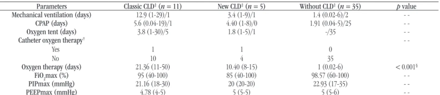
The “classic” CLD group received oxygen therapy for the longest period of time (p < 0.0001). The risk factors/therapeutic
proile of the study sample, including oxygen therapy parameters, gender, gestational age, maternal age and birth weight are shown
in Table 2, 3 and 4.
a decreased in the number and in the perimeter of the alveoli in the “new” CLD group, p = 0.033 and 0.018, respectively
(Table 1 and Figure).
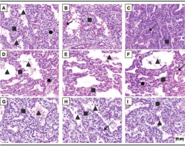
FIGURE −Photomicrograph of groups
A, B and C) photomicrograph of group 1 (classic BPD), here we observe areas of hyperinflation (▲) interspersed with areas of atelectasis (●) associated with marked fibrotic septal thickening and chronic inflammatory process (■). In other areas hyaline membranes (arrow) are observed as well as septal arterioles muscle layer thickening (dashed arrow); D, E and F) photomicrograph of group 2 (new BPD), here we observe areas of hyperinflation (▲) interspersed with areas of atelectasis (●) associated with minimal septal thickening with mild edema and mild chronic inflammatory process (■). There are no hyaline membranes and the septal arterioles muscle layer thickening (dashed arrow) is very mild; G, H and I) photomicrograph of group 3 (without BPD), in which we observed few areas of hyperinflation (▲) associated with discrete septal thickening with mild edema (■). There are no hyaline membranes and septal arterioles muscle layer thickening (dashed arrow) are minimal. The alterations found in group 3 are more associated with pulmonary immaturity than neonatal hypoxic injury.
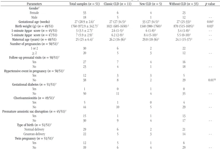
TABLE 2 − Clinical data for the three groups
Parameters Total samples (n = 51) Classic CLD (n = 11) New CLD (n = 5) Without CLD (n = 35) p value Gender† Female Male 33 18 6 5 4 1 23
12
-Gestational age (weeks) 27 (28.9 ± 2.6)* 27 (27-34.5)‡ 33 (27-34.5)‡ 27 (25-33)‡ 0.04§ Birth weight (g) (n = 49/51) 1760 (972.9 ± 342.7)* 1021 (685-1650) ‡ 1340 (900-1760)‡ 870 (515-1695)‡ 0.03§ 1-minute Apgar score (n = 45/51) 3 (3.5 ± 2.7)* 2.6 (1-5)‡ 6 (1-8)‡ 3.4 (1-8)‡ -5-minute Apgar score (n = 47/51) 7 (5.9 ± 2.9)* 6.2 (2-9)‡ 8.4 (5-10)‡ 5.5 (0-10)‡ -Maternal age (years) (n = 48/51) 25 (25 ± 6.4)* 26.2 (16-36)‡ 29.8 (18-36)‡ 24.1 (15-37)‡ -Number of pregnancies (n = 50/51)†
1 or 2
> 2
30 20 6 5 2 3 22
12
-Follow-up prenatal visits (n = 50/51)†
Yes No 27 23 7 4 4 1 16
18
-Hypertensive event in pregnancy (n = 50/51)†
Yes No 12 38 3 8 3 2 5
29 0.01§§ Gestational diabetes (n = 51/51)†
Yes No 1 50 0 11 1 4 0
35
-Chorioamnionitis (n = 49/51)†
Yes No 5 44 1 10 0 5 4
29
-Premature amniotic sac disruption (n = 45/51)†
Yes No 15 30 1 9 1 4 13
17
-Type of birth (n = 51/51)†
Normal delivery Cesarean delivery 29 22 6 5 2 3 21
14
-Twin pregnancy (n = 51/51)†
Yes No 12 39 5 6 1 4 6
29
-CLD: chronic lung disease; *:median (average ± standard deviation); †: number of cases; ‡: median (min-max); §: Kruskal-Wallis non-parametric test (p < 0.05); §§: Pearson correlation; - -: not statistically significant data.
Immunohistochemical data
The neutrophil inlux was much higher in “classic” CLD group than in “new” CLD or the “without” CLD groups; this difference was statistically signiicant (p < 0.001) (Table 5).
There were more CD25+ lymphocytes in the group with “new” CLD than in the other groups (p = 0.0400). There were
more CD74+ cells and CD4+ T lymphocytes in “classic” CLD group (p = 0.0500 and 0.0160, respectively). The CD45RO+ T
cell count was signiicantly higher in “classic” CLD group than in “without” CLD and “new” CLD groups (p = 0.0004). The CD14+
cell count was higher in the alveolar septum in “new” CLD group than in “classic” CLD or “without” CLD groups, with a trend toward signiicance (p = 0.0523), as shown in Table 5. There was no statistically signiicant difference between the groups in terms of CD20+ cell and CD8+T cell counts.
Whereas ICAM-1 expression was higher in the “new” CLD group than in the other groups (p = 0.0200), there were no
statistically signiicant differences in VCAM expression between the groups.
DISCUSSION
The pathogenesis of BPD/CLD is complex and remains unclear despite numerous studies on the subject. The lungs of very premature neonates have immature distal airways with a thick blood-air barrier and a small area for gas exchange. Furthermore, they are deicient in surfactant because of the predominance of undifferentiated epithelial cells and the scarcity of type II alveolar cells(25).
TABLE 3 − The postnatal factors for the three groups
Parameters Total samples (n = 51) Classic CLD(n = 11) New CLD (n = 5) Without CLD (n = 35) p value Patent ductus arteriosus†
Yes No 15 36 10 1 2 3 3 32
-Cardiopulmonary resuscitation†
Yes No 40 11 8 3 3 2 29 6
-Antibiotic therapy†
Yes No 33 18 10 1 4 1 19 16
-Surfactant replacement therapy†
Yes No 15 36 7 4 0 5 8 27
-Necrotizing enterocolitis†
Yes
No 1140 74 23 332 0.0001
§§ Bronchopneumonia† Yes No 12 39 6 5 0 5 6 29 0.0165§§
Pulmonary hemorrhage†
Yes No 9 42 3 8 1 4 5 30
-Pulmonary hypertension†
Yes
No 447 29 14 314
-Choking† Yes No 28 23 6 5 1 4 21 14
-Intracranial hemorrhage†
Yes No 11 40 4 7 2 3 5 30 -Pneumothorax† Yes
No 483 101 05 332
-Sepsis† Yes No 24 27 11 0 4 1 9 26 0.0001§§
Corrected gestational age (weeks)* 29 (29.7 ± 3.2)* 32 (29-39)‡ 34 (28-36)‡ 27 (25-33)‡ 0.0002§ Survival time (days) 3 (7.6 ± 10.7)* 23.4 (11-50)‡ 12.4 (8-21)‡ 1 (0-6)‡ 0.0001§
Cause of death†
Sepsis Intracranial hemorrhage Perinatal asphyxia Others 24 4 11 22 11 0 0 0 4 1 0 0 9 3 11 22
-CLD: chronic lung disease; *: median (average ±standard deviation); ‡: median (min-max); †:number of cases; §: Kruskal-Wallis non-parametric test (p < 0.05); §§:Pearson correlation; - -:not statistically significant data.
TABLE 4 − The parameters of oxygen therapy for the three groups
Parameters Classic CLD‡ (n = 11) New CLD‡ (n = 5) Without CLD‡ (n = 35) p value
Mechanical ventilation (days) 12.9 (1-29)/1 3.4 (1-9)/1 1.4 (0.02-6)/2
-CPAP (days) 5.6 (0.04-19)/1 4.40 (1-8)/0 1.91 (0.04-5)/25
-Oxygen tent (days) 3.8 (1-30)/5 1.8 (1-5)/1 -/35
-Catheter oxygen therapy†
Yes No 1 10 1 4 0 35
-Oxygen therapy (days) 21.36 (11-50) 10.40 (8-15) 1 (0.02-6) < 0.001§
FiO2max (%) 95 (40-100) 85 (40-100) 98.57 (60-100)
-PIPmax (mmHg) 21.16 (18-30) 20 (20-20) 22.93 (17-35)
-PEEPmax (mmHg) 4.78 (4-5) 5 (5-5) 5 (5-6)
-CLD: chronic lung disease; ‡: mean (max-min)/median; †: number of cases; CPAP: continuous positive airway pressure; FiO
2max: maximum fraction of inspired oxygen; PIPmax:
TABLE 5 − The median neutrophils count, the positive cells per high power field for each marker and the mean VCAM and ICAM-1 positive scores for each group
Marker Classic CLD (n = 11) New CLD (n = 5) Without CLD (n = 33) p value§
Neutrophils count 3.11 1.49 0.59 < 0.001
CD20+ 1.5 2.12 1.25 0.938
CD25+ 0.3214 0.95 0.3839 0.04
CD45RO+ 5.25 1.36 2 0.0004
CD14 (alveolar septum) 3 341.5 15 0.0523
CD14 (alveolar space) 2 61 11 0.138
CD4+ 10.887 2.766 2.86 0.016
CD8+ 0.472 2.5 3.991 0.17
CD74+ 12.25 9.625 7.187 0.05
ICAM 0.106 0.6 0.4292 0.02
VCAM 0 0.2 1 0.076
VCAM: vascular cell adhesion molecule; ICAM-1: intercellular adhesion molecule-1; CLD: chronic lung disease; §:Kruskal-Wallis non-parametric test (p < 0.05); p value is between classic
and without CLD and new and without CLD.
arterial duct, malnutrition, vitamin A deiciency and genetic factors. Inlammation, however, is the common inal pathway for the factors that cause lung injury in CLD(26).
In this study, the “new” CLD group was distinguished from the “without” CLD group by morphometric analysis. This analysis revealed statistically signiicant differences between the two groups, with a decrease in the number and the perimeter of the alveoli in the “new” CLD group(10, 12-14, 22).
The clinical, histopathological and morphometric patterns of CLD have changed with the emergence of the concept of underdeveloped distal airways. The histopathological indings for “new” CLD were recently described and were observed in our
study(10, 12). As observed in this study, although the clinical criteria
for the diagnosis of CLD should depend on respiratory support at 28 days of postnatal life, the pathological changes seem to precede this clinical concept based on the pathological changes found in the infants with 7-28 days of oxygen therapy.
The imbalance between pro-inlammatory and anti-inlammatory activity and its close relationship with cellular apoptosis and proliferation may affect alveolar formation and pulmonary vascular growth, eventually leading to pulmonary hypoplasia(19, 26, 27).
Furthermore, neutrophils may produce recruiting factors for other leukocytes, such as interleukin-8 (IL-8), as well as for tumor necrosis factor (TNF), which is responsible for prolonging neutrophil survival by inhibiting the activation of the apoptotic cascade. Some authors have shown that there is increased evidence of CD4+ and CD8+ T lymphocyte involvement in the mobilization and maintenance of neutrophils at the injury site, which may be a key factor, particularly in CLD, bronchial hyper-reactivity and bronchiectasis(27-30).
Our indings show that the neutrophils inlux was higher in the “classic” CLD group than in the “without” CLD and “new” CLD” groups (p < 0.001). Furthermore, the number of CD4+ T lymphocytes was higher in the “classic” CLD group than in the “new” CLD group (p = 0.016), corroborating the hypothesis that
the inlammatory response is more intense in the “classic” CLD
form(20, 28, 29, 31).
The physiopathological relationship between the accumulation of neutrophils and the presence of CD4+ T lymphocytes has not been fully clariied. There is some evidence that interleukin-17 (IL-17) plays a signiicant role in the action orchestrated by these two cells(31-33).
IL-17 released by the CD45RO+ T lymphocytes may activate T cells, which act as mediators in the increased neutrophil recruitment and activation in the damaged tissues, particularly
the lung tissue(34, 35). This inding was conirmed in studies in
which azithromycin was used as an inhibitor of IL-17 secretion, and a reduction in tissue damage was observed(35-38).
In our study, the CD45RO+ T lymphocyte count was higher in the “classic” CLD than in the “new” CLD and “without” CLD groups (p = 0.0004). It was followed by an increased neutrophil
count in the same group (p < 0.001), corroborating the hypothesis
of an initial inlammatory response mediated by neutrophils and maintained by T lymphocytes in this form of CLD(20, 28, 29, 31).
CD14 is a glycoprotein expressed on the myelomonocytic lineage cells, including monocytes, macrophages and Langerhans cells, and CD14 acts as an opsonin receptor to promote the release of pro-inlammatory cytokines, particularly those associated with Toll-like receptors (TLR)(39, 40). In our study, the number of CD14+
septum, for which the results showed a trend toward a statistically signiicant difference (p = 0.0523). Some studies have shown that CD14 expression in peripheral blood is lower in premature infants
than in term neonates(41-43).
The anti-CD74 antibody detects a major histocompatibility complex (MHC) class II receptor on the macrophage membrane that is involved in macrophage migration inhibitory factor (MIF) induction. CD74+ cells are usually macrophages, histocytes or monocytes, as well as type II pneumocytes, and they are correlated with the induction of type II pneumocyte proliferation in alveolar epithelium repair(44).
Furthermore, Kevin and Bandhari found elevated levels of CD74+ cells and MIF in the lungs of premature rats with respiratory distress syndrome and in tracheal aspirate from premature neonates. Both factors are related to a slight probability of developing CLD. This inding may be explained by the role played by CD74+ cells in pneumocyte proliferation, exacerbation of angiogenesis, increase in leukocyte migration, and therefore the maintenance of inlammatory response(45).
We found more CD74+ cells in the “classic” CLD group than in the other groups, which may be explained by the fact that during the process of alveolar damage that is observed in “classic” CLD, there is an extensive damage to type I pneumocytes, which are replaced by hyaline membranes. The acute phase is followed by the subacute phase, in which the area is cleaned by abundant macrophages and the alveolar lining is regenerated by type II pneumocyte proliferation. The macrophages and type II pneumocytes have the MIF receptor (CD74), and therefore they reacted with the anti-CD74 antibody
used in this study(45).
CD25+ T-cells were more abundant in the “new” CLD group. This inding can be explained by the fact that regulatory T cells modulate the inlammatory response, resulting in less inlammation and ibrosis in this form of the disease(46).
Neutrophil migration across the endothelium and epithelial cell barrier depends on the ICAM-1 adhesion receptor, a molecule stimulated in inlammatory disorders that can be induced by cytokines and detected in soluble form in plasma, which has been proposed as a marker of inlammatory activity. The endothelium plays a fundamental role in acute lung injury, particularly in the activation of pro-inlammatory cytokines. Some authors have proposed that the presence of interleukin-1 (IL-1) and TNF supports the presence of activated macrophages, which stimulate the production of other cytokines and increase the expression of adhesion molecules such as ICAM-1(39, 46, 47).
Little et al. showed that plasmatic levels of ICAM-1 in premature neonates at 14 days may be correlate with chronic
neonatal lung disease and its severity(46). Ballabh et al. reported
similar indings of elevated ICAM-1 levels in neonates who developed CLD on approximately the 28th day of life(47). In our
study, the lung samples of premature neonates from the “new” CLD group had higher levels of ICAM-1 than the lung samples from the other groups. It reasonable to hypothesize that ICAM-1 is an early biomarker for CLD because it appears before the 28th day of oxygen supplementation, i.e., before a clinical diagnosis of CLD. These results contrast with the indings of other authors regarding the plasma levels of ICAM-1(39).
In our study, high levels of ICAM-1 in “new” CLD may have been the determining factor for the increased number of CD14+ cells in the alveolar septum. Wether this conclusion is correct, it is reasonable to assume that the increased number of CD14+ cells can influence the increase in apoptotic cascade activity, which seems to be a major cause of the reduced alveolar formation that occurs in “new” CLD. Further immunohistochemical and molecular studies are required to clarify this hypothesis(46, 47).
Few studies have correlated B-lymphocytes and VCAM with CLD, and no statistically signiicant differences in the number of CD20+ cells and VCAM immunoexpression were found between the groups in our study(44).
Our indings seems to corroborate the hypothesis that the initial inlammatory response is mediated by neutrophils and sustained by CD45RO+ and CD4+ T lymphocytes in the “classic” CLD form. Furthermore, the results suggest that CD14 may be a biomarker to connect inlammatory and apoptosis process, particularly in the lung injury associated with decreased alveolar volume observed in “new” CLD. Further studies are needed to better elucidate the pathogenesis of CLD, especially regarding the apoptotic process of the “new” CLD.
ETHICAL CONDUCT OF RESEARCH
REFERENCES
1. Northway WH, Rosan RC, Porter DY. Pulmonary disease following respiratory therapy of hyaline membrane disease. Bronchopulmonary dysplasia. N Engl J Med. 1967; 276: 357-68.
2. Bancalari E, Claure N. Deinitions and diagnostic criteria for bronchopulmonary dysplasia. Semin Perinatol. 2006; 30: 164-70. 3. Rebello CM, Mascaretti RS. A “nova” displasia broncopulmonar. In: Manual da atualização em Neonatologia. 1st ed. Rio de Janeiro, RJ: Panamericana; 2004.
4. Sankar MJ, Agarwal R, Deorari AK, et al. Chronic lung disease in newborns. J Pediatr. 2008; 75(4): 369-76.
5. Bancalari E. Bronchopulmonary dysplasia: old problem, new presentation. J Pediatr. 2006; 82(1): 2-3.
6. Jobe AH. The new bronchopulmonary dysplasia. Curr Opin Pediatr. 2011; 23(2): 167-72.
7. Jobe AH, Bancalari E. Bronchopulmonary dysplasia. Am J Respir Crit Care Med. 2001; 163: 1723-9.
8. Ehrenkranz RA, Walsh MC, Vohr BR, et al. Validação da deinição de displasia broncopulmonar do consenso do Instituto Nacional de Saúde. Pediatrics. 2006; 10(6): 413-22.
9. Walsh MC, Szeler S, Davis J, et al. Summary proceedings from the bronchopulmonary dysplasia group. Pediatrics. 2006; 117: 52-5. 10. Okamoto CT, Bahr JA, Silva LLG, et al. Análises histopatológica e morfométrica no diagnóstico da “nova” displasia broncopulmonar e comparação clinicopatológica com a forma clássica da doença. J Bras Patol Med Lab. 2009; 45(2): 147-52.
11. Tapia JL, Agost D, Alegria A, et al. Bronchopulmonary dysplasia: incidence, risk factors and resource utilization in a population of South American very low birth weight infants. J Pediatr. 2006; 82: 15-20. 12. Coalson JJ, Winter V, de Lemos RA. Decreased alveolarization in baboon survivors with bronchopulmonary dysplasia. Am J Respir Crit Care Med. 1995; 152: 640-6.
13. Coalson JJ, Winter VT, Siler-Khodr T, et al. Neonatal chronic lung disease in extremely immature baboons. Am J Respir Crit Care Med. 1999; 160: 1333-46.
RESUMO
Introdução: A doença pulmonar crônica (DPC), conhecida clinicamente como displasia broncopulmonar, é uma das maiores causas de morbidade em neonatos que nasceram prematuros e foram submetidos à oxigenioterapia. Objetivo: Identificar moléculas inflamatórias em tecido pulmonar de recém-nascidos prematuros que morreram com DPC por meio do método de imuno- -histoquímica. Métodos: Análise imuno-histoquímica de 51 amostras de pulmões de recém-nascidos prematuros – formando os grupos: sem DPC, DPC “nova” e DPC “clássica”. Resultados: O influxo de neutrófilos e o número de células CD4+ e CD45RO+ foram maiores no grupo DPC “clássica” (p < 0,001). Conclusão: Os resultados sugerem que o processo inflamatório é mediado por neutrófilos e linfócitos CD45RO+ e CD4+ na DPC “clássica”.
Unitermos: displasia broncopulmonar; imuno-histoquímica; nascimento prematuro; oxigenoterapia; neonatologia.
14. Mascaretti RS, Mataloun MM, Dolhnikoff M, et al. Lung morphometry, collagen and elastin content: changes after hyperoxic exposure in preterm rabbits. Clinics. 2009; 64(11): 1099-04.
15. Bose CL, Dammann CEL, Laughon MM. Bronchopulmonary dysplasia and inlammatory biomarkers in the premature neonate. Arch Dis Child Fetal Neonatal. 2008; 93: 455-61.
16. Bourbon J, Boucherat O, Chailley-Heu B, et al. Control mechanisms of lung alveolar development and their disorders in bronchopulmonary dysplasia. Pediatr Res. 2005; 57: 38-46.
17. Santos CC, Slutsky A. The contribution of biophysical lung injury to the development of biotrauma. Annu Rev Physiol. 2006; 68: 585-618. 18. Santos CC, Zhang H, Liu M, et al. Bench-to-bedside review: biotrauma and modulation of the innate immune response. Critical Care. 2005; 9(3): 280-6.
19. Speer CP. Inlammation and bronchopulmonary dysplasia. Semin Neonatol. 2003; 8: 29-38.
20. Urlichs F, Speer CP. Neutrophil function in preterm and term infants. Neo Rev. 2004; 5: 417-29.
21. Ballabh P, Simm M, Kumari J, et al. Lymphocyte subpopulations in bronchopulmonary dysplasia. Am J Perinatol. 2003; 20(8): 465-75. 22. Willet KE, Jobe AH, Ikegami M, et al. Antenatal endotoxin and glucocorticoid effects on lung morphometry in preterm lambs. Pediatr Res. 2000; 48: 782-8.
23. Baurakiades E, Martins APC, Moreschi VN, et al. Histomorphometric and immunohistochemical analysis of infectious agents, T-cell subpopulations and inlammatory adhesion molecules in placentas from HIV-seropositive pregnant women. Diagn Pathol. 2011; 6: 101-8. 24. Costa Jr VHS, Baurakíades E, Azevedo MLV, et al. Immunohistochemistry analysis of pulmonary iniltrates in necropsy samples of children with non-pandemic lethal respiratory infections (RSV; ADV; PIV1; PIV2; PIV3; FLUa; FLUB). J Clin Virol. 2014; 61: 211-5.
25. Ambalavanan N, Carlo WA, D’Angio CT, et al. Cytokines associated with bronchopulmonary dysplasia or death in extremely low birth weight infants. Pediatrics. 2009; 123: 1132-41.
27. Turato G, Zuin R, Saetta M. Pathogenesis and pathology of COPD. Respiration. 2001; 68: 117-28.
28. Gaga M, Bentley AM, Humbert M, et al. Increases in CD4+ T lymphocytes, macrophages, neutrophils and interleukin 8 positive cells in the airways of patients with bronchiectasis. Thorax. 1998; 53: 685-91. 29. Bochner BS. Road signs guiding leukocytes along the inlammation superhighway. J Allergy Clin Immunol. 2000; 106(5): 817-28.
30. O’Byrne PM. Airway inlammation and airway hyperresponsiveness. Chest. 1986; 90: 575-7.
31. Lindén A, Laan M, AndersonGP. Neutrophils, interleukin-17A and lung disease. Eur Respir J. 2005; 25: 159-72.
32. Lindén A. Interleukin-17 and airway remodeling. Pulm Pharmacol Ther. 2006; 19: 47-50.
33. Schmidt-Weber C, Akdis M, Akdis CA. Th17 cells in the big picture of immunology. J Allergy Clin Immunol. 2007; 120: 247-54.
34. Laan M, Linén A. IL-17 as a potential target for modulating airway neutrophilia. Curr Pharm Des. 2002; 8(20): 1855-61.
35. Laan M, Prause O, Miyamoto M, et al. A role of GM-CSF in the accumulation of neutrophils in the airways caused by IL-17 and TNF-alpha. Eur Respir J. 2003; 21(3): 387-93.
36. Ballard HO, Bernard P, Qualls J, et al. Azithromycin protects against hyperoxic lung injury in neonatal rats. J Invest Med. 2007; 55(6): 299-05. 37. Aghai ZH, Kode A, Saslow JG, et al. Azithromycin suppresses activation of nuclear factor-kappa B and synthesis of pro-inlammatory cytokines in tracheal aspirate cells from premature infants. Pediatr Res. 2007; 62(4): 483-8.
38. Ballard HO, Anstead M, Shook LA. Azithromycin in the extremely low birth weight infant for the prevention of bronchopulmonary dysplasia: a pilot study. Respir Res. 2007; 8: 41.
39. Bessler H, Komlos L, Punsky I, et al. CD14 receptor expression and lipopolysaccharide-induced cytokine production in preterm and term neonates. Biol Neonate. 2001; 80: 186-92.
40. Pedreira PR, Garcia-Prieto E, Albaiceta GM, et al. Respuesta inlamatória y apoptosis em La lesion pulmonar aguda. Med Intensiva. 2006; 30: 268-75.
41. Levy O. Innate immunity of the human newborn: distinct cytokine responses to LPS and other toll-like receptor agonists. J Endotoxin Res. 2005; 11(2): 113-6.
42. Henneke P, Osmers I Bauer K, et al. Impaired CD14-dependent and independent response of polymorphonuclear leukocytes in preterm infants. J Perinat Med. 2003; 31(2): 176-83.
43. Forster-Waldi E, Sadeghi K, Tamandl D, et al. Monocyte toll-like receptor 4 expression and LPS induced cytokine production increase during gestational aging. Pediatr Res. 2005; 58: 121-4.
44. Marsh LM, Cakarova L, Kwapiszewska G, et al. Surface expression of CD74 by type II alveolar epithelial cells: a potential mechanisms for macrophage migration inhibitory factor-induced epithelial repair. Am J Physiol Lung Cell Mol Physiol. 2009; 296(3): 442-52.
45. Kevill KA, Bhandari V, Kettunen M. A role for macrophage migration inhibitory factor in the neonatal respiratory distress syndrome. J Immunol. 2008; 180: 601-8.
46. Little S, Dean T, Bevin S, et al. Role of elevated plasma soluble ICAM-1 and bronchial lavage luid IL-8 levels as markers of chronic lung disease in premature infants. Thorax. 1995; 50: 1073-9.
47. Ballabh P, Kumari J, Krauss AN, et al. Soluble E-selectin, soluble L-selectin and soluble ICAM-1 in bronchopulmonary dysplasia, and changes with dexamethasone. Pediatrics. 2003; 111(3): 461-8.
CORRESPONDING AUTHOR
Lúcia de Noronha