Dermoid cyst of the pancreas: a case report
Cisto dermoide de pâncreas: relato de caso
Carla Kellen da Silva Menezes1; Luciana Botinelly Mendonça Fujimoto2; Leonardo Simão Coelho Guimarães3; Silvana de Albuquerque Silva Damasceno4; Jenifer Morais de Melo5
First submission on 07/05/13; last submission on 04/07/13; accepted for publication on 04/07/13; published on 20/10/13
1. Resident in Surgical Pathology at Hospital Universitário Getúlio Vargas (HUGV) of Universidade Federal do Amazonas (UFAM).
2. Doctorate in Biotechnology from UFAM; associate professor at the Department of Pathology and Legal Medicine of the Medical School of UFAM.
3. Specialist in Gastrointestinal Surgery; full professor of Colégio Brasileiro de Cirurgia Digestiva (TCBCD); gastrointestinal surgeon at HUGV-UFAM; professor of Clinical Surgery at Universidade do Estado do Amazonas (UEA).
4. Physician of the Department of Pathology and Legal Medicine of the Medicine School of UFAM. 5. Graduating student of Medicine at UFAM.
ABSTRACT
Dermoid cysts or mature cystic teratomas are mesenchymal neoplasms most commonly found in the ovaries, but which may occur in any location along the pathways of ectodermal cell migration. They are rarely seen in the pancreas, where they show a slight preference for the pancreatic head. We report a case of dermoid cyst of the pancreas in a 69-year-old male patient, discussing the epidemiology, clinical presentation, diagnosis and treatment of this neoplasm. Since preoperative diagnosis is dificult, given its rarity in this site, it is usually diagnosed by histopathology of the specimen.
Key words: dermoid cyst; mature teratoma; pancreas.
INTRODUCTION
Dermoid cyst of the pancreas (mature cystic teratoma of the pancreas) is a very rare mesenchymal neoplasm, with 35 cases described in the world literature(7).
Dermoid cysts, in general, occur in all ages, have no sex preference, and are commonly found in ovaries, but may occur in any pathway of ectodermal cell migration. The pancreas is extremely rare as a primary site(1-5, 7, 8, 12, 13, 15), where there is a slight
preference for the pancreatic head(3, 11, 15).
The clinical picture is non-speciic(3, 4, 7, 8, 12, 13, 15), and the
preoperative diagnosis is dificult. Many times it is suggested by imaging studies, but only conirmed by histological evaluation(9).
Macroscopically, the cyst generally presents a well-delimited and thick wall, with pasty yellow content of caseous aspect, rarely clear and serous. Microscopically, it contains differentiated
elements from one or more germ layers(13).
The objective of this report is to present a case of dermoid cyst of the pancreas, located at the tail of pancreas, of dificult preoperative diagnosis, and to review epidemiological, clinical, diagnostic and histopathological aspects of this rare neoplasm.
CASE REPORT
A 69-year-old man, from Manaus, Amazonas, was admitted in December, 2010, to treat a urinary tract infection. A computed tomography (CT) of abdomen was ordered, for evaluation of renal lithiasis.
Other imaging and laboratory exams were performed. The patient had a history of social drinking, ex-smoking, osteoarthrosis of knee and hip, eventually on use of nonsteroidal anti-inlammatory drugs. He referred a car accident 18 years before, with femur fracture and closed blunt abdominal trauma. He denied abdominal pain and previous pancreatitis. The physical examination was normal.
Laboratory investigation revealed serum dosages of amylase, lipase and tumor markers (carcinoembryonic antigen [CEA], carbohydrate antigen 19-9 [CA19-9], cancer-associated antigen 125 [CA 125] and alpha-fetoprotein) within normal ranges, as well as the other hematological and biochemical investigations.
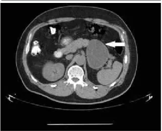
In January, 2010, the patient underwent a new total abdominal CT, which revealed a lesion with the same characteristics as the previous one, but with a discrete increase, measuring 8.8 × 7 cm
(Figure 1).
cystic degeneration, intrapapillary mucinous neoplasm and lymphoepithelial cyst of the pancreas.
The patient underwent laparoscopic enucleation of the lesion in June, 2010, and the material was sent for histopathological examination, which macroscopically revealed a thick cyst wall, with several micronodular formations on the surface, and abundant pasty homogeneous yellowish content (Figure 2).
FIGURE 1 – Total abdominal CT: hypodense expansive lesion of irregular
contours, with internal septations, situated in the distal portion of the pancreatic body/tail, measuring 8.8 × 7 cm (APxT), with discrete parietal and septal enhancement (arrow)
CT: computed tomography; APxT: anteroposterior × transverse diameters.
FIGURE 2 – Pancreatic dermoid cyst: thickened cystic wall, with several
micronudulations on surface and pasty homogeneous yellowish content of caseous aspect
In March, 2010, a magnetic resonance imaging (MRI) was ordered. It demonstrated a multiloculated cystic lesion, in contact with the gastric wall; the pancreatic tail measured 7.2 × 6.4 × 7.3 cm.
No image showed invasion of adjacent organs, distance metastases or lymphadenopathy.
The possibilities of pseudocyst and malignant neoplasm were eliminated by the surgery team, and the suggested differential diagnoses were mucinous cystic neoplasm, solid pseudopapillary tumor (Frantz’s tumor), non-functioning endocrine tumor with
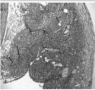
Microscopic examination depicted cystic formations lined by keratinizing stratiied squamous epithelium, with the presence of sebaceous glands, surrounded by mature lymphoid tissue, which contains lymphoid follicles and germinal centers. The cystic content was composed by laminated strands of keratin (Figures 3 and 4).
The patient had a favorable evolution, with no complications, being discharged two days after surgery. He remains without complaints.
DISCUSSION
The dermoid cyst of the pancreas, also called mature cystic teratoma of the pancreas, is an uncommon mesenchymal neoplasm, irstly described in 1918(4, 5, 10-13), with 35 cases reported
in the world literature(7). This is the 36th reported case up to now,
according to a bibliographic survey.
It can affect all age groups, but according to Lane et al.(7),
(ranging from 4 months to 74 years). Among patients, 20 were male and 15, female.
Degrate et al.(3), in a review of 33 cases, afirms that the
average size of lesions was 7.5 cm (ranging from 2.2 to 20 cm), similarly to the observed in this case.
Dermoyd cysts are frequently found in the ovaries, but may occur in any pathway of ectodermal cell migration, typically in the midline, as in testes, skull, brain, mediastinum, retroperitoneum, omentum, and bladder. Occurrence in the pancreas, though, is
very rare(1-5, 7, 8, 11-13, 15).
They may appear in any site in the pancreas, being lightly more common in the pancreas head(3, 7, 11, 15), what differs from
the case presented here, in which the lesion was found in the pancreatic tail.
Clinical presentation is non speciic. The most common signs and symptoms are palpable mass in the upper quadrant of the abdomen, abdominal pain, back pain, nausea, vomits, anorexia, weight loss, fatigue and fever. However, there are some cases with
nosymptoms (18.3%)(6), only incidentally discovered at diagnostic
imaging(3, 4, 8, 12, 13, 15), as it happened in our study.
Preoperative diagnosis, due to the rarity of the lesion, is dificult(9), but it may be suspected after image exams, like
ultrasonography (USG), CT, and ultrasound-guided ine-needle aspiration (FNA). The radiologic appearance of these lesion is variable, depending on the proportions of the diverse tissues of which they are composed(2-4, 12, 14).
There is also report of the conduction of endoscopic ultrasound-guided FNA(8), but there are no pathognomonic
data to recognize a dermoyd cyst, and many times preoperative diagnosis is inconclusive(9, 11, 13), as well as in this case, in which the
pancreatic dermoyd cyst was not a differential diagnosis according to the surgery team.
Daghfous et al.(2) performed a CT-guided FNA of a pancreatic
mass whose content was sebaceous secretion, being the diagnosis of pancreatic teratoma suggested preoperatively. It was later conirmed as mature cystic teratoma of the pancreas, after distal pancreatectomy and histopathological examination, what suggests a good alternative for a preoperative diagnosis.
Tumor markers CEA and CA19-9 are generally lower than those in other pancreatic cystic neoplasms(13), but no consensus exists and
further studying is necessary. In the present case, the patient presented normal levels of these and other markers, as previously cited.
The treatment is based on surgical removal, through either open surgery (distal pancreatectomy, cystectomy, external drainage and cystogastrectomy)(13) or laparoscopy, as in this case.
This seems to be a good alternative for the treatment of benign pancreatic cyst lesions.
Dermoid cysts are true cysts; thus, their wall consists of stratiied squamous epithelium and underlying connective
FIGURE 3 – Pancreatic dermoid cyst (HE 4×): cystic formation lined by
keratinizing stratiied squamous epithelium, with the presence of sebaceous glands, surrounded by mature lymphoid tissue
HE: hematoxylin and eosin.
FIGURE 4 – Pancreatic dermoid cyst (HE 20×): closer view of Figure 3
tissue(3, 5). This characteristic is important to distinguish them
from pancreatic pseudocysts, which correspond to 90% of cystic lesions of the pancreas and are collections of pancreatic secretions enclosed in a ibrous wall with no epithelium lining(6).
Macroscopically, the cyst is composed by a well-limited thick wall, which may contain micronodulations; its content is pasty pale yellow, of caseous aspect, as in this case. Rarely it may be light
and serous(13) and sometimes may contain other elements, such as
teeth, hair, bone, cartilage and skin appendices(3).
Microscopically, the cyst contains differentiated elements of one or more germ cell layers(13) as, for example, the cystic wall,
formed commonly by a stratiied squamous epithelium with the presence of sebaceous glands, and immediately adjacent, lymphoid tissue that may contain germinal centers. This layer of lymphoid tissue corresponds to the micronodulations observed in the cystic wall at macroscopy.
Frequently, there is the presence of mucin-producing ciliated columnar epithelium. Hair may also be found inside the cystic wall(11).
Yu et al.(14) reported a dermoid cyst of the pancreas in a
2-year-old child. The cystic wall contained smooth muscle, pancreatic tissue, lymphoid tissue, intestinal tissue and glial tissue.
The cystic content corresponds to the great quantity of keratin, sometimes disposed in laminated formation, possibly containing sebaceous secretion and cholesterol clefts(15). There is the report of a
carcinoid tumor arising in a mature pancreatic cystic teratoma(8).
Histopathologically, the differential diagnosis must include all cystic lesions of the pancreas: pseudocysts; neoplastic cysts, as the serous and mucinous cystadenoma; intraductal papillary mucinous neoplasm; and solid pseudopapillary tumor. However, one of the main differential diagnoses is the lymphoepithelial cyst, different from the dermoid cyst for the absence of skin annexes(7, 11, 13).
Although dermoid cysts are benign neoplasms, a small percentage may develop into malignant forms. Hence, a complete sampling of the lesion is necessary to exclude the presence of immature foci(3, 10, 11). Up to now, all pancreatic teratomas reported
were mature, that is, of benign behavior(3).
Pancreatic dermoid cysts are benign neoplasms, of good prognosis, and have been little reported in literature. They must be remembered during preoperative investigation, to enable a more conservative treatment, like the laparoscopic enucleation performed in this patient, which may be used as a good alternative. Besides, they seem to be of easy histopathological diagnosis, but may confound pathologists because they rarely present in this location.
RESUMO
Cistos dermoides ou teratomas císticos maduros são neoplasias mesenquimais comumente encontradas nos ovários, mas que podem ocorrer em qualquer via de migração das células ectodérmicas. No pâncreas, a ocorrência é rara, sendo mais comum na cabeça pancreática. Relata-se caso de cisto dermoide do pâncreas em paciente masculino de 69 anos, discutindo-se epidemiologia, clínica, diagnóstico e tratamento dessa neoplasia, pouco suspeitada no pré-operatório devido à sua raridade nessa topograia; geralmente, é diagnosticada apenas pelo exame histopatológico da peça cirúrgica.
Unitermos: cisto dermoide; teratoma maduro; pâncreas.
REFERENCES
1. BADIA, A. C. et al. Teratoma quístico de páncreas. Cir Esp, v. 88, n. 6, p. 419-21, 2010.
2. DAGHFOUS, A. et al. Mature teratoma of the pancreas diagnosed by ine-needle aspiration. Arab J Gastroenterol, v. 12, n. 2, p. 92-3, 2011.
Available at: <http://www.ncbi.nlm.nih.gov/pubmed/21684481>.
Accessed on: 16 Aug. 2012.
3. DEGRATE, L. et al. Mature cystic teratoma of the pancreas. Case report and review of the literature of a rare pancreatic cystic lesion. JOP J
Pancreas, v. 13, n. 1, p. 66-72, 2012. Available at: <http://www.ncbi.nlm.
nih.gov/pubmed/22233950>. Accessed on: 13 Aug. 2012.
4. FEUERLEIN, S. et al. Pancreatic teratoma: case report of a rare cystic neoplasm. Eur J Radiol Extra, v. 71, n. 2, p. e71-e72, 2009. Available
at: <http://intl.elsevierhealth.com/journals/ejrex>. Accessed on: 13 Aug.
2012.
MAILING ADDRESS
Carla Kellen da Silva Menezes
Rua 4, 19, apto 2; Residencial Luiza; Parque 10 de novembro; CEP: 69054-735; Manaus-AM, Brazil; e-mail: [email protected].
7. LANE, J. et al. Dermoid cyst of the pancreas: a case report with literature review. J Radiol Case Rep, v. 12, n. 6, p. 17-25, 2012.
8. MATEOS, E. A. et al. Mature cystic teratoma of the pancreas diagnosed by endoscopic ultrasound-guided ine needle aspiration. A case report. Rev Esp Patol, v. 43, n. 2, p.94-7, 2010. Available at: <http://www.elsevier.
es/patologia>. Accessed on: 13 Aug. 2012.
9. RIVKINE, E. et al. Affections rares du pancréas et dês voies biliaires: tératome kystique du pancréas. Gastroenterol Clin Biol, v. 31, n. 11, p. 1016-9, 2007.
10. SCHEELE, J. et al. Dermoid cyst of the pancreas. Int J Colorectal Dis, v. 25, n. 3, p. 415-6, 2010.
11. SEKI, M. et al. Image-diagnostic features of mature cystic teratomas
of the pancreas: report on two cases dificult to diagnose preoperatively. J Hepatobiliary Pancreat Surg, v. 12, n. 4, p. 336-40, 2005.
12. STRASSER, G. et al. Mature teratoma of the pancreas: CT and MR indings. Eur Radiol, v. 12, n. 3, p. 56-8, 2002.
13. TUCCI, G. et al. Dermoid cyst of the pancreas: presentation and
management. World J Surg Oncol, v. 85, n. 5, 2007. Available at: <http://
www.biomedcentral.com/info/about/charter/>. Accessed on: 13 Aug.
2012.
14. YU, C. H. et al. Mature cystic teratoma of the pancreas in a child. Pediatr Radiol, v. 33, n. 4, p. 266-8, 2003.