J Bras Pneumol. 2010;36(1):148-151
In this report, we present the case of a tool sharpener with diffuse lung disease who secondarily developed spontaneous bilateral pneumothorax.
Case report
A 27-year-old male patient with dry cough and progressive dyspnea for 1 year sought treat-ment in the Pulmonology Clinic of the Federal University of Goiás Hospital das Clínicas in the city of Goiânia, Brazil. He reported worsening dyspnea on minimal exertion in the last two weeks and weight loss (10 kg) in the last six months. The patient denied having a history of
Introduction
Hard metal pneumoconiosis is a diffuse disease caused by the inhalation of cobalt particles.(1) Hard metal is an alloy of cobalt and tungsten carbonate, occasionally added with small quantities of other metals, such as titanium, tantalum, nickel and chrome. The remaining metals other than cobalt are consi-dered inert and do not cause lung injury.(2)
Exposure to hard metal can cause occu-pational asthma and diffuse lung disease.(2) Occupations related to the manufacture and refining of hard metal, as well as those related to the use of cobalt-coated disks to polish diamonds and sharpen tools, are associated with these diseases.(3)
Hard metal pneumoconiosis with spontaneous
bilateral pneumothorax*
Pneumoconiose por exposição a metal duro com pneumotórax bilateral espontâneo
Maria Auxiliadora Carmo Moreira, Amanda da Rocha Oliveira Cardoso, Daniela Graner Schuwartz Tannus Silva,
Maria Conceição de Castro Antonelli Monteiro de Queiroz, Albino Alegro Oliveira, Tiago Marinho Almeida Noleto
Abstract
Hard metal pneumoconiosis, first described in 1964, is a diffuse disease caused by the inhalation of cobalt particles. The disease can manifest as occupational asthma, interstitial disease or allergic alveolitis. We report the case of a young male, working as a tool sharpener, who presented with dry cough and progressive dyspnea for one year, as well as with spontaneous bilateral pneumothorax at admission. The diagnosis was confirmed by open lung biopsy.
Keywords: Lung diseases, interstitial; Pneumoconiosis; Pneumothorax.
Resumo
A pneumoconiose por metal duro, descrita pela primeira vez em 1964, é uma doença difusa causada por inalação de partículas de cobalto. A doença pode se manifestar de três formas diferentes: asma ocupacional, doença inters-ticial e alveolite alérgica. Relata-se um caso de um jovem do sexo masculino, afiador de ferramentas, com quadro de tosse seca e dispnéia progressiva há um ano, apresentando-se à admissão com pneumotórax espontâneo bila-teral. O diagnóstico foi confirmado através de biópsia pulmonar a céu aberto.
Descritores: Doenças pulmonares intersticiais; Pneumoconiose; Pneumotórax.
* Study carried out at the Federal University of Goiás Hospital das Clínicas, Goiânia, Brazil.
Correspondence to: Maria Auxiliadora Carmo Moreira. Primeira Avenida s/n, Setor Universitário, CEP 74605-020, Goiânia, GO, Brasil.
Tel 55 62 269-8385. E-mail: [email protected] Financial support: None.
Submitted: 31 August 2009. Accepted, after review: 18 December 2009.
Hard metal pneumoconiosis with spontaneous bilateral pneumothorax
J Bras Pneumol. 2010;36(1):148-151 149
A pulmonary function test performed six months after discharge revealed severe restric-tive lung disease (VC = 34% of predicted), with negative bronchodilator test, and a moderate reduction in DLCO (60% of predicted).
At this writing, one year after diagnosis, the patient continued on prednisone (10 mg/ day), had occasional dry cough, had dyspnea on moderate exertion and presented an SpO2 of 93% at rest.
Discussion
Hard metal pneumoconiosis was first described in 1969 by Liebow & Carrington, being included among idiopathic interstitial pneumonias. In 2001, the American Thoracic lung disease. He had a two-year history of
occa-sional smoking and had discontinued the habit 5 years prior. The patient had been a hard metal tool sharpener (saws and knives) for eight years. He worked eight hours a day, did not wear indi-vidual protective equipment and used a synthetic diamond grinding wheel in an enclosed space, measuring approximately 5.5 m2, where he was the only worker.
Physical examination revealed weight loss, dyspnea, an SpO2 of 86% on room air and diffusely decreased vocal fremitus in both hemithoraces, primarily in the apices, where tympanism could be observed. No other altera-tions were found.
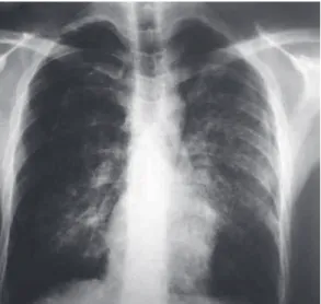
A chest X-ray performed one month prior to admission revealed bilateral pneumothorax (Figure 1). An HRCT scan showed bilateral pneu-mothorax, paraseptal emphysema in the apices, some interposed bullae, diffuse ground-glass opacities, perilymphatic nodules, mediastinal lymph nodes and a calcified halo (Figure 2).
The patient was submitted to bilateral closed chest tube drainage with complete re-expansion of the left lung and partial re-expansion of the right lung, where a bronchopleural fistula with medium output persisted. During the same hospital stay, the fistula was sutured and the patient underwent right parietal pleurectomy, left chemical pleurodesis with tetracycline and open lung biopsy (middle lobe).
The anatomopathological findings revealed interstitial pulmonary fibrosis, predominantly peribronchial and periseptal, containing nume-rous giant cells, alveolar histiocytes and areas of honeycombing (Figure 3). Examination of the pleural tissue showed granulation tissue proliferation, fibrosis, lymphohistioplasmocytic infiltrate, containing some interposed neutro-phils, and deposition of fibrin on the surface.
Based on the patient occupational history, as well as on imaging studies and on the anatomo-pathological examination, a diagnosis of hard metal pneumoconiosis was made.
The patient was started on systemic corticos-teroid therapy, initially with methylprednisolone and subsequently with prednisone.
During the follow-up period, the patient had repeated episodes of bilateral pneumothorax and required additional drainage and pleurodesis. He was discharged on prednisone (10 mg/day) and home oxygen therapy.
Figure 1 - Chest X-ray revealing diffuse, heterogeneous opacities and bilateral pneumothorax. “BJP” e “080208” por “020808”.
150 Moreira MAC, Cardoso ARO, Silva DGST, Queiroz MCCAM, Oliveira AA, Noleto TMA
J Bras Pneumol. 2010;36(1):148-151
The clinical profile of giant cell pneumonia includes dyspnea on exertion, hypoxemia, cough, weight loss, fatigue, wheezing and rales at the end of inhalation.(8) With the progression of fibrosis, there can be digital clubbing, cyanosis, signs of right heart failure, pulmonary hyperten-sion and cor pulmonale.(6,8) Pneumothorax has been reported in one case.(9)
Pulmonary function can show a restrictive pattern, with reduced lung volumes. Occasionally, an obstructive pattern can be found.(4,6) The DLCO tends to decrease in the same proportion as TLC.(7,8)
Chest X-rays can be normal or reveal mild nodular or reticulonodular infiltrates. Being more sensitive in the detection of alterations, HRCT can reveal ground-glass opacities and irregular linear opacities, as well as parenchymal distortion, traction bronchiectasis, bronchiolec-tasis and reticulation.(4,5) Other isolated findings include large peripheral cysts, which can repre-sent honeycombing, as well as centrilobular nodules and emphysema.(8)
If there is no contraindication, bronchial lavage and transbronchial biopsy should be performed to confirm the diagnosis. If the trans-bronchial biopsy sample is insufficient or the examination is inconclusive, open lung biopsy should be performed. If there are multinucle-ated giant cells in the bronchoalveolar lavage fluid accompanied by a clinical and radiological profile consistent with the disease, biopsy in not indicated.(7)
The histopathological pattern comprises mononuclear cell infiltrate, predominantly in regions of the peribronchiolar interstitium, and accumulation of macrophages and multinu-cleated giant cells within the alveoli,(7,8) with alveolar wall thickening.(7) With the progression of the disease, honeycombing can be observed. Being highly soluble in tissues, cobalt is rarely found in biopsy specimens.(1)
The treatment of hard metal lung disease involves complete removal from exposure and high-dose corticosteroid therapy. When there already is extensive pulmonary fibrosis, there is no significant response to treatment.
A peculiarity of the case reported here was the presence of bilateral recurrent pneumo-thorax, which required multiple interventions. This is an unusual presentation,(9) which made the management of the case difficult. In addi-Society and the European Respiratory addi-Society
recognized this entity as pneumoconiosis caused by the inhalation of cobalt or of an alloy of cobalt and other hard metals, and, therefore, it was excluded from the original classification.(4) This entity is also known as hard metal lung, giant cell pneumonitis and cobalt lung.
Hard metal is an alloy of tungsten carbonate, cobalt and small quantities of other metals, such as titanium and tantalum.(3,4) Since it is hard and resistant to high temperatures, it is used for polishing diamonds, galvanizing armaments, drilling oil wells and cutting tools.(5,6)
Although the hard metal alloy has other components, cobalt is the one responsible for inducing lung disease.(5) One group of authors stated that interstitial disease develops only when exposure to cobalt occurs in associa-tion with exposure to tungsten carbonate or diamond powder.(1) A small number of exposed workers develop the disease, usually after ten to twelve years of exposure, although the disease can occur early.(4)
Currently, the scientific community recog-nizes three pathological entities related to the inhalation of hard metal dust: occupational asthma; interstitial lung disease, which occurs in two varieties (non-specific form and giant cell intra-alveolar pneumonitis); and allergic alve-olitis or hypersensitivity pneumonitis. Allergic alveolitis or hypersensitivity pneumonitis occurs in the acute phase of exposure, being considered an early (potentially reversible) inflammatory phase of fibrosis, although it can evolve to fibrosis as a result of long-term exposure.(2,4,5,7,8)
Hard metal pneumoconiosis with spontaneous bilateral pneumothorax
J Bras Pneumol. 2010;36(1):148-151 151
correlation of the initial findings and demonstration of interval improvement.. J Thorac Imaging. 2005;20(4):301-4.
5. Gotway MB, Golden JA, Warnock M, Koth LL, Webb R, Reddy GP, et al. Hard metal interstitial lung disease: high-resolution computed tomography appearance. J Thorac Imaging. 2002;17(4):314-8.
6. Maier LA. Clinical approach to chronic beryllium disease and other nonpneumoconiotic interstitial lung diseases. J Thorac Imaging. 2002;17(4):273-84.
7. Cugell DW. Cobalt-Related Lung Diseases. Clin Pulm Med. 1998; 5(3): 158-64.
8. Enriquez LS, Mohammed TL, Johnson GL, Lefor MJ, Beasley MB. Hard metal pneumoconiosis: a case of giant-cell interstitial pneumonitis in a machinist. Respir Care. 2007;52(2):196-9.
9. Wahbi ZK, Arnold AG, Taylor AJ. Hard metal lung disease and pneumothorax. Respir Med. 1997;91(2):103-5.
tion, the case was aggravated by the use of corticosteroids, and this might have affected the results of pleurodesis.
References
1. Davis GS. Mineral-Induced Lung Disease in Modern Industry. Part 2: Sensitizing Metals. Clin Pulm Med. 2006;13(2):103-10.
2. Capitani EM, Algranti E. Outras Pneumoconioses. J Bras Pneumol. 2006;32(Suppl 2): S72-S77.
3. Davison AG, Haslam PL, Corrin B, Coutts II, Dewar A, Riding WD, et al. Interstitial lung disease and asthma in hard-metal workers: bronchoalveolar lavage, ultrastructural, and analytical findings and results of bronchial provocation tests. Thorax 1983;38(2):19-28. 4. Dunlop P, Müller NL, Wilson J, Flint J, Churg A. Hard
metal lung disease: high resolution CT and histologic
About the authors
Maria Auxiliadora Carmo Moreira
Head of the Department of Pulmonology. Federal University of Goiás Hospital das Clínicas, Goiânia, Brazil.
Amanda da Rocha Oliveira Cardoso
Resident in Pulmonology. Federal University of Goiás Hospital das Clínicas, Goiânia, Brazil.
Daniela Graner Schuwartz Tannus Silva
Physician in the Department of Pulmonology. Federal University of Goiás Hospital das Clínicas, Goiânia, Brazil.
Maria Conceição de Castro Antonelli Monteiro de Queiroz
Physician in the Department of Pulmonology. Federal University of Goiás Hospital das Clínicas, Goiânia, Brazil.
Albino Alegro Oliveira
Physician in the Department of Cardiothoracic Surgery. Federal University of Goiás Hospital das Clínicas, Goiânia, Brazil.
Tiago Marinho Almeida Noleto