Correlation between inflammatory mediators in the
nasopharyngeal secretion and in the serum of children
with lower respiratory tract infection caused by respiratory
syncytial virus and disease severity*
Correlação entre mediadores inflamatórios na secreção nasofaríngea e no soro de crianças com infecção do trato respiratório inferior por vírus
sincicial respiratório e a gravidade da doença
Renata Amato Vieira, Edna Maria de Albuquerque Diniz, Maria Esther Jurfest Rivero Ceccon
Abstract
Objective: To determine whether the concentrations of inflammatory mediators (CCL5, soluble intercellular adhesion molecule type 1 [sICAM-1], TNF-α, IL-6 and IL-10) in the nasopharyngeal secretion and in the serum of children with lower respiratory tract infection (LRTI) caused by respiratory syncytial virus (RSV) correlate with the clinical markers of disease severity. Methods: Between July of 2004 and December of 2005, 30 children less than three months of age, diagnosed with LRTI caused by RSV and admitted to a neonatal ICU, were included in this study. Results: The severity of disease at hospital admission, as determined with a modified clinical scoring system, presented a significant positive correlation with sICAM-1 and IL-10 concentrations in the nasopharyngeal secretion, as well as with IL-6 concentrations in the serum, of the patients. In addition, serum IL-6 concentrations presented a significant positive correlation with the duration of oxygen therapy and with the length of hospital stay. Conclusions: At hospital admission, the concentrations of sICAM-1 and IL-10 in the nasopharyngeal secretion, as well as the concentration of IL-6 in the serum, could be used as markers of severity in patients with LRTI caused by RSV. The serum levels of IL-6 determined at admission could also be used to predict prolonged oxygen supplementation and hospital stay.
Keywords: Respiratory syncytial virus, human; Chemokine CCL5; Intercellular adhesion molecule-1; Interleukin-6; Interleukin-10; Tumor necrosis factor-alpha.
Resumo
Objetivo: Avaliar se as concentrações dos mediadores inflamatórios (CCL5, soluble intercellular adhesion molecule type 1 [sICAM-1], TNF-α, IL-6 e IL-10) na secreção nasofaríngea e no soro de crianças com infecção do trato respiratório inferior (ITRI) por vírus sincicial respiratório (VSR) apresentam correlação com os marcadores clínicos de gravidade da doença. Métodos: Entre julho de 2004 e dezembro de 2005, 30 crianças com idade inferior a três meses, diagnosticadas com ITRI por VSR e admitidas em uma UTI neonatal foram incluídas neste estudo.
Resultados: Houve uma correlação positiva significante entre a gravidade da doença na admissão hospitalar, determinada por um sistema de escore clínico modificado, e as concentrações de sICAM-1 e de IL-10 na secreção nasofaríngea e de IL-6 no soro dos pacientes. Houve também uma correlação positiva significante entre a concen-tração de IL-6 no soro e o tempo de oxigenoterapia e a duração da internação. Conclusões: As concentrações de sICAM-1 e IL-10 na secreção nasofaríngea e de IL-6 no soro determinadas na admissão poderiam ser usadas como marcadores de gravidade da ITRI por VSR. Os níveis de IL-6 determinados no soro na admissão também poderiam ser usados para predizer o prolongamento da oxigenoterapia e da duração da internação.
Descritores: Vírus sincicial respiratório humano; Quimiocina CCL5; Molécula 1 de adesão intercelular; Interleucina-6; Interleucina-10; Fator de necrose tumoral alfa.
* Study carried out in the Unidade de Cuidados Intensivos Neonatal – UCINE, Neonatal Intensive Care Unit – University of São Paulo School of Medicine Hospital das Clínicas Institute for Children, São Paulo, Brazil.
Correspondence to: Renata Amato Vieira. Praça Irmãos Karmann, 111, apto. 154B, Sumaré, CEP 01252-000, São Paulo, SP, Brasil. Tel 55 11 3872-3149. Fax 55 11 3872-3149. E-mail: [email protected]
Financial support: None.
pathogenesis of LRTI caused by RSV in the first three months of age.
The objective of this study was to determine whether the concentrations of inflammatory mediators (CCL5, sICAM-1, TNF-α, IL-6 and IL-10) in the nasopharyngeal secretion and in the serum of children less than three months of age and diagnosed with LRTI caused by RSV correlate with disease severity.
Methods
In the period between July of 2004 and December of 2005, 30 children less than three months of age, diagnosed with LRTI caused by RSV, without comorbidities and admitted to the Unidade de Cuidados Intensivos Neonatal (UCINE, Neonatal Intensive Care Unit) of the Hospital das Clínicas da Faculdade de Medicina da Universidade de São Paulo (HC-FMUSP, University of São Paulo School of Medicine Hospital das Clínicas) Institute for Children, were included in this cross-sectional study. Those chil-dren presented with bronchiolitis or pneumonia, or a combination of the two. During the study period, 39 children were admitted to the UCINE for LRTI. In our study, there was no control group of healthy children in the same age bracket due to ethical implications, such as the question of whether samples should be collected from suck-ling infants without respiratory symptoms. All parents or legal guardians agreed to their chil-dren’s participation in the study.
Nine patients with LRTI and negative or inconclusive tests for RSV in the airways were excluded, as were those with RSV infection associated with cyanotic heart disease, bron-chopulmonary dysplasia, gastroesophageal reflux disease, bacterial sepsis or a family history of atopy, as well as those with RSV infection receiving bronchodilators or corticosteroids, either alone or in combination. The diseases mentioned above, as well as the use of bron-chodilators or anti-inflammatory agents, can alter the respiratory pattern and the levels of inflammatory mediators.
Clinical and radiological criteria were used in order to define LRTI. The clinical criteria included findings of tachypnea, cyanosis, chest retrac-tions, rhonchi or diffuse or localized wheezing or crackles on physical examination of the chest, which was performed by one of the authors. The radiological criteria included diffuse lung
hyper-Introduction
Lower respiratory tract infections (LRTIs) caused by respiratory syncytial virus (RSV) constitute one of the most common and most severe types of diseases presenting in the first months of life,(1) especially in children less than six weeks of age and in those who are born prematurely.(2) The RSV accounts for 50-90% of the cases of bronchiolitis and for approximately 50% of all childhood pneumonias, principally in the autumn and winter months.(3-5)
In vitro studies and studies of children with RSV infection have demonstrated that the epithelial cells of the airways and the alveolar macrophages produce various inflammatory mediators, such as prostaglandins, leukotrienes, cytokines (TNF-α, IFN-, IL-1, IL-2, IL-6 and IL-10), chemokines (IL-8 and CCL5), soluble intercellular adhesion molecule type 1 (sICAM-1) and growth factors.(6,7) Lower concentrations of Th1 cytokines, such as TNF-α, and higher concentrations of Th2 cytokines, such as IL-6, have been described in the acute phase of severe disease caused by RSV.(8)
The cytokines TNF-α and IL-1, synthesized by respiratory epithelial cells infected with RSV, acti-vate the cascade of pro-inflammatory mediators, as well as promoting recruitment, migration and adhesion of specific types of leukocytes (mono-cytes, neutrophils and T lymphocytes) to the tissues affected by the viral activity, with subse-quent degranulation and an increase in tissue damage. Inflammatory cell activation initiates the production of new pro-inflammatory and anti-inflammatory mediators.(9) The increase in inflammatory mediators has a significant effect on the initial inflammatory response and on late immunological events.
immunologically relevant to the entire respira-tory tract.
With the patient in the supine position, head on the midline, a disposable silicone tube (number six or eight) was introduced up to the posterior nasopharynx. In order to prevent any dilution-related differences among the speci-mens studied, we did not employ the previous administration of saline solution. This tube was connected to a plastic vial, and this vial was attached to a plastic extension tube connected to a vacuum pump. The entire procedure was performed in a sterile way, and we collected a quantity of nasopharyngeal secretion that was sufficient for processing. The vials containing the specimens collected were labeled, stored on ice and immediately taken to the laboratory.
A vial containing approximately 1 mL of nasopharyngeal aspirate was used in the testing for respiratory viruses by direct immunofluo-rescence with specific monoclonal antibodies (Respiratory Viruses Panel I Viral Screening & Identification Kit; Chemicon International Inc., Temecula, CA, USA) for rapid viral antigen detection (sensitivity of 80-90% and specificity > 95%) and viral isolation in culture of HEp-2 (human epidermoid carcinoma) cells, NCI-H292 (human lung mucoepidermoid carcinoma) cells, MDCK (Madin-Darby canine kidney) cells and Vero (African green monkey kidney) cells, with a sensitivity of 60-90% and a specificity of 100%, when the rapid test result was negative for respi-ratory viruses.(11) Therefore, we attempted to reduce the number of false-negative results to a minimum. Real-time RT-PCR was used in order to identify metapneumovirus and rhinovirus, as well as to confirm the presence of RSV.
Another vial, this one containing 2 mL of nasopharyngeal aspirate, and a tube with silicone gel containing 2 mL of peripheral blood were inflation or interstitial/alveolar opacification, or
a combination of the two.
The following clinical markers were used in order to evaluate the severity of the respira-tory disease: a clinical scoring system (Table 1), adapted from De Boeck et al.(10); duration of oxygen therapy; duration of mechanical ventila-tion; and length of hospital stay.
For each participant, we used specific proto-cols for pulmonary diseases, including evaluation of comorbidities, testing for respiratory viruses (RSV, influenza, parainfluenza, metapneumo-virus and rhinometapneumo-virus) and blood culture. Patients who had conjunctivitis prior to or concomi-tantly with the respiratory condition, as well as those with LRTI and whose mothers had leuk-orrhea at the time of delivery, were submitted to Chlamydia trachomatis serology. The study was approved by the Research Ethics Committee of the Department of Pediatrics and by the HC-FMUSP Research Ethics Committee. The parents or legal guardians of the children gave written informed consent after receiving infor-mation regarding the objectives of the study and the procedures to which their children would be submitted.
In order to measure the concentrations of the inflammatory mediators CCL5; sICAM-1; TNF-α; IL-6; and IL-10, nasopharyngeal aspirates and peripheral blood were collected in the first 24 h of the hospital stay, during routine respiratory therapy. From the children involved in the study, even from those who were intubated, we did not collect any secretions other than nasopharyngeal secretions. We did so in order to standardize the method for collecting respiratory secretion, since the collection of nasopharyngeal aspiration is a low-risk procedure, as well as being a procedure in which it is easy to obtain specimens that are
Table 1 - Modified clinical scoring system. Score Respiratory rate,
breaths/min
Wheezing Oxygen
saturation
Accessory muscle recruitment
0 ≤ 30 None ≥ 95% None
1 31-45 End of exhalation (stethoscope auscultation) 90-94% Minimal 2 46-60 Total exhalation and inhalation (stethoscope
auscultation)
85-89% Moderate
of significance was set at p < 0.05. The database was created using Excel 2007, and the statistical program used was the Statistical Package for the Social Sciences, version 11.0 (SPSS Inc., Chicago, IL, USA).
Results
Of the 30 patients with LRTI caused by RSV included in the study, 20 (67%) were male and 22 (73%) were born at term. The mean weight at hospital admission was 3,593 g (range, 2,000-4,820 g). The mean age at the onset of signs and symptoms was 24.3 days (range, 11-49 days), and the mean age at admission was 27.7 days (range, 12-50 days). Regarding diagnosis, 9 children (30%) had bronchiolitis, 3 (10%) had pneumonia and 18 (60%) had bronchiolitis accompanied by pneumonia. At hospital admission, as determined with the modified clinical scoring system, 16 patients (53%) had a normal to mild respiratory condition and 14 (47%) had a moderate to severe respira-tory condition. The mean score was 6.4 (range, 1-12). During the UCINE stay, 26 patients (87%) required oxygen therapy, and the mean duration of oxygen supplementation was 8.5 days (range, 1-43 days). Mechanical ventilation was required in 10 children (33%), and the mean duration was 9.7 days (range, 3-32 days). The mean length of hospital stay was 11.9 days (range, 4-50 days). At hospital admission, 2 children (7%) with RSV infection had concomitant infection with the following etiologic agents: Chlamydia trachomatis (one case) and human metapneu-movirus (one case). Two children (7%) developed sepsis by oxacillin-resistant, coagulase-negative Staphylococcus sp. during the hospital stay, and sent to the laboratory for immediate
centrifuga-tion at 3,000 rpm for 5 min. The supernatant of nasopharyngeal secretion and the serum were fractioned into five plastic Eppendorf tubes,
each containing 200 µL, and stored at −70°C
for later determination of the concentration of inflammatory mediators.
The supernatant of nasopharyngeal secretion and the serum were defrosted, and the concen-trations of CCL5, sICAM-1, TNF-α, IL-6 and IL-10 were determined by enzyme-linked immu-nosorbent assay using Quantikine kits (R&D Systems, Minneapolis, MN, USA), in accord-ance with the manufacturer instructions. The analysis of the levels of inflammatory mediators was performed in duplicate. The reading was performed by spectrophotometry with a 450-nm filter, and the lower limits of detection were as follows: CCL5, 8 pg/mL; sICAM-1, 15.6 pg/mL; TNF-α, 4.4 pg/mL; IL-6, 4.7 pg/mL; and IL-10, 3.9 pg/mL. The immunoenzymatic assays used presented intra-assay precision of 1-4% and inter-assay precision of 2-7%.
The concentrations of inflammatory media-tors in the nasopharyngeal secretion and in the serum were compared using the nonpara-metric Mann-Whitney test. The presence of a correlation between the concentrations of inflammatory mediators (nasopharyngeal secre-tion and serum) and the clinical markers of the severity of the respiratory disease caused by RSV was investigated using Spearman’s correlation coefficient.(12) The study was designed to iden-tify clinically significant associations. The sample size (n = 30) was calculated based on the values obtained for the concentrations of CCL5, TNF-α and IL-6 (mean and standard deviation) by Chung & Kim(13) and by Wang et al.(14) The level
Table 2 - Comparison of the concentrations of inflammatory mediators in the nasopharyngeal secretion and in the serum of children less than three months of age determined at admission and lower respiratory tract infection caused by respiratory syncytial virus.a
Inflammatory mediator
Concentrations, pg/mL p
Nasopharyngeal secretiona
Serum
(n = 30) (n = 30)
CCL5 240.8 (98.8-554.4) 842.7 (764.8-878.4) < 0.001 sICAM-1 774.7 (529.0-1.062.0) 1.573.3 (1.513.0-1.631.0) < 0.001
TNF-α 41.1 (12.5-258.1) 4.4 (4.4-4.4) < 0.001
IL-6 209.4 (138.9-325.0) 11.6 (7.5-41.4) < 0.001
IL-10 11.9 (11.9-57.4) 53 (53-53) < 0.001
Discussion
The prevalence of RSV bronchiolitis and RSV pneumonia is high in the pediatric population in all continents, there being a broad spec-trum of clinical manifestations and pulmonary involvement of varying severity.(15-18) Studies of the response of inflammatory mediators during RSV infection have contributed to furthering the understanding of the pathogenesis of the disease caused by the virus and the understanding of the immune response.
Levels of CCL5, sICAM-1, TNF-α, IL-6 and IL-10 were detected in all of the nasopharyngeal secretion and serum specimens from the children with LRTI caused by RSV admitted to the UCINE, confirming the role of these inflammatory medi-ators in the pathogenesis of the disease. Our results were similar to those reported by other authors(19,20) and suggest that the pro-inflam-matory mediators CCL5, sICAM-1, TNF-α and IL-6, as well as the regulatory cytokine IL-10, play a fundamental role in the local inflamma-tory response and in the RSV-induced systemic response, although mucosal immunity can be independent from the systemic response. We chose to study the inflammatory mediators CCL5, sICAM-1, TNF-α, IL-6 and IL-10 due to the importance of these cytokines in the 3 (10%) developed urinary tract infection, two
of which were cases of infection with Escherichia coli and one of which was a case of infection with Enterococcus faecalis. There were no deaths among the patients involved in the study. None of the children received ribavirin, bronchodila-tors or corticosteroids during the UCINE stay.
At hospital admission, the median concen-trations of CCL5, sICAM-1 and IL-1 in the serum of the children with RSV infection were higher than were those in their nasopharyngeal secretion, in a statistically significant manner, whereas the median concentrations of IL-6 and TNF-α in their nasopharyngeal secretion were significantly higher (Table 2).
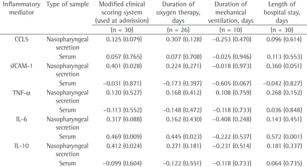
The analysis of the correlations between disease severity at hospital admission, as deter-mined with the modified clinical scoring system, and the concentrations of inflammatory media-tors in the nasopharyngeal secretion and in the serum (first specimens) of the patients with LRTI caused by RSV revealed that disease severity presented a significant positive correlation with the concentrations of sICAM-1 (r = 0.401; p = 0.028) and IL-10 (r = 0.412; p = 0.024) in the nasopharyngeal secretion, as well as with the concentrations of IL-6 (r = 0.469; p = 0.009) in the serum (Table 3).
Table 3 - Correlations of high concentrations of inflammatory mediators in the nasopharyngeal secretion and in the serum with clinical markers of the severity of the disease caused by respiratory syncytial virus.a
Inflammatory mediator
Type of sample Modified clinical scoring system (used at admission)
Duration of oxygen therapy,
days
Duration of mechanical ventilation, days
Length of hospital stay,
days
(n = 30) (n = 26) (n = 10) (n = 30)
CCL5 Nasopharyngeal secretion
0.325 (0.079) 0.307 (0.128) −0.253 (0.470) 0.096 (0.614)
Serum 0.057 (0.765) 0.077 (0.708) −0.025 (0.946) 0.113 (0.553) sICAM-1 Nasopharyngeal
secretion
0.401 (0.028) 0.224 (0.271) −0.018 (0.973) 0.360 (0.051)
Serum −0.031 (0.871) −0.173 (0.397) −0.605 (0.067) −0.042 (0.827) TNF-α Nasopharyngeal
secretion
0.120 (0.527) 0.168 (0.412) 0.108 (0.759) 0.268 (0.152)
Serum −0.113 (0.552) −0.148 (0.472) −0.118 (0.733) 0.036 (0.848) IL-6 Nasopharyngeal
secretion
0.317 (0.088) 0.162 (0.430) −0.408 (0.248) 0.143 (0.451)
Serum 0.469 (0.009) 0.445 (0.023) −0.222 (0.537) 0.572 (0.001) IL-10 Nasopharyngeal
secretion
0.412 (0.024) 0.271 (0.181) −0.231 (0.514) 0.181 (0.337)
therapy and hospital stay. Some authors(25-27) have also demonstrated that the increase in the concentrations of pro-inflammatory and anti-inflammatory mediators at the onset of the infection is related to the greater severity of the disease caused by RSV.
The association between high IL-10 levels in the nasopharyngeal secretion and the severity of the respiratory condition possibly occurred due to the immunosuppressive behavior of this cytokine.(28,29) The higher morbidity of the children with a moderate to severe respiratory condition can be partially explained by the more intense activity of the inflammatory cascade in some individuals, with an increase in the damage to the respiratory epithelium already damaged by RSV activity, and by the presence of coinfection with other etiologic agents at hospital admis-sion. Despite this, there were no deaths in our sample, partly due to the absence of specific risk factors for severe disease caused by RSV, such as bronchopulmonary dysplasia and congenital heart disease, and also due to the fact that most of our patients were born at term.
One of the limitations of the present study was the fact that we did not evaluate the asso-ciation between the severity of the respiratory condition caused by RSV and the genetic poly-morphisms of the cytokines. This will be the object of new studies involving larger samples. Real-time RT-PCR was not performed for all of the most common respiratory viruses due to the difficulty in collecting nasopharyngeal samples from suckling infants and also due to the attempt at reducing the risk of hypoxia and trauma caused by prolonged aspirations.
In conclusion, we believe that this study serves as a basis to suggest that, at hospital admission, the concentrations of the pro-inflam-matory mediators sICAM-1 in the nasopharyngeal secretion and IL-6 in the serum, as well as the concentration of the regulatory cytokine IL-10 in the nasopharyngeal secretion, constitute good parameters for evaluating the inflammatory and immune response in LRTI caused by RSV. These concentrations can be used as markers of disease severity. These observations could contribute to the development of new therapeutic strategies aimed at the immunomodulation of the disease caused by RSV.
genesis of the inflammatory and immune proc-esses caused by RSV and the absence of studies correlating the levels of these mediators with the severity of the respiratory disease caused by RSV in children less than three months of age.
The importance of cytokines and chemok-ines in the severity of LRTIs caused by RSV has yet to be fully elucidated. Previous studies have demonstrated that specific inflammatory media-tors and their gene polymorphisms, as well as the imbalance in the immune response,(21) can contribute to the severity of the viral disease.(22) The proper production of pro-inflammatory and anti-inflammatory mediators promotes a potent antiviral activity, reducing the pathogenesis, the morbidity and the mortality of the respiratory disease caused by RSV. Younger age translates to a greater difficulty in regulating the produc-tion of pro-inflammatory and anti-inflammatory mediators.(23,24)
In Brazil, this is the first study to evaluate whether the concentrations of inflammatory mediators (CCL5, sICAM-1, TNF-α, IL-6 and IL-10) in the nasopharyngeal secretion and in the serum correlate with the clinical markers of the severity of LRTI caused by RSV in patients less than three months of age. In our study, the patients with higher concentrations of sICAM-1 and IL-10 in the nasopharyngeal secretion and higher concentrations of IL-6 in the serum (Table 3) presented with a more severe respiratory condition at hospital admission, as determined with the clinical scoring system adapted from De Boeck et al.(10) The patients with higher concen-trations of IL-6 at hospital admission required prolonged oxygen therapy and hospital stays.
in hospitalized and ambulatory children. J Infect Dis. 1990;162(6):1283-90.
12. Armitage P, Berry G. Statistical methods in medical research. 3rd ed. Oxford: Blackwell Scientific Publications; 1994.
13. Chung HL, Kim SG. RANTES may be predictive of later recurrent wheezing after respiratory syncytial virus bronchiolitis in infants. Ann Allergy Asthma Immunol. 2002;88(5):463-7.
14. Wang CM, Tang RB, Chung RL, Hwang BT. Tumor necrosis factor-alpha and interleukin-6 profiles in children with pneumonia. J Microbiol Immunol Infect. 1999;32(4):233-8.
15. Anderson LJ, Parker RA, Strikas RL. Association between respiratory syncytial virus outbreaks and lower respiratory tract deaths of infants and young children. J Infect Dis. 1990;161(4):640-6.
16. Avendaño LF, Larrañaga C, Palomino MA, Gaggero A, Montaldo G, Suárez M, et al. Community- and hospital-acquired respiratory syncytial virus infections in Chile. Pediatr Infect Dis J. 1991;10(8):564-8.
17. Miyao CR, Gilio AE, Vieira S, Hein N, Pahl MM, Betta SL, et al. Viral infections in hospitalized children affected by acute lower respiratory tract disease [Article in Portuguese]. J Pediatr (Rio J). 1999;75(5):334-44. 18. Weber MW, Dackour R, Usen S, Schneider G, Adegbola
RA, Cane P, et al. The clinical spectrum of respiratory syncytial virus disease in The Gambia. Pediatr Infect Dis J. 1998;17(3):224-30.
19. Bermejo-Martin JF, Garcia-Arevalo MC, De Lejarazu RO, Ardura J, Eiros JM, Alonso A, et al. Predominance of Th2 cytokines, CXC chemokines and innate immunity mediators at the mucosal level during severe respiratory syncytial virus infection in children. Eur Cytokine Netw. 2007;18(3):162-7.
20. Jafri HS, Carubelli CM, Sheeran P, Saavedra J, Sanchez PJ, Ramilo O. Systemic IL-6, IL-8, and RANTES response in children with respiratory syncytial virus disease. Pediatr Res. 1999;45(4):A164.
21. Legg JP, Hussain IR, Warner JA, Johnston SL, Warner JO. Type 1 and type 2 cytokine imbalance in acute respiratory syncytial virus bronchiolitis. Am J Respir Crit Care Med. 2003;168(6):633-9.
22. Garofalo RP, Patti J, Hintz KA, Hill V, Ogra PL, Welliver RC. Macrophage inflammatory protein-1alpha (not T helper type 2 cytokines) is associated with severe forms of respiratory syncytial virus bronchiolitis. J Infect Dis. 2001;184(4):393-9.
23. Bennett BL, Garofalo RP, Cron SG, Hosakote YM, Atmar RL, Macias CG, et al. Immunopathogenesis of respiratory syncytial virus bronchiolitis. J Infect Dis. 2007;195(10):1532-40.
24. McNamara PS, Flanagan BF, Selby AM, Hart CA, Smyth RL. Pro- and anti-inflammatory responses in respiratory syncytial virus bronchiolitis. Eur Respir J. 2004;23(1):106-12.
25. Hornsleth A, Klug B, Nir M, Johansen J, Hansen KS, Christensen LS, et al. Severity of respiratory syncytial virus disease related to type and genotype of virus and to cytokine values in nasopharyngeal secretions. Pediatr Infect Dis J. 1998;17(12):1114-21.
26. Hornsleth A, Loland L, Larsen LB. Cytokines and chemokines in respiratory secretion and severity of
Acknowledgments
The authors would like to thank the techni-cians of the Virology Laboratory of the Adolfo Lutz Institute in the city of São Paulo, Brazil, and Professor Edison Luiz Durigon, Head of the Virology Laboratory of the USP Institute of Biomedical Sciences II, for performing the specific tests for respiratory viruses. We are also grateful to Lídia Yamamoto and to Professor Thelma Suely Okay, from the Laboratório de Investigação Médica 36 (LIM 36, Laboratory for Medical Research 36) of the HC-FMUSP Institute for Children, for the analysis of inflammatory mediators.
References
1. Macedo SE, Menezes AM, Post P, Albernaz E, Knorst M. Infecção pelo vírus respiratório sincicial em crianças menores de um ano de idade internadas por doença respiratória aguda em Pelotas, RS. J Pneumol. 2003;29(1):4-8.
2. Berman S. Epidemiology of acute respiratory infections in children of developing countries. Rev Infect Dis. 1991;13 Suppl 6:S454-62.
3. Filippell MB, Rearick T. Respiratory syncytial virus. Nurs Clin North Am. 1993;28(3):651-71.
4. Vieira RA, Diniz EM, Vaz FA. Clinical and laboratory study of newborns with lower respiratory tract infection due to respiratory viruses. J Matern Fetal Neonatal Med. 2003;13(5):341-50.
5. Diniz EM, Vieira RA, Ceccon ME, Ishida MA, Vaz FA. Incidence of respiratory viruses in preterm infants submitted to mechanical ventilation. Rev Inst Med Trop Sao Paulo. 2005;47(1):37-44.
6. Abbas AK, Lichtman AH. Cytokines. In: Abbas AK, Lichtman AH, editors. Cellular and molecular immunology. Philadelphia: Saunders; 2005. p. 243-74. 7. Mariscalco MM. Integrins and cell adhesion molecules.
In: Polin RA, Fox WW, Abman SH, editors. Fetal and neonatal physiology. Philadelphia: Saunders; 2004. p. 1572-91.
8. Gill MA, Long K, Kwon T, Muniz L, Mejias A, Connolly J, et al. Differential recruitment of dendritic cells and monocytes to respiratory mucosal sites in children with influenza virus or respiratory syncytial virus infection. J Infect Dis. 2008;198(11):1667-76.
9. McNamara PS, Smyth RL. The pathogenesis of respiratory syncytial virus disease in childhood. Br Med Bull. 2002;61(1):13-28.
10. De Boeck K, Van der Aa N, Van Lierde S, Corbeel L, Eeckels R. Respiratory syncytial virus bronchiolitis: a double-blind dexamethasone efficacy study. J Pediatr. 1997;131(6):919-21.
virus infection in young children. Pediatr Allergy Immunol. 2007;18(2):94-9.
29. Helminen M, Nuolivirta K, Virta M, Halkosalo A, Korppi M, Vesikari T, et al. IL-10 gene polymorphism at -1082 A/G is associated with severe rhinovirus bronchiolitis in infants. Pediatr Pulmonol. 2008;43(4):391-5.
disease in infants with respiratory syncytial virus (RSV) infection. J Clin Virol. 2001;21(2):163-70.
27. Oda K, Yamamoto Y. Serum interferon-gamma, interleukin-4, and interleukin-6 in infants with adenovirus and respiratory syncytial virus infection. Pediatr Int. 2008;50(1):92-4.
28. Chung HL, Park HJ, Kim SY, Kim SG. Age-related difference in immune responses to respiratory syncytial
About the authors
Renata Amato Vieira
Attending Physician. Unidade de Cuidados Intensivos Neonatal – UCINE, Neonatal Intensive Care Unit – University of São Paulo School of Medicine Hospital das Clínicas Institute for Children, São Paulo, Brazil.
Edna Maria de Albuquerque Diniz
Associate Professor. University of São Paulo School of Medicine, São Paulo, Brazil.
Maria Esther Jurfest Rivero Ceccon