J Vasc Bras. 2012;11(3):256-262.
Abstract
Introduction: Despite the high mortality rates of hemodialysis patients from cardiovascular diseases, diagnostic tests for peripheral arterial occlusive disease (PAOD) and poorly compressible arteries are not routinely performed.
Objectives: To analyze the prevalence of peripheral arterial disease and poorly compressible arteries in hemodialysis patients, by comparing them to a Control Group.
Methods: his is a cross-sectional study of 78 hemodialysis patients and 88 non-hemodialysis patients with at least two risk factors for peripheral arterial disease (Control Group). Arterial blood pressure of both lower limbs at the ankle was evaluated by portable vascular Doppler and sphygmomanometer. he arterial blood pressure of the upper limb without arteriovenous istula was measured. he ankle-brachial index was calculated for each artery of the lower limb. Values from 0.9 to 1.3 were considered normal.
Results: PAOD and poorly compressible arteries were diagnosed in 26.9 and 30.8% of hemodialysis patients and in 33 and 22.7% of the Control Group. In hemodialysis patients, we found abnormal ankle-brachial index in 75% of symptomatic patients (p=0.005), in 67.3% of men and 31% of women (p<0.005), in 78.6% of the elderly, 34.8% of young adults (p<0.01), and 76.9% of diabetics (p<0.005) versus nondiabetics. Peripheral arterial disease was more common among these patients than those from Control Group.
Conclusions: Abnormal ankle-brachial index was common in the groups studied; however, hemodialysis patients had more severe alterations when compared to the Control Group. Diabetes mellitus, male gender, and advanced age were important risk factors for abnormal ankle-brachial index in hemodialysis patients. he ABI is a good method for the diagnosis of the studied changes; therefore, we suggest it to be routinely measured in hemodialysis patients.
Keywords: ankle-brachial index; dialysis; peripheral vascular diseases.
Resumo
Introdução: Apesar da elevada mortalidade dos pacientes em hemodiálise devido às doenças cardiovasculares, é incomum a realização de exames diagnósticos para doença arterial obstrutiva periférica e artérias pouco compressíveis.
Objetivos: Analisar a prevalência de doença arterial obstrutiva periférica e artérias pouco compressíveis em hemodialisados, comparando-os com o Grupo Controle.
Métodos: Tratou-se de um estudo transversal, com 78 hemodialisados e 88 pacientes que não faziam hemodiálise com, pelo menos, dois fatores de risco para doença arterial obstrutiva periférica (Grupo Controle). Para aferição da pressão arterial sistólica, utilizou-se Doppler vascular portátil e esigmomanômetro. Esta foi aferida somente nos membros que não possuíam fístula arteriovenosa. O índice tornozelo-braço foi calculado utilizando cada artéria do membro inferior. Foram considerados normais os valores de 0,9 a 1,3.
Resultados: Diagnosticou-se doença arterial obstrutiva periférica e artérias pouco compressíveis em 26,9 e 30,8%, dos hemodialisados, e em 33 e 22,7%, do Grupo Controle. Nos hemodialisados, veriicou-se o índice tornozelo-braço alterado em 75% dos sintomáticos (p=0,005), em 67,3% dos homens e 31% das mulheres (p<0,005), em 78,6% dos idosos, 34,8% dos adultos jovens (p<0,01) e em 76,9% dos diabéticos (p<0,005 versus não diabéticos). Esses pacientes apresentaram maior prevalência de doença arterial obstrutiva periférica grave do que o Grupo Controle (p<0,01).
Conclusões: O índice tornozelo-braço anormal foi muito prevalente nos grupos estudados; entretanto, os hemodialisados apresentaram alterações mais graves quando comparados ao Grupo Controle. Diabetes melito, sexo masculino e idade avançada foram fatores de risco importantes para a alteração do índice tornozelo-braço nos hemodialisados. O índice tornozelo-braço foi um bom método de rastreio para alterações pesquisadas. Portanto, a utilização deste na rotina de manejo de pacientes em hemodiálise é sugerida.
Palavras-chave: índice tornozelo-braço; diálise; doenças vasculares periféricas.
Ankle-brachial index in hemodialysis patients
Índice tornozelo-braço em pacientes hemodialíticos
Mariane Torres Uchôa1, Diego Nunes de Albuquerque Oliveira2, Maria Eliete Pinheiro3,
Daniella Bezerra Duarte4, Jairo Calado Cavalcante5, Glauber Schettino Silva6, Marcos Mota Gomes7
From the Hospital Universitário of Universidade Federal de Alagoas, at Clinicor, and at the Hospital Ortopédico de Maceió – Maceió (AL), Brazil.
1 Physician, Universidade Federal de Alagoas (Ufal) – Maceió (AL), Brazil. 2 Physician, Universidade Federal de Alagoas (Ufal) – Maceió (AL), Brazil.
3 Nephrologist; Professor (PhD) at Universidade Federal de Alagoas (Ufal) – Maceió (AL), Brazil. 4 Nephrologist; Professor at Universidade Federal de Alagoas (Ufal) – Maceió (AL), Brazil.
5 Physician; Specialist of Epidemiology; Professor (MD) at Universidade Federal de Alagoas (Ufal) – Maceió (AL), Brazil. 6 Physical herapist at Centro de Pesquisas Clínicas da Clinicor – Maceió (AL), Brazil.
7 Cardiologist; Coordinator of the Centro de Pesquisas Clínicas da Clinicor – Maceió (AL), Brazil.
Submitted on: 19.12.11. Accepted on: 18.06.12. Financial source: none
of Nephrology, and 88 patients not on hemodialysis from a private service of Cardiology (Control Group).
he hemodialysis group included patients older than 18 years on renal replacement therapy by hemodialysis, patients with a functioning arteriovenous istula in upper limb, patients that were not on dialysis for acute renal failure and had no amputations of both lower limbs or the upper limb contralateral to the istula.
he control group included patients older than 18 years with at least two risk factors for PAOD, without amputations of upper or lower limbs, and no istulas or chronic kidney disease. Patients from three dialysis centers meeting the inclusion criteria were invited to participate in the study. Two patients who refused to be examined, one that complained of severe pain at lower limb blood pressure measurement, and one patient with undetectable blood pressure in the upper limb were excluded from the sample.
he research project was approved by the Ethics Committee of the University to which the study is related (procedure 009707/2010-11). All patients signed an informed consent form before data collection. Demographic data were collected through interviews with individuals in control group, which included questions on drugs taken and presence of comorbidities.
In the hemodialysis group, we collected the same information plus signs and/or symptoms related to PAOD (rest pain, intermittent claudication, signs of hypoperfusion, history of lower extremity revascularization, or amputation and necrosis of the ingers, feet or one of the lower limbs).
Patients whose medical record had the diagnosis of systemic arterial hypertension, those with blood pressure measurement greater than 140 × 90 mmHg on the day of examination, and those using anti-hypertensive drugs were classiied as hypertensive17.
Patients with diagnosis of diabetes in medical records or in regular use of antidiabetic drugs were classiied as diabetics18.
Ater the interview, physical examination was performed and anthropometric data was collected.
he patient was placed in supine position for at least 5 minutes before the hemodialysis session (in the case of Hemodialysis Group) in order for the systolic blood pressure of the lower limbs’ arteries (posterior tibial and dorsalis pedis arteries) to be measured, as well as both upper limbs’ arteries (Control Group) or only the limb that was contralateral to the arteriovenous istula (hemodialysis patients).
Systolic blood pressure was measured twice in each artery by diferent examiners previously trained, using portable vascular Doppler ultrasound (model Introduction
Despite the high mortality rates from cardiovascular diseases reported in hemodialysis patients, diagnostic tests for poorly compressible arteries and peripheral arterial occlusive disease (PAOD) are not commonly performed, even though these entities are frequent in patients with chronic kidney disease1-9. Cardiovascular mortality in
hemodialysis patients is 5-30 times higher than in the general population10, and PAOD is an independent
predictor of heart failure11.
Some scientiic associations disagree that chronic kidney disease implies PAOD risk12, but most authors conirm and
emphasize the importance of this relationship2,13.
Regardless of the controversy over the classiication of chronic kidney disease as a high-risk factor for PAOD, there are very few studies to allow the resolution of this impasse and to evaluate for such diseases in hemodialysis patients. he recommendations for PAOD diagnosis in patients with chronic renal failure by the Kidney Disease Outcomes Quality Initiative (K/DOQI), for instance, are based on studies with pre-dialysis patients and on experts’ opinions1,13.
Both PAOD and poorly compressible arteries are associated with increased mortality rates (from cardiovascular or other causes) in patients with chronic renal disease14,15. In hemodialysis patients, this increase is
present even in cases of asymptomatic PAOD1. he search
for symptoms and pulse palpation in the lower limb arteries are inefective for the diagnosis of these diseases.
Ankle-brachial index (ABI) is a low-cost and non-invasive method8 which has 95% sensitivity and 99%
speciicity for PAOD compared to angiography (gold-standard pattern)2. Abnormal ABI values are associated
with higher morbimortality rates7,16, hence it is considered
to be a reliable prognosis predictor. ABI measurement in hemodialysis patients is not routinely performed, so the prevalence of PAOD and poorly compressible arteries are not properly documented.
he aim of this study was to analyze the prevalence of PAOD and poorly compressible arteries in a group of patients on hemodialysis and to compare the indings with a control group at high risk for both diseases. he ABI values were then correlated with risk factors or signs and symptoms found in patients with chronic kidney disease.
Methods
he results with error probability inferior to 5% (p<0.05) were considered statistically signiicant.
Results
In the hemodialysis group, mean age was 49.07 ± 14.89 (mean -X ± standard deviation - SD) and mean time on dialysis was 36.90 ± 35.65 months (X ± SD).
Males and caucasian patients were more common in the dialysis group, with 49 and 34, respectively. his group had 76 (97.4%) patients with hypertension, 10 (12.8%) smokers, 11 (14.1%) former smokers and 24 (30.8%) patients with coronary disease.
In the control group, the mean age was 61.09 ± 12.07 years, and there were 52 (59.1%) male patients, 42 (47.7%) caucasians, 57 (64.8% ) patients with hypertension, 14 (15.9%) smokers, 34 (38.6%), former smokers and 54 (61.4%) patients with coronary artery disease, which characterizes a group at high risk for peripheral arterial disease.
he prevalence of diabetes mellitus and cerebrovascular diseases were similar in both groups, which allocated nine patients with cerebrovascular disease and a similar number of diabetic patients: 26 in the hemodialysis group and 25 in the control group.
Systolic blood pressure measurements were higher than the values recommended by the Brazilian Society of Hypertension17 (Table 1).
Age groups showed to be statistically signiicant in the analysis, as shown in Figure 1.
Twenty-eight patients on hemodialysis (35.9%) presented the signs and/or symptoms searched for; however, only 12 (42.9%) patients had an ABI < 0.9, and 11 (39.3%) had an ABI > 1.3. Twenty-one (75%) symptomatic patients had abnormal ABI, with a relative risk of 1.79 (p = 0.005).
Five patients who had tissue necrosis or amputation of ingers, feet or limbs (6.4% of hemodialysis patients) had abnormal ABI (p<0.05), and no patient reported history of peripheral revascularization.
2001, Medpej® in São Paulo, Brazil) and a calibrated sphygmomanometer (aneroid mechanical model, BD®, Germany).
he ABI was obtained by simple division of the higher systolic blood pressure obtained in each artery of the lower limb by the values obtained in the upper limbs (Control Group) of those not presenting arteriovenous istula (hemodialysis patients).
In order to increase the method’s sensitivity, up to two ABI values were obtained from each lower limb of each patient. he indexes were then included in the database, because considering only the highest blood pressure values in a certain extremity would underestimate the poorly compressible arteries, since some patients present both alterations in the same limb.
ABI was considered abnormal when lower than 0.9 (indicative of PAOD)1-3,8,11,15,19-26 or higher than 1.3
(indicative of poorly arterial compressibility)1,3,8,19,25. ABI
between 0.7 and 0.9 were considered to indicate mild PAOD; between 0.4 and 0.7, moderate PAOD; and below 0.4, severe PAOD24.
Poorly compressible arteries were those that could not be compressed, that is, not showing bruit fading (Korotkof Phase V), even when inlating the cuf at 300 mmHg.
Arteries with inaudible signals were those that could not be identiied by portable vascular Doppler ultrasound performed by both examiners.
Ater gathering of information and patients’ examination, the data was typed in a database program (Epi-Info Windows, version 3.4.3) for statistical analysis.
Qualitative variables were measured by the χ2 test and
Odds Ratio.
Numerical variables were assessed by means of ANOVA test and showed the rang homogeneity by Bartlett’s test. Aterwards, the non-parametric Kruskal-Wallis test was used when necessary.
Table 1. Systolic blood pressure values and ankle-brachial index in he-modialysis and control patients
Variables HP Control Group P value
BP UL – mmHg 155.79±29.21 144.13±25.64 0.007a
BP RUE – mmHg 181.11±61.50 155.26±38.91 0.001k
BP RLE – mmHg 177.9±53.82 151.01±32.19 0.000k
ABI RLE 1.17±0. 37 1.09±0.26 0.066k
ABI LLE 1.15±0.90 1.06±0.50 0.014k
HP: hemodialysis patients; BP: blood pressure; ABI: ankle-brachial index; UE: upper extre-mity; RLE: right lower extreextre-mity; LLE: left lower extreextre-mity; aANOVA test; kKruskal-Wallis
test; results expressed in mean±standard deviation. HP: hemodialysis patients; CG: Control Group.
only the hemodialysis group was found to be statistically signiicant as to this diference (p<0.005), where ABI was abnormal in 33 men and 9 women, with a relative risk of 1.77.
In the group of patients on hemodialysis, a statistically signiicant diference was observed in elderly and young adults with altered ABI (p <0.01), unlike the control group, in which only four individuals were considered to be young adults. Out of the 14 elderly patients in the hemodialysis group, 11 (78.6%) had abnormal ABI, as well as 8 out of the 23 young adults in this group (34.8%).
hus, patients over 65 years of age were found to be more likely to present abnormal ABI compared to those aged 20 to 39 years and relative risk of 2.26.
In addition, 21 patients (half of the hemodialysis group) presenting abnormal ABI were asymptomatic, and eight of them presented ABI < 0.9.
No statistical diference was found as to the prevalence of altered ABI between the groups, although poorly compressible arteries were more prevalent in patients on hemodialysis, and PAOD afected more patients in the control group (Table 2).
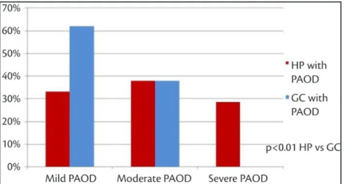
Figures 2 and 3 show that the most signiicant alterations afected more patients from the hemodialysis group than those from the control group. hus, no subjects in the control group showed ABI < 0.4, while 29% of patients on hemodialysis with PAOD (6 patients) had a more severe form of the disease.
More than half of the patients diagnosed with PAOD in the control group had the disease in its mild form (25 patients), whereas only 15 patients in the hemodialysis group were classiied as such.
Also, 10 (23.8%) hemodialysis patients with abnormal ABI had poorly compressible arteries versus 3 (6.8%) from
the other group presenting altered ABI and and relative risk of 3.49 (p<0.05).
No patient in the control group presented inaudible arterial low, while this inding was observed in 7 (16.7%) hemodialysis patients with abnormal ABI (p<0.005).
As one may note from Figure 4, altered ABI was more common among males than females in both groups, but
Table 2 - Prevalence of normal and abnormal ABI values
ABI interpretation
HP (n – %)
Control Group
(n – %) P value
Normal ABI 36-46.2 44-50.0 NS
PAOD 21-26.9 29-33.0 NS
Poorly compressible arteries 24-30.8 20-22.7 NS
Both alterations 3-3.8 5-5.7 NS
HP: hemodialysis patients; ABI: ankle-brachial index; PAOD: peripheral arterial occlusive disease; NS: non signiicant; qχ2 test.
PAOD: peripheral arterial occlusive disease; HP: hemodialysis patients; CG: Control Group. Figure 2. Degrees of PAOD in hemodialysis and control patients.
HP: hemodialysis patients; CG: Control Group; ABI: ankle-brachial index.
Figure 3. Prevalence of noncompressible and inaudible arteries in he-modialysis and control patients.
HP: hemodialysis patients; CG: Control Group; ABI: ankle-brachial index.
which was also observed by De Loach and Mohler1 and is
highlighted in the K/DOQI guidelines13 and Transatlantic
Inter-Society Consensus Document on Management of Peripheral Arterial Disease (TASC II)2.
Liew et al.24 stated that patients with chronic kidney
disease and PAOD are at a signiicantly higher risk of death than patients with either disease alone. his inding justiies the continuous searching for PAOD diagnosis and poorly compressible arteries in patients on hemodialysis.
he inclusion of end-stage chronic kidney disease as a signiicant risk factor for cardiovascular diseases can be explained both by the traditional and nontraditional cardiovascular risk factors, the latter acting the development of atherosclerosis. Among the traditional risk factors, the most common are diabetes, hypertension, sedentary lifestyle, hypertriglyceridemia, smoking, and low HDL, the former being the most signiicant factor related to the onset of cardiovascular diseases and PAOD1,10,30-33.
As it was noted, eight hemodialysis patients who were diagnosed with PAOD by ABI were asymptomatic, which proves the eicacy of ABI in PAOD diagnosis compared to the method used as routine in clinical practice for this purpose (search for symptoms and pulses of lower limb arteries). Moreover, it makes one relect on the large number of patients with PAOD that go unnoticed in hemodialysis services that do not measure ABI routinely.
A high prevalence of asymptomatic patients with abnormal ABI was also described by O’Hare3, Carmo et al.19
and Makdisse et al.11, which underscores the importance of
ABI as a method for PAOD diagnosis.
Seven symptomatic patients on hemodialysis (25%) had normal ABI. However, patients presenting with tissue necrosis or that had been submitted to some type of amputation had alterations on ABI.
he high rate of amputations in hemodialysis patients is attributed to chronic kidney disease and to the high prevalence of diabetes mellitus1,2,34. he absence of ABI
measurement in patients at the services included in this study is another factor contributing to high amputation rates.
As expected, old age increased the risk of abnormal ABI in both groups, but most frequently among hemodialysis patients. he studies of Ono et al.14, De Loach and Mohler1,
Cheung et al.6 and Resnick et al.15 also showed that advanced
age and the presence of diabetes mellitus are associated with alterations in ABI.
Ater the ith year of hemodialysis, a decrease in the prevalence of the studied variables was observed. his may be related to the high mortality rates in this population, which is in agreement with De Loach and Mohler1, Sesso35
Another important risk factor analyzed is diabetes mellitus. he prevalence of alterations in ABI among diabetic patients is higher than that of non-diabetics, a statistically signiicant relation in the hemodialysis group (p <0.005). In this group, 20 (76.9%) patients with diabetes presented abnormal ABI, with a relative risk of 2.86.
he diabetic patients of both groups showed a prevalence of poorly compressible arteries slightly higher than individuals without the disease, but only in hemodialysis patients were considered statistically signiicant as to this feature (p<0.01). herefore, seven patients on hemodialysis with poorly compressible arteries were identiied.
Among patients included in an hemodialysis program for up to ive years, the frequency of abnormal ABI increased gradually. From this period on, a decrease of this frequency was noted.
hus, we found alterations in ITB in 51.6% of patients on hemodialysis for up to one year, in 60% of patients on the program for one to three years, in 64.3% those being treated for three to ive years, and in 50% of patients on hemodialysis for more than ive years.
Discussion
he predominance of males in the hemodialysis group was similar to that of other studies conducted with patients undergoing hemodialysis1,10,14, but it difers from indings
in studies with pre-dialysis patients19. One explanation for
this is that men go on to stage 5 of chronic kidney disease more oten than women, in which the estrogen hormone functions as protective factor, favoring angiogenesis and slowing the progression of the disease to the terminal stage27,28.
Smoking, male gender, diabetes mellitus, advanced age, coronary and cerebrovascular diseases are risk factors for ABI alterations and high mortality1,6,9,14. As indicated,
the prevalence of these factors in patients from the control group was higher than in the hemodialysis group, which sets up a group at high risk for PAOD.
he high prevalence of hypertension and diabetes mellitus in patients undergoing hemodialysis, which is compatible with the indings of studies by Ono et al.14 and
Longenecker et al.10, was expected because these are the
leading causes of chronic kidney disease29, besides being
3. O’Hare AM. Peripheral arterial disease in chronic kidney disease. [internet]. Up to date. 2009 [cited 2010 Feb 02]. Available from: http://www.utdol.com/patients/content/topic. do?topicKey=~ROSOn9Yr_DYNx3l.
4. Canziani MEF. Doenças Cardiovasculares na Doença Renal Crônica. J Bras Nefrol. 2004;26(3):20-1.
5. Martin LC, Franco RJS. Renal disease as a cardiovascular risk factor. Arq Bras Cardiol. 2005;85(6):1-5. http://dx.doi.org/10.1590/S0066-782X2005001900011
6. Cheung AK, Sarnak MJ, Yan G, et al. Atherosclerotic cardiovascular disease risks in chronic hemodialysis patients. Kidney Int. 2000;58(1):353-62. PMid:10886582. http://dx.doi.org/10.1046/ j.1523-1755.2000.00173.x
7. Canziani MEF, Moysés RMA. Calciicação Vascular na DRC. J Bras Nefrol. 2008;30(Suppl 2):23-6. http://dx.doi.org/10.1590/S0101-28002011000200017
8. Vinuesa SG, Ortega M, Martinez P, Goicoechea M, Campdera FG, Luño J. Subclinical peripheral arterial disease in patients with chronic kidney disease: Prevalence and related risk factors. Kidney Int Suppl. 2005;(93):S44-7. PMid:15613068.
9. O’Hare AM, Hsu CY, Bacchetti P, Johansen KL. Peripheral Vascular Disease Risk Factors among Patients Undergoing Hemodialysis. J Am Soc Nephrol. 2002;13(2):497-503. PMid:11805180.
10. Longenecker JC, Coresh J, Powe NR, et al. Traditional cardiovascular disease risk factors in dialysis patients compared with the general population: the CHOICE study. J Am Soc Nephrol. 2002;13(7):1918-27.
11. Makdisse M, Pereira AC, Brasil DP, et al. Prevalência e fatores de risco associados à doença arterial periférica no projeto Corações do Brasil. Arq Bras Cardiol. 2008;91(6):402-14. http://dx.doi. org/10.1590/S0066-782X2008001800008
12. Hirsch AT, Haskal ZJ, Hertzer NR, et al. ACC/AHA 2005 Practice Guidelines for the management of patients with peripheral arterial disease (lower extremity, renal, mesenteric, and abdominal aortic): A collaborative report from the American Association for Vascular Surgery/Society for Vascular Surgery, Society for Cardiovascular Angiography and Interventions, Society for Vascular Medicine and Biology, Society of Interventional Radiology, and the ACC/AHA Task Force on Practice Guidelines. Circulation. 2005;113:1474-547. PMid:16990459.
13. National Kidney Foundation. K/DOQI Clinical practice guidelines for chronic kidney disease: Evaluation, classiication and stratiication. Am J Kidney Dis. 2002;39(2 Suppl 1):S1-266.
14. Ono K, Tsuchida A, Kawai H, et al. Ankle-brachial blood pressure index predicts all-cause and cardiovascular mortality in hemodialysis patients. J Am Soc Nephrol. 2003;14(6):1591-8. PMid:12761260. http://dx.doi.org/10.1097/01.ASN.0000065547.9825
15. Resnick HE, Lindsay RS, McDermott MM, et al. Relationship of high and low ankle brachial index to all-cause and cardiovascular disease mortality: the strong heart study. Circulation. 2004;109(6):733-9. http://dx.doi.org/10.1161/01.CIR.0000112642.63927.54
16. Chen S, Chang J, Hwang S, et al. Signiicant correlation between ankle-brachial index and vascular access failure in hemodialysis patients. Clin J Am Soc Nephrol. 2009;4(1):128-34. PMid:19141657. e Silva et al.36, which state that the probability of survival
in diabetic patients undergoing hemodialysis for more than ive years are 25%, 23% and 50%, respectively1,35,36.
he prevalence of PAOD in hemodialysis patients was 26.9%, and is in agreement with the indings by O’Hare3,
which states that this value varies from 12 to 32%.
In the hemodialysis group, 30.8% of patients had poorly compressible arteries, a value slightly higher than those found by a Finnish study, in which the prevalence of poorly compressible arteries was estimated to be 24%3.
he higher prevalence of poorly compressible arteries among hemodialysis patients is consistent with data presented by Ono et al.14 and is attributed to the fact that
patients with end-stage renal disease have secondary severe bone dystrophy7,37,38. he authors suggest that the prevalence
of PAOD is likely to be underestimated, for patients with poorly compressible arteries may present it regardless of abnormal ABI25.
Patients in the group of hemodialysis presented with more severe disease compared to those in the control group, as the latter did not present inaudible arteries or severe PAOD. his is very important data, as ABI < 0.5 in symptomatic patients is a strong predictor of amputation2.
Conclusions
PAOD and poorly compressible arteries were frequent indings in both groups. However, hemodialysis patients presented with more severe alterations such as noncompressible or inaudible arteries, or amputation, even when compared with patients who were not on hemodialysis but presented risk factors, i.e. advanced age, smoking and coronary heart disease.
Abnormal ABI in hemodialysis patients, whether indicating PAOD or poorly compressible arteries, was strongly correlated with the variables: male gender, diabetes and advanced age. In half of patients diagnosed with PAOD by ABI, the disease was asymptomatic, so the use of this index as a method of search for vascular alterations should be considered. herefore, we suggest the use of ABI in the routine assessment of patients undergoing hemodialysis.
References
1. De Loach SS, Mohler ER. Peripheral arterial disease: a guide for nephrologists. Clin J Am Soc Nephrol. 2007;2:839-46. PMid:17699501. http://dx.doi.org/10.2215/CJN.04101206
problema. Rev Assoc Med Bras. 2007;53(5):446-50. http://dx.doi. org/10.1590/S0104-42302007000500022
31. Belkin M, Owens CD, Whittemore AD, Donaldson MC, Conte MS, Gravereaux E. Doença Oclusiva Arterial Periférica. In: Townsend CM, Beauchamp RD, Evers BM, Mattox KL, editores. Sabiston Tratado de Cirurgia: A Base Biológica da Prática Cirúrgica Moderna, 18ª ed. Rio de Janeiro: Elsevier; 2010. p. 1823-58. 32. Keane WF, Brenner BM, Zeeuw D, et al. he risk of developing
end-stage renal disease in patients with type 2 diabetes and nephropathy: he RENAAL Study. Kidney Int. 2003;63(4):1499-507. PMid:12631367. http://dx.doi.org/10.1046/j.1523-1755.2003.00885.x
33. Sarnak MJ, Levey AS, Schoolwerth AC, et al. Kidney Disease as a Risk Factor for Development of Cardiovascular Disease: A Statement From the American Heart Association Councils on Kidney in Cardiovascular Disease, High Blood Pressure Research, Clinical Cardiology, and Epidemiology and Prevention. Hypertension. 2003;42(5):1050-65. PMid:14581387.
34. Romão Junior JE. Tratamento de substituição da insuiciência renal crônica. In: Lopes AC, editor. Tratado de clínica médica. São Paulo: Roca; 2006. p. 2810-3.
35. Sesso R. Epidemiologia da doença renal crônica no Brasil e sua prevenção [Internet]. Secretaria de Estado da Saúde; Coordenadoria de Controle de Doenças; Centro de Vigilância Epidemiologia. São Paulo, 2007 [cited 2010 Jan]. Available from: ftp://ftp.cve.saude.sp.gov.br/doc_tec/cronicas/irc_prevprof.pdf 36. Silva LAM, Mezzomo NF, Pansard HM, et al. Sobrevida em
hemodiálise crônica: estudo de uma coorte de 1.009 pacientes em 25 anos. J Bras Nefrol. 2009;31(2):190-7.
37. Boaz M, Weinstein T, Matas Z, Green MS, Smetana S. Peripheral vascular disease and serum phosphorus in hemodialysis: A nested case-control study. Clin Nephrol. 2005;63(2):98-105. PMid:15730051.
38. McCullough PA, Agrawal V, Danielewicz E, Abela GS. Accelerated Atherosclerotic Calciication and Mönckeberg’s Sclerosis: A Continuum of Advanced Vascular Pathology in Chronic Kidney Disease. Clin J Am Soc Nephrol. 2008;3(6):1585-98. PMid:18667741.
Correspondence:
Mariane Torres Uchôa Loteamento Jardim Petrópolis I, quadra CH, 207 – Jardim Petrópolis CEP: 57080-535 – Maceió (AL), Brazil E-mail: [email protected]
Authors’ contribution
Study conception and design: MTU, DNAO, MEP, DBD Data analysis and interpretation: MTU, DNAO, MEP, JCC Data collection: MTU, DNAO, GSS Writing: MTU, DNAO, MEP Critical analysis: MTU, DNAO, MEP, MAMG Final approval*: MTU, DNAO, MEP, DBD, JCC, GSS, MAMG Statistical analysis: MTU, DNAO, JCC Overall responsibility: MEP *All authors have read and approved the inal version of the paper submitted to J Vasc Bras.
17. Sociedade Brasileira de Cardiologia; Sociedade Brasileira de Hipertensão; Sociedade Brasileira de Nefrologia. V Diretrizes Brasileiras de Hipertensão Arterial. Hipertensão. 2006;9(4):121. 18. Sociedade Brasileira de Endocrinologia e Metabologia.
Diabetes Mellitus: Classiicação e Diagnóstico. Projeto Diretrizes da Associação Médica Brasileira e Conselho Federal de Medicina. 2004;1-8.
19. Carmo WB, Pinheiro HS, Bastos MG. Doença Arterial Obstrutiva de Membros Inferiores em Pacientes com Doença Renal Crônica Pré-Dialítica. J Bras Nefrol. 2007;29(3):127-34.
20. Sociedade Brasileira de Angiologia e Cirurgia Vascular. Diagnóstico da Doença Arterial Obstrutiva Periférica (DAOP). J Vasc Bras. 2005;4(3 Suppl 4):222-38.
21. Wittke EI. Associação entre diferentes parâmetros de variabilidade da pressão sistólica fornecidos pela monitorização ambulatorial de pressão arterial (MAPA) e o índice tornozelo-braquial. [dissertação]. Porto Alegre (RS): Universidade Federal do Rio Grande do Sul; 2009.
22. Aragão JA, Reis FP, Borges RR, Aragão MECS, Nunes MAP, Feitosa VLC. Prevalência da doença arterial obstrutiva periférica em doentes com insuiciência renal crônica. J Vasc Bras. 2009;8(4):301-6. http://dx.doi.org/10.1590/S1677-54492009000400004
23. Santos RA, Boas LGCV, Osiro PM, Costa GM, Cordeiro JA, Martins JFV. A importância do índice tornozelo-braquial no diagnóstico da doença carotídea em pacientes hipertensos. Rev Soc Bras Clin Med. 2009;7(5):299-303.
24. Liew YP, Bartholomew JR, Demirjian S, Michaels J, Schreiber Junior MJ. Combined Efect of Chronic Kidney Disease and Peripheral Arterial Disease on All-Cause Mortality in a High-Risk Population. Clin J Am Soc Nephrol. 2008;3(4):1084-9. PMid:2440260. http:// dx.doi.org/10.2215/CJN.04411007
25. Miguel SJB. Calciicações vasculares em pacientes hemodialisados: Correlação entre os achados ultra-sonográicos, radiológicos e clínico-laboratoriais. [dissertação]. Niterói (RJ): Universidade Federal Fluminense; 2009.
26. Gabriel AS, Seraim IPH, Freitas CEM, et al. Doença arterial obstrutiva periférica e índice tornozelo-braço em pacientes submetidos à angiograia coronariana. Rev Bras Cir Cardiovasc. 2007;22(1):49-59. http://dx.doi.org/10.1590/S0102-76382007000100011
27. Kang D, Yu ES, Yoon K, Johnson RL. he impact of gender on progression of renal disease: potential role of estrogen-mediated vascular endothelial growth factor regulation and vascular protection. Am J Pathol. 2004;164(2):679-88. http://dx.doi. org/10.1093/eurheartj/ehp007
28. Stenvinkel P, Wanner C, Metzger T, et al. Inlammation and outcome in end-stage renal failure: Does female gender constitute a survival advantage? Kidney Int. 2002;62(5):1791-8. PMid:12371981. 29. Takaoka HH. Peril de pacientes diabéticos em diálise. J Bras
Nefrol. 2009;31(2):100-4.