A rare venous anatomic variation of the lower limb
Variação anatômica venosa rara em membros inferiores
Melissa Andreia de Moraes Silva1
*
, Hanna Fatima Paranaíba Mesquita1, Iara Gabriel Carneiro1, Arturo Eduardo Krupa1, Seleno Glauber de Jesus Silva1, Rodolfo Souza Cardoso1
Abstract
he anatomy of the venous system of the lower limbs is among the most complex in the human body. In view of this, it is extremely important to know how to identify variations that can afect it, such as congenital malformations, for example. In cases of a rare vascular malformation such as agenesis of deep veins, clinical status may manifest with chronic venous insuiciency, which can progress with edema, hyperpigmentation, and lower limb ulcers. his is very often therefore an incapacitating disease that is diicult to treat. his article describes a case of agenesis of a segment of femoropopliteal vein in the right lower limb of a 36-year-old patient who had edema and large caliber varicose veins in the afected limb.
Keywords: femoral vein; popliteal vein; congenital abnormalities; anatomy; anatomic variant.
Resumo
A anatomia do sistema venoso dos membros inferiores é uma das mais complexas no corpo humano. Devido a essa condição, é de extrema importância saber identiicar variações que possam acometê-la, como as malformações congênitas. Em casos de agenesia de veias profundas, como uma malformação vascular rara, o quadro clínico pode manifestar-se com insuiciência venosa crônica, que pode evoluir com edema, hiperpigmentação e úlcera de membro inferior. Assim, em muitos casos, torna-se uma doença incapacitante e de difícil tratamento. Apresenta-se um caso de agenesia de segmento venoso femoropoplíteo no membro inferior direito em paciente de 36 anos de idade, que cursou com edema e varizes de grosso calibre no membro acometido.
Palavras-chave: veia femoral; veia poplítea; anormalidades congênitas; anatomia; variação anatômica.
1 Faculdade de Medicina de Itajubá – FMIt, Cirurgia Vascular, Itajubá, MG, Brazil.
Financial support: None.
Conlicts of interest: No conlicts of interest declared concerning the publication of this article. Submitted: September 04, 2016. Accepted: November 10, 2016.
INTRODUCTION
The venous anatomy of the lower limbs is highly variable because of venous malformations that occur during later development of the embryo, particularly
during the inal phase of embryogenesis.1 It is believed
that a generalized defect of the mesoderm can cause vascular abnormalities, such as agenesis of deep
veins. Venous malformation is the most common
congenital vascular disorder and is generally a single
lesion.2 However, in 15-20% of cases it will present
as a mixed lesion, combined with other congenital vascular malformations, such as lymphatic and
arteriovenous malformations.3
The femoral vein may be duplicated partially or
throughout its entire length. Occasionally it will
pass through the adductor canal, above the femoral artery, running parallel to it until it joins the deep
vein, forming the common femoral vein.4 A study
of malformations of the femoropopliteal venous segment described four distinct categories of variants:
(1) agenesis, in 0.3% of cases; (2) multiplications,
the femoral vein in isolation in 21% of cases, the popliteal vein in isolation in 2%, and both veins
in 6%; (3) anatomic variant of the paths followed by veins, in 8%; and (4) high junction of the tibial veins, seen in 7% of cases. The principal variation
was seen in the femoral vein, where 6-46% of the
patients exhibited duplicated or multiple vessels.
The same study reported an incidence of agenesis
of the femoropopliteal venous segment of 0.2% in the right lower limb (RLL), 0.4% in the left lower
limb (LLL), and no cases whatsoever in both lower
limbs simultaneously.5
In cases of agenesis of deep veins, clinical status
may manifest with chronic venous insuficiency (CVI), which is a clinical syndrome comprising
varicose veins of lower limbs, dermatosclerosis, edema, hyperpigmentation (ochrodermatitis) and lower limb ulcers and is very often an incapacitating
disease that is dificult to treat. The principal causes of CVI are incompetence of perforating, supericial, and/or deep veins, in the primary form of the disease;
and proximal venous obstruction, arteriovenous
istulas, dysfunction of the musculature in the calf,
and congenital venous malformations, which are all
secondary causes of CVI.6
Venous malformations can often be diagnosed
on the basis of a careful patient history and physical
examination. Invasive examination with phlebography
is often unnecessary for routine diagnosis, but it is
essential for planning treatment.7 Furthermore, the
initial baseline assessment should include an active
search for acute complications related to venous
malformations, such as supericial and deep venous
thromboses and pulmonary embolisms, and for chronic complications and sequelae, such as problems walking and scoliosis with pelvic tilt, which are
common associated disorders. Once a diagnosis of CVI probably caused by agenesis of a deep vein has been conirmed, treatment is founded on lifestyle
changes, compressive stockings, and medications,
since surgery is contraindicated in these cases.8
The objective of this study is to describe a rare case with few reports in the literature of a patient with
CVI, probably caused by agenesis of a femoropopliteal
venous segment in the RLL, who developed large caliber
varicose veins in the affected limb. This project was
approved by the Plataforma Brasil Ethics Committee
in 2015, under hearing number 1.361.567.
CASE DESCRIPTION
The patient (MJB), was a 36-year-old, white, male
production assistant. He presented for a consultation
on 24 May 2014 complaining of varicose veins in the RLL, associated with asymmetrical edema with onset 10 years previously and progressive
deterioration. He stated that he was free from itching, skin lesions, and previous traumatisms. He had no
history of thromboembolic events, venous surgery, or other comorbidities and had no family history of
venous disease.
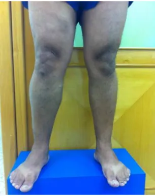
On clinical examination, he exhibited large caliber
tortuous veins in the RLL (Figures 1, 2 and 3) and
mild edema (+/4+). No active or healed lesions of the skin were observed.
A vascular ultrasound examination of the lower limbs with Doppler was unable to detect the popliteal or femoral veins in the RLL, but showed the great and small saphenous veins, which did not have signs
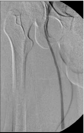
of valvular relux (Figure 4). The investigation was continued with phlebography of the RLL, which
conirmed the absence of the femoral and popliteal
veins (Figures 5, 6, and 7).
After conirmation of the diagnosis, the patient was
prescribed elastic stockings and palliative medication
to control the varicose veins. Countless attempts
were made to contact the patient for follow-up, but he never attended a follow-up consultation so his
progress is unknown.
DISCUSSION
Anatomic abnormalities of the deep vein system are a rare entity caused by disorders of embryonic
In many cases, diagnosis is dificult and patients remain
for long periods being erroneously treated for other vascular diseases that can exhibit similar symptoms
to anatomic anomalies. Physical examination offers the tools needed to construct a diagnosis of CVI, but is insuficient to detect the structures involved or the extent of lesions. The initial work-up examination
is therefore vascular ultrasound with Doppler, as was employed in this case, offering anatomic and hemodynamic analysis of the vascular structures involved, aiding with diagnosis and differentiation of
Figure 2. Lower limbs with patient standing upright, front view,
showing asymmetry of right lower limb.
Figure 1. Lower limbs with patient standing upright, front view,
showing asymmetry of right lower limb.
Figure 3. Lower limbs with patient standing upright, rear view,
showing varicose veins in proximal leg.
Figure 4. Doppler ultrasound showing varicose veins with atypical
types of malformations.9 In this case, the examination
revealed a probable agenesis of the deep vein system in
the RLL, which was later conirmed by phlebography.
A retrospective study conducted with 445 patients (890 limbs), using venous angiotomography to analyze the venous anatomy of patients with varicose veins and edema, found evidence of agenesis of deep femoropopliteal venous segments (as described in
this study) in just 0.3% of cases (three limbs).5
This condition is one of the causes of CVI, which
is currently one of the most frequent diseases affecting
both the Brazilian and the global populations. Since
this is a chronic and progressive disease, around 3-11% of patients with varicose veins may reach advanced stages at which skin complications are
irreversible.10 In the case described here, the patient
presented with burning pains combined with edema of the limb involved, but did not exhibit clinical signs
suggestive of a severe process.
Clinical presentation is variable, ranging from asymptomatic patients to cases with severe venous
ulcers.11 In the case described, the patient exhibited
moderate symptoms of disease unilaterally, with
edema and large caliber varicose veins.
Figure 7. Ascending phlebography showing deep veins of the
leg with anomalous drainage to the great saphenous vein. he popliteal vein was not visible.
Figure 5. Ascending phlebography showing iliac vein and inferior
vena cava with no abnormalities.
Figure 6. Ascending phlebography showing great saphenous
Almost all vascular malformations will beneit
from correctly prescribed and monitored compressive treatment started as soon as possible, preventing
progressive deterioration of clinical status.12 In rarer
cases in which varicose veins are caused by a congenital venous agenesis, it may be erroneously concluded that the deep vein system is patent and that the case
is merely a primary venous insuficiency. However,
these cases are problematic when it comes to surgical
treatment. If an inappropriate strategy is employed, with removal of the supericial varicose veins, the
patient may suffer exacerbation due to overload of the
remaining supericial vein system, and it should be remembered that the deep vein system is incomplete.13
In cases of hypoplasia or agenesis of the deep vein system, surgical treatment for varicose veins is
therefore contraindicated. More appropriate measures
include lifestyle changes, wearing elastic stockings, and prescription of palliative medications,14 as were
adopted in the case described here.
CONCLUSIONS
Lower limb venous anatomy is highly variable, but agenesis of elements of the deep vein system is
rare. Early diagnosis is not always achieved, and the
correct therapeutic choice is of fundamental importance
in these cases. Management should be founded on conservative approaches and resection of supericial veins should be avoided in these limbs.
REFERENCES
1. Uhl JF, Gillot C, Clahim M. Anatomic variations of the femoral vein. J Vasc Surg. 2010;52(3):714-9. PMid:20598472. http://dx.doi. org/10.1016/j.jvs.2010.04.014.
2. Uhl JF, Gillot C. Embryology and three-dimensional anatomy of the superficial venous system of the lower limbs. Phlebology. 2007;22(5):194-206. PMid:18269070. http://dx.doi.org/10.1258/ 026835507782101717.
3. Lee BB, Laredo J, Kim YW, Neville R. Congenital vascular malformations: general treatment principles. Phlebology. 2007;22(6):258-63. PMid:18274333. http://dx.doi.org/10.1258/02683550778265518 2.
4. Sangari SK. Veins of the lower limb, in bergman’s comprehensive encyclopedia of human anatomic variation. In: Tubbs RS, Shoja MM; Loukas M, editores. Hoboken: John Wiley & Sons, Inc.; 2016.
5. Park EA, Chung JW, Lee W, et al. Three-dimensional evaluation of the anatomic variations of the femoral vein and popliteal vein in relation to the accompanying artery by using ct venography. Korean J Radiol. 2011;12(3):327-40. PMid:21603292. http://dx.doi. org/10.3348/kjr.2011.12.3.327.
6. Bergan JJ, Schmid-Schönbein GW, Smith PD, Nicolaides AN, Boisseau MR, Eklof B. Chronic venous disease. N Engl J Med. 2006;355(5):488-98. PMid:16885552. http://dx.doi.org/10.1056/ NEJMra055289.
7. Liu GC, Ferris EJ, Reifsteck JR Jr, Baker ME. Effect of anatomic variations on deep venous thrombosis of the lower extremity. AJR Am J Roentgenol. 1986;146(4):845-8. PMid:3485360. http:// dx.doi.org/10.2214/ajr.146.4.845.
8. Quinlan DJ, Alikhan R, Gishen P, Sidhu PS. Variations in lower limb venous anatomy: implications for US diagnosis of deep vein thrombosis. Radiology. 2003;228(2):443-8. PMid:12821771. http:// dx.doi.org/10.1148/radiol.2282020411.
9. Coleridge-Smith P, Labropoulos N, Partsch H, Myers K, Nicolaides A, Cavezzi A. Duplex ultrasound investigation of the veins in chronic venous disease of the lower limbs--UIP consensus document. Part I. Basic principles. Vasa. 2007;36(1):53-61. PMid:17323300. http:// dx.doi.org/10.1024/0301-1526.36.1.53.
10. Sociedade Brasileira de Angiologia e Cirurgia Vascular. Insuficiência venosa crônica/Varizes dos membros inferiores. São Paulo: SBACV; 2016 [citado 2016 jan 4]. Disponível em: http://sbacvsp.com.br/ index.php/homepage/doencas-vasculares/170-insuficiencia-venosa-cronica-varizes-dos-membros-inferiores.html. 11. Chiesa R, Marone EM, Limoni C, Volontè M, Petrini O. Chronic
venous disorders: correlation between visible signs, symptoms, and presence of functional disease. J Vasc Surg. 2007;46(2):322-30. PMid:17600668. http://dx.doi.org/10.1016/j.jvs.2007.04.030. 12. Tau AP, Martins DM. Classificação das anomalias vasculares
congênitas. In: Lopes AC, editor. Diagnóstico e tratamento. São Paulo: Manole; 2006. vol. 2, p. 13-17.
13. Sales CM, Bilof ML, Petrillo KA, Luka NL. Correction of lower extremity deep venous incompetence by ablation of superficial venous reflux. Ann Vasc Surg. 1996;10(2):186-9. PMid:8733872. http://dx.doi.org/10.1007/BF02000764.
14. Somerville JJ, Brow GO, Byrne PJ, Quill RD, Fegan WG. The effect of elastic stockings on superficial venous pressures in patients with venous insufficiency. Br J Surg. 1974;61(12):979-81. PMid:4441819. http://dx.doi.org/10.1002/bjs.1800611213.
*
Correspondence
Melissa Andreia de Moraes Silva Av. BPS, 492/1001 CEP 37500-177 - Itajubá (MG), Brazil Tel.: +55 (35) 99904-7400 E-mail: [email protected]
Author information
MAMS - MD, board certiied in Vascular Surgery and Vascular Ultrasound, professor of Vascular Surgery at Faculdade de Medicina de Itajubá (FMIt). HFPM and IGC - Medical students (6th year) at Faculdade de Medicina de Itajubá (FMIt). AEK - MD, board certiied in Vascular Surgery, professor of Vascular Surgery at Faculdade de Medicina de Itajubá (FMIt). SGJS and RSC - MDs, board certiied in Vascular Surgery and Interventional Radiology, and professors of Introduction to Minimally Invasive Procedures at Faculdade de Medicina de Itajubá (FMIt).
Author contributions
Conception and design: MAMS, AEK Analysis and interpretation: MAMS, IGC, HFPM Data collection: IGC, HFPM Writing the article: MAMS, IGC, HFPM Critical revision of the article: MAMS, AEK, IGC, HFPM, SGJS, RSC Final approval of the article*: MAMS, AEK, IGC, HFPM, SGJS, RSC Statistical analysis: N/A. Overall responsibility: MAMS