Patterns of saphenous relux in men with chronic venous
insuiciency
Padrões de refluxo nas veias safenas em homens com insuficiência venosa crônica
Carlos Alberto Engelhorn1, Francisco Eduardo Coral1, Isabela Chaves Monteiro Soares1, Gabriel Fernando de Araújo Corrêa1, Jaqueline Pozzolo Ogeda1, Larissa Yuri Hara1, Luisa Saemi Murasse1
Abstract
Background: Chronic venous insuiciency (CVI) is frequent and predominantly afects women, but there is a lack of information about saphenous vein relux in the male population. Objective: To identify diferent patterns of relux in the great and small saphenous veins of men and correlate them with clinical presentation graded according to the Clinical-Etiology-Anatomy-Pathophysiology (CEAP) classiication. Methods: A total of 369 lower limbs in 207 men with a clinical diagnosis of primary CVI of the lower limbs were evaluated using vascular ultrasound (VU). he variables analyzed were CEAP clinical classiication, patterns of relux in the great and small saphenous veins, and the correlations between them. Results: A total of 369 limbs were evaluated and in 72.9% of them the great saphenous vein (GSV) had relux, predominantly the segmental pattern (33.8%), while in 16% of the lower limbs analyzed the small saphenous vein (SSV) had relux, among which the most frequent pattern was distal (33.9%). All limbs classiied as C4, C5, or C6 had GSV relux, predominantly proximal (25.64%), while 38.46% had SSV relux compatible with distal and proximal relux patterns (33.3%). Relux was detected at the saphenofemoral junction (SFJ) in 7.1% of limbs graded C0 and C1, in 35.6% of C2 and C3 limbs and in 64.1% of C4 to C6 limbs. Conclusion: he predominant relux patterns are segmental at the GSV and distal at the SSV. he frequency of SFJ relux is higher in patients with more advanced CVI.
Keywords: varicose veins; ultrasonography; relux.
Resumo
Contexto: A insuiciência venosa crônica (IVCr) é frequente e predomina nas mulheres, mas ainda há poucas informações sobre o reluxo nas veias safenas na população masculina. Objetivos: Identiicar os diferentes padrões de reluxo nas veias safenas magnas (VSMs) e parvas (VSPs) em homens, correlacionando esses dados com a apresentação clínica conforme a classiicação Clínica, Etiológica, Anatômica e Fisiopatológica (CEAP). Métodos: Foram avaliados 369 membros inferiores de 207 homens pela ultrassonograia vascular (UV) com diagnóstico clínico de IVCr primária. As variáveis analisadas foram a classiicação CEAP, o padrão de reluxo nas VSMs e VSPs e a correlação entre os dois.
Resultados: Nos 369 membros avaliados, 72,9% das VSMs apresentaram reluxo com predominância do padrão segmentar (33,8%). Nas VSPs, 16% dos membros inferiores analisados apresentaram reluxo, sendo o mais frequente o padrão distal (33,9%). Dos membros classiicados como C4, C5 e C6, 100% apresentaram reluxo na VSM com predominância do reluxo proximal (25,64%), e 38,46% apresentaram reluxo na VSP com equivalência entre os padrões distal e proximal (33,3%). Reluxo na junção safeno-femoral (JSF) foi detectado em 7,1% dos membros nas classes C0 e C1, 35,6% nas classes C2 e C3, e 64,1% nas classes C4 a C6. Conclusões: O padrão de reluxo segmentar é predominante na VSM, e o padrão de reluxo distal é predominante na VSP. A ocorrência de reluxo na JSF é maior em pacientes com IVCr mais avançada.
Palavras-chave: varizes; ultrassonograia; reluxo.
1 Pontifícia Universidade Católica do Paraná – PUC-PR, Curitiba, PR, Brazil.
Financial support: None.
Conlicts of interest: No conlicts of interest declared concerning the publication of this article. Submitted: June 13, 2016. Accepted: September 21, 2016.
INTRODUCTION
Chronic venous insuficiency (CVI) is a very common condition in the young and middle-aged population, primarily in women, and its prevalence increases progressively with age.1
According to the Edinburgh study, telangiectasias and reticular veins affect up to 85% of women, while one third of the population of both sexes aged 18 to 64 years have varicose veins.2
Maffei et al. assessed 1,755 adults over the age of 15 years (443 men and 1,312 women) and demonstrated a 47.6% prevalence of varicose veins: 37.9% in men and 50.9% in women.3
In the lower limbs (LL), CVI manifests with pain, edema, varicose veins, eczema, hyperpigmentation, athrophie blanche, lipodermatosclerosis, and ulcers resulting from venous hypertension caused by relux in supericial, perforating and/or deep veins. The clinical severity of CVI can be graded according to the Clinical-Etiology-Anatomy-Pathophysiology classiication (CEAP).4-6
According to the CEAP classification, CVI clinical status is graded as follows: C0 – no visible or palpable signs of venous disease; C1 – telangiectasias and reticular veins; C2 – varicose veins; C3 –edema; C4a – pigmentation or eczema; C4b – lipodermatosclerosis or athrophie blanche; C5 – healed venous ulcer; or C6 – active venous ulcer.7
Vascular ultrasonography (VU) is the imaging exam of choice for evaluation of patients with CVI, enabling anatomic and hemodynamic assessment of the deep, saphenous, tributary, and perforating veins, and detection and location of venous relux.8
Once sources of relux in the great saphenous veins (GSVs) and small saphenous veins (SSVs) and the points at which the relux drains into the deep vein system have been identiied and located anatomically using VU, it is possible to deine the patterns of saphenous vein relux, offering an individual assessment of each extremity.9
Venous relux patterns have been studied in female patients,8 but there is no detailed evidence
in the literature describing indings speciic to the male population.
The objective of the present study was to identify the different patterns of relux in the saphenous veins of male patients and to correlate these patterns with clinical presentation graded according to the CEAP classiication.
METHODS
A cross-sectional study based on clinical diagnoses of CVI and VU assessments was conducted with a consecutive series of male patients.
Inclusion criteria were male patients over the age of 18 years, with primary CVI and no previous varicose vein surgery. Exclusion criteria were secondary and congenital CVI, recent or long-term thrombophlebitis of the saphenous veins, and being female.
Patients were assessed consecutively over a 4-month period at a vascular laboratory with ISO 9001 certiication by experienced vascular ultrasonographers with certification from the Brazilian Society for Angiology and Vascular Surgery (Sociedade Brasileira de Angiologia e de Cirurgia Vascular).
The study was approved by the Research Ethics Committee at the Pontifícia Universidade Católica do Paraná (PUC-PR), Curitiba, PR, Brazil, under protocol number 39755314.0.0000.0020.
Patients were examined with VU while standing upright and at the same time a clinical assessment was made of each LL, which were then classiied using the CEAP. Each LL was allocated to one of three groups on the basis of CEAP grade: C0 and C1, C2 and C3, or C4 to C6, equivalent to mild, moderate and severe CVI respectively.
Ultrasonographic assessment
Patients were examined in decubitus dorsal using Siemens-Antares and Siemens-X700 equipment
(Issaquah, WA, USA), initially to rule out recent or past venous thrombosis, using transverse ultrasound slices in B mode and vein compressibility maneuvers with a low frequency transducer (5 MHz).
Great and small saphenous veins were examined with the patients standing upright, using a high frequency transducer (7-10 MHz) to acquire images of the veins on longitudinal ultrasound slices in B mode. With the aid of color low mapping, manual compression maneuvers distal of the transducer were used to screen for relux.
Quantiication of relux in the saphenous veins was based on a duration of relux greater than half a second.10,11 Peak relux velocity was not taken into
Reflux patterns
The types of relux (Figures 1 and 2) observed in GSVs and SSVs were deined according to the classiication proposed by Engelhorn et al.,9 as follows:
I. Perijunctional relux pattern - characterized by relux at the saphenofemoral junction (SFJ) or saphenopopliteal junction (SPJ) drained by tributaries, with valvular competence of the saphenous vein.
II. Proximal relux pattern - characterized by relux at the SFJ or SPJ, extending to the saphenous vein, drained by supericial tributary or perforating vein at thigh or leg level, with preservation of valvular competence in more distal segments of the saphenous vein.
III. Distal relux pattern - characterized by absence of relux in SFJ and SPJ and in the proximal saphenous vein, with relux in the great saphenous vein up to the medial
malleolus, caused by supericial tributary or perforating vein at thigh or leg level.
IV. Segmental relux pattern - characterized by relux in a single segment of the great saphenous vein at thigh, knee and/or leg level, with no involvement of SFJ or SPJ, caused and drained by a tributary or perforating vein. V. Multisegmental relux pattern - characterized
by relux in two or more segments of the great saphenous vein at thigh and/or leg level. This relux pattern is divided into multisegmental with relux at SFJ or SPJ and multisegmental without relux at SFJ or SPJ. VI. Diffuse relux pattern - characterized by relux
in the whole of the great saphenous vein, from the saphenofemoral junction up to the medial malleolus.
For the purposes of assessing relux at the SFJ or SPJ, the perijunctional relux, proximal relux, multisegmental with reflux at SFJ or SPJ and
Figure 1. Patterns of relux in the great saphenous vein: I) Medial junction relux pattern; II) Proximal relux pattern; III) Distal
relux pattern; IV) Segmental relux pattern; V1) Multisegmental pattern without relux at SFJ; V2) Multisegmental pattern with relux at SFJ; VI) Difuse relux pattern.
Figure 2. Patterns of relux in the small saphenous vein: I) Medial junction relux pattern; II) Proximal relux pattern; III) Distal
diffuse relux patterns were all grouped together in a “junctional” relux pattern.
Results for quantitative variables were expressed as means, medians, minimum and maximum values, and standard deviations. Qualitative variables were expressed as frequencies and percentages. The chi-square test was used to analyze factors associated with CEAP classiications. Results with p values < 0.05 were considered statistically signiicant. Data were analyzed using IBM SPSS Statistics v20.
RESULTS
A total of 395 LL were evaluated, but 26 limbs were excluded because of saphenectomies. The study sample therefore comprised 369 LL from 207 patients aged 23 to 85 years, with a mean age of 48 years. Of these 207 patients, 165 had both LL examined and 39 had a unilateral examination, resulting in 184 left LL and 185 right LL.
Clinical assessment of the LL resulted in 108 (29.3%) LL classiied as C0 or C1; 222 (60.2%) as C2 or C3; and 39 (10.6%) as C4, C5 or C6.
With regard to presence of relux in the saphenous veins, 269 (72.9%) LL exhibited relux in the GSV and 39 (16%) in the SSV.
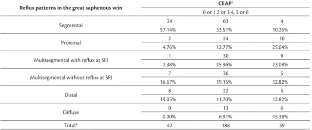
The results for relux patterns in the saphenous veins (Tables 1 and 2) showed that the most common type of relux in the GSV was the segmental relux pattern (33.8%) and the most common type in the SSV was the distal relux pattern (33.9%).
Correlating the relux patterns in saphenous veins with the CEAP classiications (Table 1) showed that 100% of GSVs in limbs classiied as C4 to C6 exhibited relux. In a similar manner, there was also a predominance of relux in the SSVs of limbs with these clinical grades and this inding was also statistically signiicant (Table 1 and Figure 3).
Correlating the different relux patterns in GSVs with clinical presentation (Table 2) showed that the segmental relux pattern was the most common in
Table 1. Incidence of relux in the saphenous veins, by clinical classiication.
C (CEAP*)
0 OR 1 2 OR 3 4, 5 OR 6 P VALUE
Relux in the great saphenous vein
NO 66 34 0
< 0.001
61.11% 15.32% 0.00%
YES 42 188 39
38.89% 84.68% 100.00%
Relux in the small saphenous vein
NO 97 189 24
89.81% 85.14% 61.54%
YES 11 33 15
10.19% 14.86% 38.46%
*CEAP: Clinical-Etiology-Anatomy-Pathophysiology classiication.
Table 2. Incidence of patterns of relux in the great saphenous vein by clinical classiication.
Reflux patterns in the great saphenous vein CEAP
†
0 or 1 2 or 3 4, 5 or 6
Segmental 24 63 4
57.14% 33.51% 10.26%
Proximal 2 24 10
4.76% 12.77% 25.64%
Multisegmental with relux at SFJ 1 30 9
2.38% 15.96% 23.08%
Multisegmental without relux at SFJ 7 36 5
16.67% 19.15% 12.82%
Distal 8 22 5
19.05% 11.70% 12.82%
Difuse 0 13 6
0.00% 6.91% 15.38%
Total* 42 188 39
limbs classiied as C0 or C1 (57.14%) and also in limbs classiied as C2 or C3 (33.51%). However, the highest incidence rates in extremities classiied as C4, C5, or C6 were for proximal reflux and multisegmental relux with SFJ relux (25.64% and 23.08% respectively).
Ninety-ive of the 369 LL assessed exhibited relux at the SFJ (25.7%). Correlation of relux at the SFJ with CEAP clinical grades (Table 3) showed that there was a higher incidence of relux (64.1%) in limbs with C4, C5, or C6 clinical status (p < 0.001).
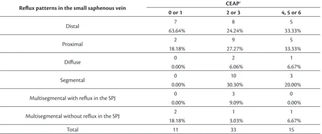
In the SSV, the distal relux pattern predominated in limbs graded C0 or C1 (63.64%). In limbs classed as C2 or C3, the distal, proximal, and segmental patterns all had very similar percentages (24.24%, 27.27%, and 30.30%, respectively). In C4, C5, and C6 limbs, the distal and proximal patterns predominated, both seen in 33.3% of cases (Table 4).
DISCUSSION
Using VU to conduct anatomic and functional assessment of the deep vein system in the LL of patients with signs or symptoms of CVI offers the possibility of conducting an individualized evaluation of each extremity and provides data to improve planning for surgery, reducing the recurrence of varicose veins.8
Table 4. Incidence of relux patterns in the small saphenous vein, by clinical classiication.
Reflux patterns in the small saphenous vein CEAP
†
0 or 1 2 or 3 4, 5 or 6
Distal 7 8 5
63.64% 24.24% 33.33%
Proximal 2 9 5
18.18% 27.27% 33.33%
Difuse 0 2 1
0.00% 6.06% 6.67%
Segmental 0 10 3
0.00% 30.30% 20.00%
Multisegmental with relux in the SPJ 0 3 0
0.00% 9.09% 0.00%
Multisegmental without relux in the SPJ 2 1 1
18.18% 3.03% 6.67%
Total 11 33 15
†CEAP: Clinical-Etiology-Anatomy-Pathophysiology classiication.
Table 3. Incidence of relux patterns in the great saphenous vein, by clinical classiication. Patterns including relux in the SFJ were
combined into a single group entitled junctional relux pattern.
Reflux patterns in the great saphenous vein C (CEAP
†)
0 or 1 2 or 3 4, 5 or 6
Junctional 3 67 25
7.14% 35.64% 64.10%
Segmental 24 63 4
57.14% 33.51% 10.26%
Multisegmental without relux in junctions 7 36 5
16.67% 19.15% 12.82%
Distal 8 22 5
19.05% 11.70% 12.82%
Total* 42 188 39
*P value: < 0.001; †CEAP: Clinical-Etiology-Anatomy-Pathophysiology classiication.
Figure 3. Incidence of relux in the saphenous veins, by clinical
Speciically with regard to the male population with CVI, there is scant literature identifying relux patterns in saphenous veins and their correlations with different phases of the disease.
This study investigated this population using quantitative analysis of relux types based on patterns deined by Engelhorn et al.,9 and analyzed their
correlations with the clinical manifestations of CVI. Our results demonstrate that relux was present in the majority (73%) of GSVs, but in just 16% of the SSVs, conirming the indings of a similar study conducted previously with women with varicose veins.12
Engelhorn et al.9 studied a population made up of
men and women with primary CVI and found that the highest incidence rate in the GSV was for the segmental relux pattern, followed by multisegmental without relux at the SFJ, distal relux, proximal, multisegmental with relux at the SFJ, and the diffuse relux pattern.
In another study by Engelhorn et al.,12 conducted with
an entirely female population with primary varicose veins in the LL (CEAP 2), it was also found that the segmental relux pattern had the highest incidence in the GSV, followed by the multisegmental pattern without relux at the SFJ.
In this study, the pattern with highest incidence was also the segmental, followed by multisegmental without junctional relux, multisegmental with relux at the SFJ, proximal relux, distal relux, and the diffuse relux pattern. These differences in relation to previous studies may have been because of the populations studied, which underscores the need to assess speciic populations. However, irrespective of study population, in the GSV, patterns in which relux is not present at the SFJ predominate.
In our study, the most common relux pattern in the SSV was the distal type, followed by proximal relux and segmental relux, demonstrating a difference in comparison to the patterns identiied in women (CEAP 2), among whom the segmental relux pattern predominated, followed by the distal and proximal relux patterns.12
Cassou et al.13 identiied the probability of different
relux patterns in the saphenous veins of women at different clinical stages of CVI, inding that 157 out of 288 GSVs (54.51%) in extremities classiied as CEAP C1 did not have relux, while 87 (30.21%) of them had segmental relux. In turn, 214 (35.97%), 104 (38.10%) and nine (42.86%) GSVs in extremities classiied as CEAP C2, C3, and C4 respectively had segmental relux. Two (50%) GSVs in extremities classiied as CEAP C5 exhibited multisegmental relux and in CEAP C6 limbs the same proportion
of absent, segmental, and diffuse patterns (33.33%) were detected in all extremities.
In our study, patients’ CVI clinical presentations were correlated with different GSV relux patterns. In common with a study by Cassou et al., among the male patients there was a greater incidence of segmental relux in classes C0 and C1 (57%) and in classes C2 and C3 (33.51%). This inding supports the hypothesis that venous disease begins in a segmental form and later degenerates into patterns that involve more segments of the vein.
In contrast, predominance of junctional relux was observed in 64% of LL with more advanced venous disease (C4 to C6). This inding is in agreement with studies that have demonstrated an association between SFJ involvement and severe forms of CVI clinical presentation.14,15
In the SSV, the distal relux pattern predominated in limbs with C0 or C1 clinical presentation. In classes C2 and C3, three different patterns, the distal, proximal, and segmental patterns, all had very similar percentages of incidence. In C4, C5, and C6 limbs the distal and proximal patterns had the highest incidence rates, all with the same percentage (33.33%), and these patterns were not observed in the female population.13
Labropoulos et al.16 compared venous relux and
clinical manifestations of CVI in 255 lower limbs from 217 patients and while they did not use exact definitions of different reflux patterns, they did report whether or not relux involved the SFJ and labeled relux as suprapatellar or infrapatellar. These authors found an association between occurrence of infrapatellar relux and presence of clinical signs of more advanced CVI.
In our study, as already explained, occurrence of relux in the SFJ and SPJ was related to clinical signs of more advanced CVI (C4 to C6). However, the possibility of associations between relux and the extent or speciic location of segmental relux was not analyzed. While there was a clear relationship in our study between junctional relux and greater intensity of clinical manifestations of disease presentation, it is possible that if a more detailed analysis had been conducted, including differentiation of the sites of involvement in segmental patterns and the extent of each relux, it would have been possible to compare our indings with those reported by Labropoulos et al. It would then be possible to determine whether there really is a relationship between segmental relux below the knee and the presence of more advanced clinical manifestations.16
pattern predominates in the SSV. Additionally, the initial clinical presentations of the disease are related to segmental relux patterns, with greater SFJ and SPJ involvement in more advanced CVI clinical presentations.
ACKNOWLEDGEMENTS
We are grateful to Prof. Marcia Olandoski, for pela competente análise estatística.
REFERENCES
1. Scuderi A, Raskin B, Al Assal F, et al. The incidence of venous disease in Brazil based on the CEAP classification. Int Angiol. 2002;21(4):316-21. PMid:12518109.
2. Evans CJ, Fowkes FGR, Ruckley CV, Lee AJ. Prevalence of varicose veins and chronic venous insuficiency in men and women in the general population : Edinburgh Vein Study. J Epidemiol Community Health. 1999;53(3):149-53. PMid:10396491. http:// dx.doi.org/10.1136/jech.53.3.149.
3. Maffei FH, Magaldi C, Pinho SZ, et al. Varicose veins and chronic venous insufficiency in Brazil: prevalence among 1755 inhabitants of a country town. Int J Epidemiol. 1986;15(2):210-7. PMid:3721683. http://dx.doi.org/10.1093/ije/15.2.210.
4. Boisseau MR, Eklof B. Chronic venous disease. N Engl J Med. 2006;355(5):488-98. PMID: 16885552. http://dx.doi.org/10.1056/ NEJMra055289.
5. Schmid-Schonbein GW, Takase S, Bergan JJ. New advances in the understanding of the pathophysiology of chronic venous insufficiency. Angiology. 2001;52(Suppl 1):S27-34. PMid:11510594. http://dx.doi.org/10.1177/000331970105200104.
6. Wittens C, Davies AH, Bækgaard N, et al. Management of chronic venous disease. clinical practice guidelines of the European Society for Vascular Surgery (ESVS). Eur J Vasc Endovasc Surg. 2015;49(6):678-737. PMid:25920631.
7. Eklof B, Rutherford RB, Bergan JJ, et al. Revision of the CEAP classification for chronic venous disorders: consensus statement. J Vasc Surg. 2004;40(6):1248-52. PMid:15622385. http://dx.doi. org/10.1016/j.jvs.2004.09.027.
8. Coleridge-Smith P, Labropoulos N, Partsch H, Myers K, Nicolaides A, Cavezzi A. Duplex ultrasound investigation of the veins in chronic venous disease of the lower limbs −UIP consensus document. Part I basic principles. Eur J Vasc Endovasc Surg. 2006;31(1):83-92. PMid:16226898. http://dx.doi.org/10.1016/j.ejvs.2005.07.019.
9. Engelhorn CA, Engelhorn AL, Cassou MF, Casagrande C, Gosalan CJ, Ribas E. Classificação anátomo-funcional da insuficiência das veias safenas baseada no eco-Doppler colorido, dirigida para o planejamento da cirurgia de varizes. J Vasc Bras. 2004;3(1):13-9.
10. van Bemmelen S, Bedford G, Beach K, Strandness DE Jr. Quantitative segmental evaluation of venous valvular reflux with duplex
ultrasound scanning. J Vasc Surg. 1989;10(4):425-31. PMid:2677416. http://dx.doi.org/10.1016/0741-5214(89)90417-5.
11. Labropoulos N, Tiongson J, Pryor L, et al. Definition of venous reflux in lower-extremity veins. J Vasc Surg. 2003;38(4):793-8. PMid:14560232. http://dx.doi.org/10.1016/S0741-5214(03)00424-5.
12. Engelhorn CA, Engelhorn AL, Cassou MF, Salles-Cunha SX. Patterns of saphenous reflux in women with primary varicose veins. J Vasc Surg. 2005;41(4):645-51. PMid:15874929. http://dx.doi.org/10.1016/j. jvs.2004.12.051.
13. Cassou MF, Gonçalves PCZ, Engelhorn CA. Probabilidade de refluxo nas veias safenas de mulheres com diferentes graus de insuficiência venosa crônica. J Vasc Bras. 2007;6(3):238-45. http:// dx.doi.org/10.1590/S1677-54492007000300007.
14. Garcia-Gimeno M, Rodriguez-Camarero S, Tagarro-Villalba S, et al. Reflux patterns and risk factors of primary varicose veins’ clinical severity. Phlebol J Venous Dis. 2013;28(3):153-61. PMid:22345327. http://dx.doi.org/10.1258/phleb.2011.011114.
15. Pittaluga P, Chastanet S, Rea B, Barbe R. Classification of saphenous refluxes: implications for treatment. Phlebol J Venous Dis. 2008;1(23):2-9. PMid:18361263. http://dx.doi.org/10.1258/ phleb.2007.007042.
16. Labropoulos N, Leon M, Nicolaides AN, Giannoukas AD, Volteas N, Chan P. Superficial venous insufficiency: correlation of anatomic extent of reflux with clinical symptoms and signs. J Vasc Surg. 1994;20(6):953-8. PMid:7990191. http://dx.doi. org/10.1016/0741-5214(94)90233-X.
Correspondence
Carlos Alberto Engelhorn Rua da Paz, 195, sala 2 - Bairro Alto da XV CEP 80060-160 - Curitiba (PR), Brazil Tel.: +55 (41) 3362-0133 E-mail: [email protected]
Author information
CAE - PhD in Vascular Surgery from Universidade Federal de São Paulo and full professor of Angiology at Pontifícia Universidade Católica do Paraná (PUC-PR). FEC - MsC in Surgery from Pontifícia Universidade Católica do Paraná and assistant professor of Angiology at Pontifícia Universidade Católica do Paraná (PUC-PR). ICMS, GFAC, JPO, LYH and LSM - Medical students at Pontifícia Universidade Católica do Paraná (PUC-PR).
Author contributions
Conception and design: CAE Analysis and interpretation: CAE, FEC, ICMS, GFAC, JPO, LYH, LSM Data collection: ICMS, GFAC, JPO, LYH, LSM Writing the article: CAE, FEC, ICMS, GFAC, JPO, LYH, LSM Critical revision of the article: CAE Final approval of the article*: CAE, FEC, ICMS, GFAC, JPO, LYH, LSM Statistical analysis: CAE Overall responsibility: CAE