Medium and long-term outcomes of endovenous treatment of
varicose veins with a 1940nm diode laser: critical analysis and
technical considerations
Resultados de médio e longo prazo do tratamento endovenoso de varizes com laser de
diodo em 1940 nm: análise crítica e considerações técnicas
Luiz Marcelo Aiello Viarengo1,2
*
, Gabriel Viarengo1, Aline Meira Martins1, Marília Wechellian Mancini2, Luciana Almeida Lopes2
Abstract
Background: Introduction of the endovenous laser technique for treatment of varicose veins triggered a eforts to identify an ideal wavelength, capable of producing the highest possible selective damage with the greatest safety and lowest incidence of adverse efects. Objectives: Assess medium to long term results of 1940nm diode laser treatment of varicose veins, correlating parameters used with durability of the anatomic outcome. Methods: his was a retrospective study of patients diagnosed with Chronic Venous Insuiciency at clinical stages CEAP C2 to C6 who underwent thermoablative treatment of trunk varicose veins using a 1940nm wavelength laser with a radial emission optical iber, from April 2012 to July 2015. A systematic review was conducted of electronic medical records to obtain demographic and clinical data, including postoperative follow-up duplex ultrasound indings. Results: he average age of the 41-patient sample was 53.3 years and 37 patients were women (90.2%). he average follow-up time was 803 days. he average caliber of the treated veins was 7.8 mm. he immediate success rate was 100% with an average LEED of 45.3 J/cm. he late success rate was 95.1%, and two recanalizations were observed around 12 months after ablation. here was no recanalization in veins treated with a LEED greater than 30 J/cm. Conclusions: he 1940nm laser proved to be safe and efective in venous segments up to 10 mm in diameter, with the parameters proposed, over medium to long term time follow-up.
Keywords: laser; varicose veins; endovenous ablation; thermoablation.
Resumo
Contexto: Desde a introdução do laser endovenoso para tratamento das varizes, há uma busca pelo comprimento de onda ideal, capaz de produzir o maior dano seletivo possível com maior segurança e menor incidência de efeitos adversos. Objetivos: Avaliar os resultados de médio e longo prazo do laser de diodo de 1940 nm no tratamento de varizes, correlacionando os parâmetros utilizados com a durabilidade do desfecho anatômico. Métodos: Revisão retrospectiva de pacientes diagnosticados com insuiciência venosa crônica em estágio clínico baseado em clínica, etiologia, anatomia e patoisiologia (CEAP) C2 a C6, submetidos ao tratamento termoablativo endovenoso de varizes tronculares, com laser com comprimento de onda em 1940 nm com ibra óptica de emissão radial, no período de abril de 2012 a julho de 2015. Uma revisão sistemática dos registros médicos eletrônicos foi realizada para obter dados demográicos e dados clínicos, incluindo dados de ultrassom dúplex, durante o período de seguimento pós-operatório.
Resultados: A média de idade dos pacientes foi de 53,3 anos; 37 eram mulheres (90,2%). O tempo médio de seguimento foi de 803 dias. O calibre médio das veias tratadas foi de 7,8 mm. A taxa de sucesso imediato foi de 100%, com densidade de energia endovenosa linear (linear endovenous energy density, LEED) média de 45,3 J/cm. A taxa de sucesso tardio foi de 95,1%, com duas recanalizações por volta de 12 meses pós-ablação. Não houve nenhuma recanalização nas veias tratadas com LEED superior a 30 J/cm. Conclusões: O laser 1940 nm mostrou-se seguro e efetivo, em médio e longo prazo, para os parâmetros propostos, em segmentos venosos com até 10 mm de diâmetro.
Palavras-chave:laser; varizes; terapia a laser; técnicas de ablação.
1 Clínica Viarengo – CV, Jundiaí, SP, Brazil.
2 Núcleo de Pesquisa e Ensino de Fototerapia nas Ciências da Saúde – NUPEN, São Carlos, SP, Brazil.
Financial support: None.
Conlicts of interest: No conlicts of interest declared concerning the publication of this article. Submitted: November 20, 2016. Accepted: February 10, 2017.
INTRODUCTION
At many different centers worldwide, endovenous laser ablation (EVLA) has emerged as a standard for
treatment of venous insuficiency and as a minimally
invasive option for treatment of trunk varicose veins. Since its appearance, there have been continuous efforts to identify the ideal wavelength that will produce the maximum possible selective damage with the greatest margin of safety and the lowest incidence of adverse events. Many different wavelengths and different types
of optical ibers have been tested in attempts to achieve
this objective.1-8 Several authors have demonstrated
that all of the wavelengths employed to treat varicose veins are equally capable of producing the desired anatomic results. The fundamental differences between different wavelengths are related to occurrence of adverse events.9-11
From the point of view of physics, the higher a tissue
or a chromophore’s coeficient of light absorption,
the greater the quantity of heat generated and the
more conined the zone in which heat is generated.
These principles indicate that a 1940nm laser would offer advantages, because it acts on water’s second largest absorption peak, theoretically increasing both
the eficacy and safety of the procedure.
Durability is an important long-term characteristic of all vascular procedures. For varicose vein treatments, recurrence rates are potentially a valuable measure for assessing medium and long-term results of different venous disease treatment modalities.12-14
The objectives of this study are to conduct a retrospective analysis of the medium and long-term results of endovenous treatment of varicose veins using a 1940nm diode laser and to correlate the parameters used during treatment with durability of anatomic
outcome (ibrotic occlusion or recanalization), clinical improvement according to the CEAP classiication,
and adverse events.
METHODS
A retrospective review was conducted of patients
diagnosed with chronic venous insuficiency at clinical
stages C2 to C6 on the clinical, etiology, anatomical, and pathophysiology scale (CEAP), who had been treated endovenously for trunk varicose veins at a single center, using a laser with a wavelength of
1940nm (Medilaser, DMC, São Carlos, SP, Brazil),
National Agency for Sanitary Vigilance [ANVISA] registration number 80030810129), with a radial emission optical fiber, from April 2012 to July
2015. The objective was to analyze medium and
long-term results in terms of anatomic outcome
(ibrotic occlusion, partial recanalization, and total recanalization), clinical improvement on the CEAP classiication, and adverse events (pigmentation,
thrombophlebitis, venous thrombosis, paresthesias,
ibrous cord, and others). All data were anonymized.
The members of the study population were initially
identiied by searching electronic records for patients diagnosed with varicose veins, venous insuficiency,
or venous hypertension (with or without ulceration) who had been treated with endovenous laser ablation of trunk varicose veins at least 12 months previously and had presented for the service’s routine clinical and ultrasonographic control examinations (at 7 days, 30 days, 3 months, 6 months, 9 months, 12 months, 18 months, 24 months, and annually thereafter). Patients with postthrombotic syndrome prior to
surgical treatment or with relux in the deep vein
system were excluded.
Once approval had been granted by the Research Ethics Committee (ruling number 1.693.514), a systematic review was conducted of electronic medical records to obtain demographic data and
clinical data, including duplex ultrasound indings
during the postoperative follow-up period.
The demographic data analyzed included age, sex,
race, and comorbidities. Clinical data included date of procedure, preoperative clinical diagnosis (CEAP
classiication), venous segment treated with laser
ablation, extension and mean caliber of the venous segment treated, associated procedures, postoperative pain measured on an analog visual scale (AVS), intraoperative intercurrent conditions, immediate postoperative intercurrent conditions (within 30 days),
late intercurrent conditions (with date of identiication
and progression), laser parameters employed (power, total energy and linear endovenous energy density
[LEED], CEAP clinical classiication at last clinical
follow-up within study period, date of last follow-up for calculation of follow-up in days) and, additionally,
information on the ultrasonographic indings from
the last follow-up appointment attended within the study period, covering appearance of the ablated
vein (ibrotic, ibroelastic, thrombotic, completely recanalized, partially recanalized, extension of recanalized segment, and competence or incompetence
of saphenopopliteal and saphenofemoral junctions).
Statistical analysis
Data were compiled in a table on Excel 2011, version 14.3.6 (Microsoft, Redmond, WA, USA),
and analyzed using Bioestat, version 5.3 (Instituto Mamirauá, Belém, PA, Brazil). Categorical variables
arranged in contingency tables. Quantitative variables
were analyzed using descriptive statistics and expressed
as means, standard deviations (SD), and maximum and minimum values. Intragroup differences were
analyzed using Student’s t test. Results with p values
< 0.05 were considered statistically signiicant.
RESULTS
A total of 152 patients were identiied who had been
treated with thermoablation of trunk varicose veins between April of 2012 and July of 2015 using lasers of a number of different wavelengths. All procedures were conducted by the same surgeon, who has a great deal of experience with EVLA, in an ambulatory, extra-hospital setting with ultrasound-guided perivenous tumescent anesthesia combined with femoral nerve block, also guided with ultrasound. Of these, 50 limbs in 50 patients were treated with a 1940nm laser.
Nine of these 50 cases that it the study objective
were excluded, two because follow-up records were available for less than 12 months, four because of
postthrombotic syndrome prior to treatment, and three because of incomplete follow-up and data.
There were 37 (90.2%) female and 4 (9.8%) male patients and mean age was 53.3 years (range: 30 - 74 years; SD: 12.4 years). Mean number of gestations among the women was 2.2 (range 0 - 9; SD: 1.71). All demographic
data are summarized in Table 1.
A total of 34 great saphenous veins (GSVs) (82.9%) and seven small saphenous veins (SSVs) (17.1%) were treated in 41 limbs of 41 patients, 19 right limbs (16 GSVs and 3 SSVs) and 22 left limbs (18 GSVs and 4 SSVs). Preoperative CEAP
clinical classiications are summarized in Table 2. The anatomic parameters of the venous segments treated
and the laser parameters employed are summarized
in Tables 3 and 4 respectively.
Primary ablation was achieved in 100% of cases. In all patients, phlebectomy of tributaries and varicose branches was conducted as a supplementary procedure. There were no intraoperative complications.
The mean length of postoperative follow-up was 803 days, varying from 467 to 1,360 days (SD: 291.3).
Intraoperative pain was rated on an AVS ranging from 0 to 10 (where 0 indicated “free from pain” and 10 “intense pain”). Twenty-three patients reported “free from pain” (56.1%), 17 patients (41.5%) chose “mild pain” (up to 3 points on the AVS), and one patient (2.4%) reported “moderate pain” (4 to 6 points on the AVS). In all cases, pain was related to phlebectomy.
Two recanalizations were observed during the follow-up period, one was a recanalization of the
entire extension of a GSV (case 1), detected 421 days
postoperatively; and the other was a recanalized
infragenicular segment of a GSV (case 2), detected 342 days postoperatively (Table 5).
No recanalizations or treatment failures were
observed in SSVs. Among the anatomically successful saphenous veins treatments (GSV + SSV), mean LEED was 46.8 J/cm (varying from 30.7 J/cm to 104.7 J/cm).
Overall success rate (permanent occlusion of trunk veins) was 95.1% over the follow-up period. In all of the cases of late post-ablation anatomic success,
the ultrasonographic indings recorded at the last
medical examination describe competence of the saphenofemoral and saphenopopliteal junctions, trunk
Table 1. Patients’ demographic data (n = 41).
Mean age (range), years 53.3 (30-74) Sex
Female 37 (90.2%)
Male 4 (9.8%)
Skin color
White 34 (82.9%)
Black 7 (17.1%)
BMI (range) 26.1 (18.6-43.1) Family history of venous disease 36 (87.8%)
Smoking 4 (9.8%)
Dyslipidemia 7 (17.1%)
Diabetes 3 (7.3%)
SAH 7 (17.1%)
Obesity 5 (12.2%)
BMI: body mass index; SAH: systemic arterial hypertension. Data are expressed as absolute values (%), except where indicated otherwise.
Table 2. Preoperative clinical classiication (CEAP).
C1 C2 C3 C4 C5 C6
Overall (n = 41) 0 19 7 10 0 5
GSV* (n = 34) 0 18 6 8 0 2
SSV** (n = 7) 0 1 1 2 0 3
CEAP: clinical, etiology, anatomical, and pathophysiology; GSV: great saphenous vein; SSV: small saphenous vein. * Patients with GSV incompetence only. ** Patients with SSV incompetence only.
Table 3. Anatomic parameters of veins ablated.
Mean Ø (mm) Range (mm) SD Extension ablated (cm) Range (cm) SD
GSV 7.82 (6.2 to 10.4) 1.08 48.1 (29 to 80) 11.9
SSV 7.14 (5.0 to 10.5) 2.03 24.3 (15 to 34) 6.2
veins occluded, with ibrotic appearance and a very reduced caliber, and dificult to identify (Figure 1).
In the two cases that progressed to recanalization (two
female patients with GSV incompetence), postoperative
ultrasonographic indings prior to identiication of the recanalization described saphenofemoral junction
incompetence, GSV occluded with predominantly hypoechogenic content, and venous retraction of less than 50% of the initial diameter.
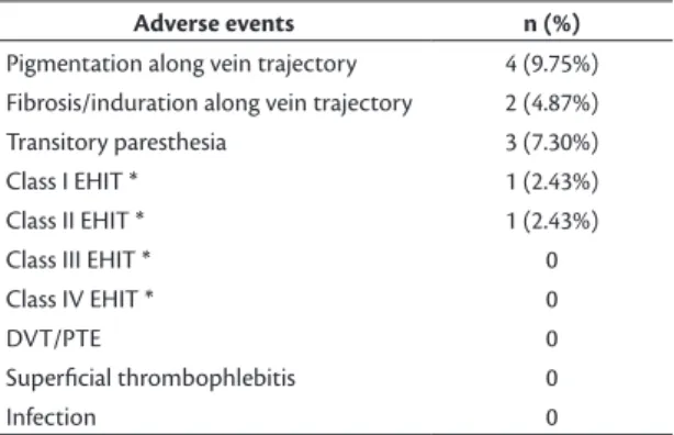
Adverse events were mild and self-limiting, with spontaneous resolution occurring from 10 days to 6 months after the procedure (Table 6). Additionally, 39 patients (95.1%) achieved and maintained to the last examination an improvement in CEAP clinical
classiication, compared with their initial clinical classiications.
DISCUSSION
Currently, EVLA is considered the gold standard
for treatment of insuficiency of supericial trunk veins
in both the United States and the United Kingdom
and is recommended as the irst-choice treatment.16-18
Many different studies have demonstrated the elevated
eficacy of endovenous thermoablation, with high rates
of technical success and low levels of complications,
particularly when a laser with a wavelength (λ) of
1470nm is used in combination with a radial emission
optical iber.19-22
In addition to the well-established diode laser with emission wavelength of 1470nm (active medium: InGaAsP), new semiconductor diode-based lasers with wavelengths of interest have been emerging, including emission wavelengths centered on 1908nm, 1920nm, and 1940nm (active medium: AlGaIn).8,23
The practical advantages of these wavelengths are also because of the high degree of absorption of laser light by the interstitial water in the vein walls, which can be illustrated by their respective
absorption coeficients (μA
water) for this chromophore
(Table 7).24 It can be considered that delivering laser
light to the vein wall via a radial iber at a λ in the
region of 1940nm is an incremental development over the current state-of-the-art technique for laser
Table 4. Laser treatment parameters.
Mean power (W) Range (W) SD Mean LEED (J/cm) Range (J/cm) SD
GSV 4.1 (3.5 to 7.0) 0.6 45.5 (15 to 104.7) 15.9
SSV 3.86 (3.0 to 4.0) 0.38 45.9 (35.8 to 54.6) 7.52
Overall 4.06 (3.0 to 7.0) 0.57 45.3 (15 to 104.7) 14.8
GSV: great saphenous vein; SSV: small saphenous vein; SD: standard deviation; LEED: linear endovenous energy density.
Table 5. Cases with late postoperative recanalization.
Time (days) CEAP Vein
treated
Mean PO diameter (mm)
Power employed (W)
Mean LEED
(J/cm) BMI
Case 1 421 C3 GSV 8.1 3.5 19.0 28.1
Case 2 342 C6 GSV 9.0 5.0 15.0 29.6
GSV: great saphenous vein; BMI: body mass index; PO: preoperative.
endovenous thermoablation, which is the 1470nm
laser with irradiation via a radial iber.
The important advantages for endovenous ablation effectively offered by wavelengths within the near infrared (NIR) region of the optical spectrum are
their high coeficients of absorption by water and
the resultant shallow depth of penetration through
tissues (which constrains the zone in which heat is
generated in the tissues exposed to laser radiation).
The higher the value of the absorption coeficient
in a chromophore (in this case, water in tissues),
which is a function of λ, the greater the quantity of
heat generated for a given amount of optical energy supplied, the shallower the penetration (increasing safety), and, therefore, the lower the quantity of energy that is needed to produce the thermal damage intended (thermoablation).25
If we compare the μA
water (λ) values offered by the
new wavelengths with the igure for 1470nm, we ind that μA
water (1940nm) is 4.83 times the μ
A
water (1470nm).
Comparing the 1940nm and 980nm wavelengths,
a laser at λ = 1940nm has an absorption in water
approximately 266 times greater than a laser at
λ = 980nm and 4.8 times greater than the absorption in water of a laser at λ = 1470nm (Table 7).24 The higher
absorption coeficient means that the LEEDs needed
when treating with a 1940nm laser are lower than those needed to achieve thermal ablation with a 980nm or even a 1470nm laser. Additionally, the values for effective optical penetration in water for 1470nm and 1940nm, based both on absorption and scattering processes, are approximately 220 µ and 48 µ respectively, in contrast with a value of approximately 3.0 mm for 980nm.24,26,27
Based on these facts, endovenous laser ablation at 1470nm, conducted by homogeneous tissue irradiation
(with a radial iber) with the vein wall as the direct
target (chromophore: interstitial water) was indeed a major breakthrough for endolaser techniques, with
impacts on minimization of the adverse effects inherent
to the technique performed with other types of lasers (808, 810, 940 and 980nm, for example).9-11,20,28
In turn, conducting the technique with a radial iber and λ = 1940nm is an incremental development of
the state-of-the-art in laser thermal ablation. In this retrospective analysis, the high rates of anatomic success (obliteration), both immediate (100%) and late (95.1%), can be explained from a theoretical perspective on the basis of the principles outlines above, which are applicable in the same way as with the 1470nm wavelength, producing similar results.10,11,20,29,30
One interesting feature that should be borne in
mind in relation to the laser with a λ of 1940nm, compared with the laser at a λ of 1470nm, is that its light is absorbed almost ive times more by water
and it has an effective depth of optical penetration that is around one quarter of the penetration of the 1470nm laser.24,26,27 This is because the greater the
absorption of the photons from a laser by a given target containing absorbent chromophores, the greater the quantity of heat generated and the more restricted
the zone of heat generation, i.e., it is absorption of
the photons from the laser by chromophores in the tissue that causes the tissues to heat up (absorptive heating). Absorptive heat (J/cm3), generated in situ, is
Table 7. Coeicient of absorption in water in the NIR band of the electromagnetic spectrum.24
λ (nm) Laser µwater (cm-1) Relative absorption:
1940nm
Relative absorption: 1910nm
Relative absorption: 1470nm
Relative absorption: 980nm
808 AlGaAs diode 0.02 5,991.50 4,517.00 1240.75 22.50
975 InGaAs diode 0.45 266.29 200.76 55.14 1
1064 Nd:YAG 0.12 998.58 752.83 206.79 3.75
1470 InGaAsP diode 24.815 4.83 3.64 1 0.018
1910 AlGaln diode 90.34 1.33 1 0.27 0.005
1940 AlGaln diode 119.83 1 0.75 0.21 0.004
2100 Ho:YAG 26.93 4.45 3.35 0.92 0.017
λ: wavelength; µwater: coeicient of absorption in water.
Table 6. Adverse events.
Adverse events n (%)
Pigmentation along vein trajectory 4 (9.75%) Fibrosis/induration along vein trajectory 2 (4.87%) Transitory paresthesia 3 (7.30%)
Class I EHIT * 1 (2.43%)
Class II EHIT * 1 (2.43%)
Class III EHIT * 0
Class IV EHIT * 0
DVT/PTE 0
Supericial thrombophlebitis 0
Infection 0
proportional to the coeficient of absorption μA (cm -1)
multiplied by the irradiance (W/cm2) and linearly
dependent on exposure time.25,31
While the heat produced in the tissue, from the optical energy absorbed and released as heat, is dependent on the optical properties of the tissues and on the parameters of irradiation, such as irradiance and exposure time, the process of conductive thermal diffusion is
responsible for transmission (low) of heat generated locally (low of heat from a higher-temperature region
to a lower temperature region).25 In other words, the
parameter that governs the entire interaction between laser and tissue, in relation to lasers that work by photothermal effects, is temperature. The greater the absorption of photons by the chromophore, the greater the quantity of heat generated and the more spatially restricted that generation of heat is; however, once generated, the heat diffuses from the site of generation to cooler areas. The result is that the nature and extent of thermal damage will depend on the optical properties of the tissue (scattering and absorption),
the thermal properties of the tissue (speciic heat and
thermal conductivity) and also, very strongly, on the parameters of exposure to the laser (power density, exposure time, and energy density).
Thermal damage to collagen plays a preeminent role in endovenous ablation, affecting short and long-term results. Biesman32 demonstrated that collagen
contracts at temperatures close to 50˚C, but that
coagulation necrosis only occurs at temperatures
from 70 to 100˚C. Only administration of a relatively high energy per unit of length results in suficiently
high temperatures to cause denaturing of collagen.33
During endovenous laser ablation, intraluminal temperatures can rise to over 100ºC,28,34-39 and these
temperature proiles are independent of wavelength, i.e. use of different wavelengths does not inluence
the endovenous temperature profile.28,36 On the
other hand, considering that the optical and thermal properties of the tissues, in this case, are the same,
the temperature is strongly inluenced by the laser
exposure parameters.
To a certain extent, these considerations provide a rational explanation for the two technical failures observed in this review study. In both cases, the power employed (Table 5) was similar to the mean power (4.1 W) employed in the other cases (Table 4). Additionally, the mean diameter of the venous
segment treated did not differ signiicantly from the
mean diameters of the venous segments for which treatment was successful over the medium and long term (Tables 3 and 5). The only parameter that was different from the successful cases was LEED, at a
mean of 17 J/cm in the recanalization cases, compared
to 46.8 J/cm in cases in which anatomic success was
achieved, which is a statistically signiicant difference (p = 0.0041).
As already mentioned, denaturing of collagen
occurs at temperatures from 70 to 100˚C; therefore, it is necessary to administer suficient energy during the
ablation process to generate high enough temperatures to make the process effective.33 In both the cases in which
there was recanalization (at approximately 12 months
after endovenous treatment), it is probable that the
quantity of energy delivered (mean LEED = 17 J/cm)
was not enough to increase the temperature to the point at which denaturing of collagen would occur.
This insuficient increase in temperature was translated into the ultrasonographic indings described, which were very different from the indings observed in
cases with anatomic success.
None of the case treated with a 1940nm λ laser
using a LEED greater than 30 J/cm (mean: 46.8 J/cm) for a maximum diameter of 10 mm resulted in treatment failure detected during the follow-up period (mean: 803 days). The incidence of adverse events was very low, and all of those that did occur were not clinically relevant and were self-limiting.
At this point, it is absolutely necessary to understand the concept of molar extinction, which is a substance’s capacity to absorb light of a given wavelength.33
Since the coeficient of molar extinction is similar for
water and blood, when a laser with a wavelength of 1470nm or 1940nm is used, it is important to empty the vein of intraluminal blood,33 because otherwise
the majority of the energy will be absorbed by the intraluminal blood, leading to thrombotic occlusion
and possible recanalization after a few months.27,33,37,38
This statement is based on a study by Vuylsteke et al.,33,40
that assessed the role played by blood in the results of endovenous treatment with a 1500nm laser, histologically evaluating the degree of destruction of the vein wall. The study concluded that the volume of intraluminal blood results in a reduction of vein
wall destruction. Tumescent iniltration of liquid
reduces the quantity of intraluminal blood, resulting in an increase in vein wall destruction, in addition to acting to dissipate the heat, preventing destruction of perivenous tissues.40 According to the authors, the
inluence of tumescence on venous diameter is more
important than the Trendelenburg position.
In summary, the inal objective of treating varicose
veins with laser ablation is to eliminate pathological
relux of blood by durable or permanent occlusion
completely or by substantial damage to the endothelium and the internal wall of the vein, leading to secondary occlusion of the lumen by a clot, in a similar manner to the effect produced by sclerosing agents. Substantial
transfer of heat to the vein wall reduces on signiicant shrinkage of its collagen ibers, with a consequent
reduction in the lumen. The magnitude of parietal shrinkage appears to be important because the lumen that remains after laser treatment is subject to occlusion by clot formation. Later, this clot may
be subject to recanalization and it can be supposed
that the greater the diameter of the clot, the greater
the risk of recanalization later.33,40 Ideally, after
laser thermal ablation, thrombotic occlusion of the
saphenous vein is substituted by a ibrous cord that
can often by detected by ultrasound, even years after the procedure (Figure 1).
CONCLUSIONS
The 1940nm laser proved to be very safe and effective over the medium and long term with the parameters employed, i.e. a mean power of 4.0 W and a LEED greater than 30 J/cm (mean of 46.8 J/cm) for GSVs and SSVs with diameters of up to 10 mm, irrespective of the region treated. It can be conducted in an ambulatory setting using tumescent local anesthesia, with a very low incidence of adverse events, which are without clinical relevance and are self-limiting.
REFERENCES
1. Pavlovic MD, Schuller-Petrovic S, Pichot O, et al. Guidelines of the first international consensus conference on Endovenous thermal ablation for varicose veins disease – ETAV Consensus Meeting 2012. Phlebology. 2015;30(4):257-73. PMid:24534341. http:// dx.doi.org/10.1177/0268355514524568.
2. Goldman MP, Mauricio M, Rao J. Intravascular 1320-nm laser closure of the great saphenous vein: a 6- to 12-month follow-up study. Dermatol Surg. 2004;30(11):1380-5. PMid:15522018.
3. Pannier F, Rabe E, Maurins U. First results with a new 1470-nm diode laser for endovenous ablation of incompetent saphenous veins. Phlebology. 2009;24(1):26-30. PMid:19155338. http://dx.doi. org/10.1258/phleb.2008.008038.
4. Vuylsteke ME, Vandekerckhove PJ, De Bo T, Moons P, Mordon S. Use of a new endovenous laser device: results of the 1,500 nm laser. Ann Vasc Surg. 2010;24(2):205-11. PMid:19748212. http:// dx.doi.org/10.1016/j.avsg.2009.06.024.
5. Kabnick LS, Caruso JA. No-wall touch laser fiber vs bare-tip laser fiber for endothermal venous ablation of great saphenous vein: are the results the same? In: Gerard JL, editor. Controversies and updates in vascular surgery. Torino: Edizioni Pan Minerva Medica; 2008. p. 401-2.
6. Viarengo LM, Meirelles GV, Potério-filho J. Tratamento de varizes com laser endovenoso: estudo prospectivo com seguimento de 39 meses. J Vasc Bras. 2006;5(3):184-93. http://dx.doi.org/10.1590/ S1677-54492006000300006.
7. Shaĭdakov EV, Bulatov VL, Iliukhin EA, Son’kin IN, Grigorian AG, Gal’chenko MI. Optimal regimes of endovenous laser obliteration at the wavelengths of 970, 1470, and 1560 nm: the multicenter retrospective longitudinal cohort study. Phlebology. 2013;1:22-9.
8. Sroka R, Pongratz T, Esipova A, et al. Endovenous laser therapy for occlusion of incompetent saphenous veins using 1940 nm. In: Lilge L, Sroka R, editors. Proceedings of the 7th Medical Laser Applications and Laser-Tissue Interactions; 2015; Munich, Germany. San Diego: Optical Society of America; 2015. vol. 9542. Paper 95420D.
9. Kabnick L. Outcome of diferente Endovenous laser wavelengths for great saphenous vein ablation. J Vasc Surg. 2006;43(1):88-93. PMid:16414394. http://dx.doi.org/10.1016/j.jvs.2005.09.033.
10. Van den Bos R, Arends L, Kockaert M, Neumann M, Nijsten T. Endovenous therapies of lower extremity varicosities: a meta-analysis. J Vasc Surg. 2009;49(1):230-9. PMid:18692348. http:// dx.doi.org/10.1016/j.jvs.2008.06.030.
11. Doganci S, Demirkilic U. Comparison of 980 nm laser and bare-tip fibre with 1470 nm laser and radial fibre in the treatment of great saphenous vein varicosities: a prospective randomised clinical trial. Eur J Vasc Endovasc Surg. 2010;40(2):254-9. PMid:20547079. http://dx.doi.org/10.1016/j.ejvs.2010.04.006.
12. Jones L, Braithwaithe BD, Selwyn D, Cooke S. Neo- vascularisation the principal cause of varicose vein recurrence: results of a randomised trial of stripping the long saphenous vein. Eur J Vasc Endovasc Surg. 1996;12(4):442-55. PMid:8980434. http://dx.doi. org/10.1016/S1078-5884(96)80011-6.
13. Blomgren L, Johansson G, Dalbergh-Akerman A, Noren A, Brundin C, Nordstrom E. Recurrent varicose veins: incidence, risk factors and groin anatomy. Eur J Vasc Endovasc Surg. 2004;27(3):269-74. PMid:14760595. http://dx.doi.org/10.1016/j.ejvs.2003.12.022.
14. Perrin MR, Guex JJ, Ruckley CV, et al. Recurrent varices after surgery (REVAS), a consensus document. Cardiovasc Surg. 2000;8(4):233-45. PMid:10950599.
15. Kabnick LS, Ombrellino M, Agis H, et al. Endovenous heat induced thrombosis (EHIT) at the superficial deep venous junction: a new post-treatment clinical entity, classification and potential treatment strategies. In: Proceedings of the 18th Annual Meeting of the American Venous Forum; 2006; Miami, FL. Miami: American Venous Forum; 2006.
16. Gloviczki P, Camerota AJ, Dalsing MC, et al. The care of patients with varicose veins and associated chronic venous diseases: clinical practice guidelines of the Society for Vascular Surgery and American Venous Forum. J Vasc Surg. 2011;53(5, Suppl):2S-48S. PMid:21536172. http://dx.doi.org/10.1016/j.jvs.2011.01.079.
17. Pavilovic MD, Schuller-Petrovic S, Pichot O, et al. Guidelines of the First International Consensus Conference on Endovenous Thermal Ablation for Varicose Vein Disease--ETAV Consensus Meeting 2012. Phlebology. 2014;30(4):257-73. PMid:24534341. http://dx.doi.org/10.1177/0268355514524568.
18. Marsden G, Perry MC, Kelly K, Davies AH. NICE guidedelines on the management of varicose veins. BMJ. 2013;347:f4279. PMid:23884969. http://dx.doi.org/10.1136/bmj.f4279.
19. Spreafico G, Piccioli A, Bernardi E, et al. Six-year follow-up of endovenous laser ablation for great saphenous vein incompetence. J Vasc Surg. 2013;1(1):20-5. PMid:26993888.
21. Schwarz T, Hodenberg E, Furtwängler C, Rastan A, Zeller T, Neumann FJ. Endovenous laser ablation of varicose veins with the 1470-nm diode laser. J Vasc Surg. 2010;51(6):1474-8. PMid:20347542. http:// dx.doi.org/10.1016/j.jvs.2010.01.027.
22. Pannier F, Rabe E, Rits J, Kadiss A, Maurins U. Endovenous laser ablation of great saphenous veins using a 1470 nm diode laser and the radial fibred follow-up after six months. Phlebology. 2011;26(1):35-9. PMid:21148467. http://dx.doi.org/10.1258/ phleb.2010.009096.
23. Somunyudan MF, Topaloglu N, Ergenoglu MU, Gulsoy M. Endovenous laser ablation with Tm- Fiber laser. In: Duco Jansen E, Thomas RJ, editors. Proceedings of the 22th SPIE Optical Interactions with tissue and cells; 2011. San Francisco, California; 2011. vol. 7897.
24. Hale GM, Querry MR. Optical constants of water in the 200 nm to 200 µm wavelength region. Appl Opt. 1973;12(3):555-63. PMid:20125343. http://dx.doi.org/10.1364/AO.12.000555.
25. Niemz MH. Biological and medical physics, biomedical enginnering - laser-tissue interactions, fundamentals and applications. 3rd ed. New York: Springer Berlin Heidelberg; 2007.
26. Roggan A, Friebel M, Dörschel K, Hahn A, Müller G. Optical properties of circulating human blood in the wavelength range 400-2500 nm. J Biomed Opt. 1999;4(1):36-46. PMid:23015168. http://dx.doi.org/10.1117/1.429919.
27. Bosschaart N, Edelman G, Aalders MC, van Leeuwen TG, Faber DJ. A literature review and a novel theoretical approach on the optical properties of whole blood. Lasers Med Sci. 2014;29(2):453-79. PMid:24122065. http://dx.doi.org/10.1007/s10103-013-1446-7. 28. Viarengo LM, Potério-Filho J, Potério GM, Menezes FH, Meirelles GV. Endovenous laser treatment for varicose veins in patients with active ulcers: measurement of intravenous and perivenous temperatures during the procedure. Dermatol Surg. 2007;33(10):1234-42, discussion 1241-2. PMid:17903157.
29. Dzieciuchowicz L, Krasinski Z, Gabriel M, Espinosa G. A prospective comparison of four methods of endovenous thermal ablation. Pol Przegl Chir. 2011;83(11):597-605. PMid:22246092. http://dx.doi. org/10.2478/v10035-011-0095-4.
30. Van den Bos R, Arends L, Kockaert M, Neumann M, Nijsten T. Endovenous therapies of lower extremity varicosities: a meta-analysis. J Vasc Surg. 2009;49(1):230-9. PMid:18692348. http:// dx.doi.org/10.1016/j.jvs.2008.06.030.
31. Malskat WS, Poluektova AA, van der Geld CW, et al. Endovenous laser ablation (EVLA): a review of mechanisms, modeling outcomes, and issues for debate. Lasers Med Sci. 2014;29(2):393-403. PMid:24366291. http://dx.doi.org/10.1007/s10103-013-1480-5.
32. Biesman BS, Khan J. Laser incisional surgery. Clin Plast Surg. 2000;27(2):213-20, x. PMid:10812521.
33. Vuylsteke ME, Mordon SR. Endovenous laser ablation: a review of mechanisms of action. Ann Vasc Surg. 2012;26(3):424-33. PMid:22305475. http://dx.doi.org/10.1016/j.avsg.2011.05.037. 34. Proebstle TM, Sandhofer M, Kargli A, et al. Thermal damage of
the inner vein wall during endovenous laser treatment: key role of energy absorption by intra-vascular blood. Dermatol Surg. 2002;28(7):596-600. PMid:12135514.
35. Proebstle TM, Lehr HA, Kargli A, et al. Endovenous treatment of the greater saphenous vein with a 940-nm diode laser: thrombotic occlusion after endoluminal thermal damage by laser. J Vasc Surg. 2002;35(4):729-36. PMid:11932671. http://dx.doi.org/10.1067/ mva.2002.121132.
36. van den Bos RR, van Ruijven PW, van der Geld CW, van Gemert MJ, Neumann HA, Nijsten T. Endovenous simulated laser experiments at 940 nm and 1470 nm suggest wavelength-independent temperature profiles. Eur J Vasc Endovasc Surg. 2012;44(1):77-81. PMid:22621979. http://dx.doi.org/10.1016/j.ejvs.2012.04.017.
37. Van den Bos RR, Kockaert M, Neumann HA, et al. Heat conduction from the exceedingly hot fiber tip contributes to the endovenous laser ablation of varicose veins. Lasers Med Sci. 2009;24(2):247-51. PMid:19219485. http://dx.doi.org/10.1007/s10103-008-0639-y.
38. Vuylsteke M, Liekens K, Moons P, Mordon S. Endovenous laser treatment of saphenous vein reflux: how much energy do we need to prevent recanalizations? Vasc Endovascular Surg. 2008;42(2):141-9. PMid:18238860. http://dx.doi.org/10.1177/1538574407311107.
39. Disselhof BC, Rem AI, Verdaasdonk RM, et al. Endovenous laser ablation: an experimental study on the mechanism of action. Phlebology. 2008;23(2):69-76. PMid:18453482. http://dx.doi. org/10.1258/phleb.2007.007038.
40. Vuylsteke ME, Martinelli T, Van Dorpe J, Roelens J, Mordon S, Fourneau I. Endovenous laser ablation: the role of intraluminal blood. Eur J Vasc Endovasc Surg. 2011;42(1):120-6. PMid:21524926. http://dx.doi.org/10.1016/j.ejvs.2011.03.017.
Correspondence
Luiz Marcelo Aiello Viarengo Clínica Viarengo Av. Nove de Julho, 1717, conj. 42 - Anhangabaú CEP 13208-056 - Jundiaí (SP), Brazil Tel.: +55 (11) 4586-4444 E-mail: [email protected]
Author information
LMAV - PhD in Surgery from Universidade Estadual de Campinas (UNICAMP); Vascular surgeon at Clínica Viarengo; Full member, Sociedade Brasileira de Angiologia e de Cirurgia Vascular (SBACV); Collaborating professor at Núcleo de Pesquisa e Ensino de Fototerapia nas Ciências da Saúde (NUPEN). GV - Assistant general surgeon at Clínica Viarengo. AMM - Assistant vascular surgeon at Clínica Viarengo. MWM - MSc and PhD in Physics from Instituto de Física de São Carlos, Universidade de São Paulo (USP); Collaborating professor at Núcleo de Pesquisa e Ensino de Fototerapia nas Ciências da Saúde (NUPEN). LAL - MSc in Biomedical Engineering from Instituto de Pesquisa e Desenvolvimento, Universidade do Vale do Paraíba; PhD in Sciences and Material Engineering (Interunidades IFSC/IQSC/EESC) from Universidade de São Paulo (USP); Collaborating professor at Curso de Maestria en Odontologia Láser, Instituto Mexicano de Técnologia Biomédica.
Author contributions
Conception and design: LMAV, MWM Analysis and interpretation: LMAV, MWM, LAL Data collection: GV, AMM Writing the article: LMAV, MWM Critical revision of the article: LAL, GV, AMM Final approval of the article*: LMAV, GV, AMM, MWM, LAL Statistical analysis: GV, LMAV Overall responsibility: LMAV