Anatomical study of the accessory axillary vein in cadavers:
a contribution to the axillary surgical approach
Estudo anatômico da veia axilar acessória em cadáveres:
uma contribuição à abordagem cirúrgica axilar
Valtuir Barbosa Felix1,2, José André Bernardino dos Santos2, Katharina Jucá de Moraes Fernandes2, Dhayanna Rolemberg Gama Cabral2
*
, Carlos Adriano Silva dos Santos3, Célio Fernando de Sousa Rodrigues3, Jacqueline Silva Brito Lima4, Antônio José Casado Ramalho2
Abstract
Background: he axillary vein is an important blood vessel that participates in drainage of the upper limb. Some individuals present a second axillary vein (accessory axillary vein), which is an important collateral drainage path.
Objectives: he goal of this study was to determine the incidence of the accessory axillary vein and to describe this vessel’s topography. Methods: In this study, axillary dissections were carried out on twenty-four (24) human cadavers of both sexes that had been ixed with 10% formaldehyde. he upper limbs of the cadavers were still attached to the bodies and the axillary structures were preserved. Data collection was carried out and the axillary structures of the cadavers were compared. Results: he incidence of accessory axillary veins was 58.3%, with no signiicant preference for sex or for side of the body. he accessory axillary vein originated from the lateral brachial vein in 39.28% of cases, from the common brachial vein in 35.71% of cases, and from the deep brachial vein in 25% of cases. Conclusions: Its high incidence and clinical relevance make the accessory axillary vein important for provision of collateral circulation in the event of traumatic injury to the axillary vein.
Keywords: axillary vein; cardiac catheterization; anatomy; axilla.
Resumo
Contexto: A veia axilar é um importante vaso que participa da drenagem sanguínea do membro superior. Porém, em alguns indivíduos, é observada uma segunda veia axilar, denominada veia axilar acessória, que é uma notável via de drenagem colateral. Objetivos: O objetivo desta pesquisa foi observar a prevalência da veia axilar acessória, bem como descrever a topograia desse vaso. Métodos: Neste estudo foram realizadas dissecações das axilas em 24 cadáveres humanos ixados em formaldeído 10%, de ambos os sexos, que apresentavam os membros superiores articulados ao tronco e com as estruturas axilares preservadas. A coleta de dados foi realizada e as estruturas axilares dos cadáveres foram comparadas. Resultados: Foi encontrada uma prevalência de 58,3% de veias axilares acessórias, não havendo predileção signiicativa por gênero nem por lado acometido. Também foi observado que, em 39,28% dos casos, a veia acessória era originada a partir da veia braquial lateral, em 35,71% a partir da veia braquial comum, e em 25% a partir da veia braquial profunda. Conclusão: Devido à sua alta prevalência e relevância clínica, a veia axilar acessória também assume grande importância no tocante à circulação colateral diante de lesão traumática da veia axilar.
Palavras-chave: veia axilar; cateterismo cardíaco; anatomia; axila.
1 Universidade Federal de Alagoas – UFAL, Hospital Universitário Prof. Alberto Antunes – HUPAA, Departamento Anatomia Humana, Maceió, AL, Brazil. 2 Centro Universitário CESMAC, Departamento Anatomia Humana, Maceió, AL, Brazil.
3 Universidade Estadual de Ciências da Saúde de Alagoas – UNCISAL, Departamento Anatomia Humana, Maceió, AL, Brazil. 4 Universidade Federal de Alagoas – UFAL, Departamento Anatomia Humana, Maceió, AL, Brazil.
Financial support: None.
Conlicts of interest: No conlicts of interest declared concerning the publication of this article. Submitted: September 17, 2016. Accepted: October 21, 2016.
INTRODUCTION
The axillary vein (AV) starts at the junction of the basilic vein with the lateral and medial brachial veins,1 or from the union of the common brachial
vein, derived from the initial junction of the two brachial veins with the basilic vein.2,3 The AV can
also originate from the direct and upward continuation
of the basilic vein, which belongs to the supericial
venous system.4
The accessory axillary vein (AAV) is a satellite axillary vein, and it can originate from the common brachial vein, the deep brachial vein, or even from the lateral brachial vein (in cases in which this vein does not join with the basilic vein or the medial brachial vein).3
Deep Venous Thrombosis of the upper limb mainly occurs in the axillary vein and subclavian vein,5 and in
cases of obstruction, the AAV can act as an important collateral drainage path.6
The objective of this paper was to determine the incidence of the accessory axillary vein and to describe the topography of this vessel in the axillary region in adult cadavers.
MATERIAL AND METHODS
This was an anatomical descriptive study of the axillary region of 24 human cadavers. A total of 48 axillae were dissected between 2013 and June 2014. The study was approved by the Human Research Ethics Committee at the Centro Universitário CESMAC, Maceió, AL, Brazil (CAAE: 17471813.5.0000.0039).
All cadavers were ixed in a 10% formaldehyde
solution, the upper limbs were still attached to the bodies, and all structures pertaining to the axilla were well preserved.
The dissection was carried out with the cadaver in supine position, while the upper limb was abducted. After making a longitudinal incision in the arm and thorax median line, the skin was removed and muscles,
vessels, and most supericial nerves were dissected
to expose the axillary vessels.
The materials used were: scalpel handles numbers 3 and 8, scalpel blades (4 number 15 blades and 4 number 24 blades), straight and curved scissors, straight and curved hemostatic tweezers, rat-tooth and anatomical forceps, a photographic camera (Canon Powershot SX400IS digital camera), Netter’s atlas of human anatomy (6th edition), and Shearer’s Manual of Human Dissection (8th Edition).
Data collection was carried out and the axillary structures of the cadavers were compared.
RESULTS
An AAV was present in 28 (58.3%) of the axillae studied, 16 (57.14%) in male and 12 (42.85%) in
female cadavers.
Accessory axillary veins were only present in the
left arms of 13 cadavers (43.42%), were only present in the right arms of 11 cadavers (39.28%), and were
present bilaterally in 2 cadavers (14.28%) (Table 1).
With regard to formation of the AAV, it was observed
that in 11 cases (39.28%) the AAV originated from the lateral brachial vein, in 10 (35.71%) cases from the common brachial vein, and in 7 (25%) cases from
the deep brachial vein (Table 2). The AAV originated at the lower margin of the teres major muscle and ended in front of the subscapularis muscle at the
level of the irst rib.
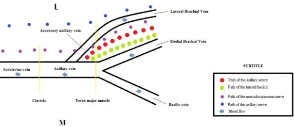
The general topography and the trajectory of AAV was as follows: the AAV was lateral to the axillary
artery in 42/48 regions (87.5%) and in 6/48 (12.5%) was
posterolateral to the artery, as illustrated in Figures 1-4. It ascended laterally to the brachial plexus in 44/48
(91.66%), while in the remaining 4/48 (8.33%) cases,
the brachial plexus was absent because it had been removed during previous dissections. The vessel can also be situated between the musculocutaneous n. (anteriorly) and the axillary n. (posteriorly), as we
observed in 21/48 (43.75%) cases. In 39/48 cases (81.25%), the AAV terminated in regions situated
along an inferior portion of the AV, in 7/48 cases
(14.58%) it terminated in the mid portion, and in 2/48 cases (4.16%) it ended at the subclavian vein.
With regard to cases with high numbers of tributary veins, 5 tributary veins of the AV were observed in
27 axillae (56.25%) and 3 tributaries of the AAV were observed in 13 axillae (46.42%).
The Langer axillary arch was not observed in any of the axillae studied.
With regard to the number of AV valves, 25 (52%) cases exhibited 7 valves, 14 (29.16%) cases had
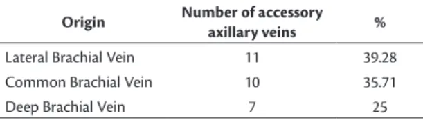
Table 2. Veins from which the accessory axillary veins originated.
Origin Number of accessory
axillary veins %
Lateral Brachial Vein 11 39.28
Common Brachial Vein 10 35.71
Deep Brachial Vein 7 25
Table 1. Incidence of accessory axillary vein in the cadavers studied.
Location Number of Cadavers %
Right axilla 13 46.42
Left axilla 11 39.28
9 valves, 5 (10.41%) cases had 4 valves, and 4 AVs (8.33%) had 6 valves. With regard to the number of AAV valves, 14 (50%) had 6, 8 (28.57%) had 7, 5 (17.85%) had 4, and 1 (3.57%) had 2 valves.
During dissection, it was observed that the axillary artery was located lateral to the AV in all of the cadavers. The lymph nodes related to upper limb drainage were situated just below the AV and the pectoralis minor muscle was located anterior to the
axillary vein and artery. However, 39 (81.25%) of the
axillae studied lacked at least one of the following structures: the intercostal brachial nerve (this is usually located medial of the axillary vessels), the long thoracic nerve (always next to the intercostal nerve), the medial cord of the brachial plexus, and the median, ulnar, and pectoral nerves (the latter are usually between the vein and the axillary artery).
That is, only 18.75% of the axillae studied exhibited
all of the structures listed above, which are usually observed in this region.
DISCUSSION
The axillary vein has been widely discussed in the literature, with an increasing number of reports regarding its use in clinical and surgical procedures.
Gray (1988) and Gusmão (1992) report that the AV is formed by the junction of the basilic vein with the lateral and medial brachial veins, or by the union of the common brachial vein, which is formed by the initial junction of the two brachial veins with the basilic vein.1,2 However, according to Hollinshead &
Rosse (1991) and later Gusmão in 2003, the AV is the direct and ascending continuation of the basilic, which
belongs to the supericial venous system. The basilic
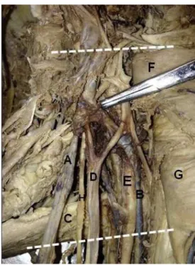
Figure 1. Right axilla, in (A) accessory axillary vein; (B) the
axillary vein; (C) teres major muscle; (D) lateral fascicle; (E) axillary a.; (F) pectoralis minor m.; (G) pectoralis major m.; (H) musculocutaneous n.
Figure 2. Left axilla, in (A) accessory axillary vein; (B) the axillary
vein; (C) teres major muscle; (F) pectoralis minor m.
Figure 3. Left axilla, in (A) accessory axillary vein; (B) the axillary
vein perforates the brachial fascia and follows a path toward the axilla, where at this deeper point, it is called the axillary vein.5 Our results showed that the
levels of the origin and termination of the axillary vein were similar to those reported by Gray (1988): it originates at the lower edge of the teres major muscles and terminates in front of the subscapularis tendon
at the lateral margin of the irst rib, at which point
the axillary vein is provided with a pair of valves. In this study, the incidence of accessory axillary
veins was 58.3%. This result is similar to the 56.7%
reported by Gusmão & Prates (1992).6 The AAV has
often been described as a satellite vein of the AV
or as a collector channel that drains the circumlex
veins, which has led some researchers to name it as
a small axillary vein, which at some point will low
into the true axillary vein and therefore should not be considered an anatomical variation, because it is
not a rare inding.
In our study, we observed that 11 (39.28%) of
the AAV found were formed by the lateral brachial
vein, 10 (35.71%) by the common brachial vein, and 7 (25%) by the deep brachial vein. Gusmão & Prates (1992) found incidence rates of 55.9% of cases of AAV formed by the lateral brachial vein, 33.4% by the common brachial vein, and 11.8% by the deep
brachial vein.6
Tributary veins were found in 27 (56.25%)
axillae, 5 of which were tributary veins of the AV,
and in 13 axillae (46.42%) there were 3 tributaries
of the AAV. This suggests that these tributaries do not perform blood drainage from the same territories and are therefore not equivalent.
The valves of the axillary veins have been widely used to replace those of patients with chronic venous
insuficiency.5,7 In our series, 25 AV (52%) had 7 valves
and 14 AAV (50%) had 6 valves.
With regard to the likelihood of using the AAV for hemodialysis purposes, we believe that due to
the dificult access and the depth of the vessel, this
is highly unlikely. However, Lin et al. (2011), used ultrasound as a guide to the axillary vein and there are no reports in the literature with regard to use of ultrasound to locate the AAV.8
There are several reports in the literature about the possibility of deep vein thrombosis involving the axillary and subclavian veins.3,9 In such cases, the AAV
would be an alternative pathway for venous drainage
and restoration of blood low to this region.3,10,11
The accessory axillary vein can also be of great importance for collateral circulation in the event of traumatic injury to the axillary vein.9 The larger the
caliber of the AAV, the better it will function as a collateral alternative.8
CONCLUSION
Our results allow us to conclude that the incidence
of AAV was 58.3%, with no predilection for sex or
body side, and the AAVs found often originated from the lateral brachial vein or the common brachial vein. There are few data in the literature regarding AAV incidence and its potential clinical and surgical applications. In view of its high incidence and clinical relevance, further studies are warranted.
REFERENCES
1. Gray H, Goss CM. Gray Anatomia. Rio de Janeiro: Guanabara; 1988. 174 p.
2. Gusmão LCB. Anatomia arterial e venosa aplicada. In: Gusmão LCB, editor. Angiologia e cirurgia vascular: guia ilustrado. Maceió: UNCISAL/ECMAL/LAVA; 2003. p. 6-7.
3. Santos CA, Figueiredo LF, Gusmão LC, et al. Estudo anatômico da veia braquial comum como via de drenagem colateral do membro superior. Rev Angiol Cir Vasc. 2010;10:40-3.
4. Santos CA, Gusmão LC. Estudo anatômico sobre a veia basílica no braço de cadáveres humanos. Rev Hosp Univer da UFAL. 1997;4:32-40.
5. Hollinshead WH, Rosse C. Anatomia. Rio de Janeiro: Inter Livros; 1991. p. 152-153.
6. Gusmão LC, Prates JC. Anatomical study of the accessory axillary vein. Surg Radiol Anat. 1992;14(2):131-6. PMid:1641737. http:// dx.doi.org/10.1007/BF01794889.
7. Mustafa S, Stein PD, Patel KC, Otten TR, Holmes R, Silbergleit A. Upper extremity deep venous thrombosis. Chest. 2003;123(6):1953-6. PMid:12796173. http://dx.doi.org/10.1378/chest.123.2003;123(6):1953-6.1953.
8. Lin CP, Wang YC, Lin FS, Huang CH, Sun WZ. Ultrasound-assisted percutaneous catheterization of the axillary vein for totally implantable venous access device. Eur J Surg Oncol. 2011;37(5):448-51. PMid:21345637. http://dx.doi.org/10.1016/j.ejso.2011.01.026.
9. Kumar V. Endovascular treatment of penetrating injury of axillary vein with via bahn endoprosthesis. J Vasc Surg. 2004;40(6):1243-4. PMid:15622382. http://dx.doi.org/10.1016/j.jvs.2004.09.022. 10. Wandderley NJ, Pitta GB. Trauma de vasos axilares. In: Pitta GBB,
editor. Angiologia e cirurgia vascular: guia ilustrado. Maceió: UNCISAL/ECMAL/LAVA; 2003. p. 1-3.
11. Kutiyanawala MA, Stotter A, Windle R. Anatomical variants during axillary dissection. Br J Surg. 1998;85(3):393-4. PMid:9529501. http://dx.doi.org/10.1046/j.1365-2168.1998.00612.x.
*
Correspondence
Dhayanna Rolemberg Gama Cabral Centro de Estudos Superiores de Maceió Rua Cônego Machado, 918 CEP 57051-160 - Maceió (AL), Brazil E-mail: [email protected]
Author information
VBF - Board certiied in Oral and Maxillofacial Surgery and Traumatology by Faculdade de Ciências Médicas, Santa Casa de Misericórdia de São Paulo (FCMSCSP); board certiied in Oral Pathology by Conselho Federal de Odontologia (CFO); MSc in Oral and Maxillofacial Surgery from Universidade de Marília (UNIMAR) and PhD in Oral Pathology from Universidade de São Paulo (USP). JABS - Board certiied in Higher Education Teaching and General, Animal, and Human Physiology by Centro Universitário CESMAC and MSc in Health Sciences from Universidade Federal de Alagoas (UFAL). KJMF - Board certiied in Higher Education Teaching by Centro Universitário CESMAC and MSc in Dentistry from Centro de Pesquisas Odontológicas São Leopoldo Mandic (SLMANDIC). DRGC - Dental student and monitor of the Discipline of Human Anatomy at Centro Universitário CESMAC. CASS - Board certiied in Anatomy of the Locomotor Apparatus by Universidade Federal de Pernambuco (UFPE); board certiied in Peripheral Vascular Surgery by Hospital Agamenon Magalhaes, Secretaria de Saúde do Estado de Pernambuco; MSc in Sciences from Universidade Federal de São Paulo (UNIFESP) and PhD in Bioethics from Centro Universitário São Camilo (CUSC). CFSR - Board certiied in Clinical Medicine by Universidade Estadual de Ciências da Saúde de Alagoas (UNCISAL); MSc and PhD in Human Anatomy from Universidade Federal de São Paulo (UNIFESP). JSBL - MSc in Human Anatomy from Universidade Federal de Pernambuco (UFPE) and MSc in Pathology from UFPE. AJCR - MSc in Surgical Gastroenterology from Universidade Federal de São Paulo (UNIFESP).
Author contributions
Conception and design: AJCR, JABS, VBF Analysis and interpretation: KJMF, CASS, VBF Data collection: DRGC, CFSR, JSBL, VBF Writing the article: JABS, KJMF, DRGC, CASS, VBF Critical revision of the article: AJCR, JABS, KJMF, VBF Final approval of the article*: AJCR, JABS, KJMF, DRGC, CASS, CFSR, JSBL, VBF Statistical analysis: AJCR, JABS, DRGC, VBF Overall responsibility: VBF