Inflammatory and immunogenetic markers in
correlation with pulmonary tuberculosis*
Marcadores inflamatórios e imunogenéticos e sua relação com tuberculose pulmonar
Beatriz Lima Alezio Muller, Daniela Maria de Paula Ramalho, Paula Fernanda Gonçalves dos Santos, Eliene Denites Duarte Mesquita,
Afranio Lineu Kritski, Martha Maria Oliveira
Abstract
Objective: To describe serum levels of the cytokines IL-10, TNF-α, and IFN-γ, as well as polymorphisms in the genes involved in their transcription, and their association with markers of the acute inflammatory response in patients with pulmonary tuberculosis. Methods: This was a descriptive, longitudinal study involving 81 patients with pulmonary tuberculosis treated at two referral hospitals. We collected data on sociodemographic variables and evaluated bacteriological conversion at the eighth week of antituberculosis treatment, gene polymorphisms related to the cytokines studied, and serum levels of those cytokines, as well as those of C-reactive protein (CRP). We also determined the ESR and CD4+ counts. Results: The median age of the patients was 43 years; 67 patients (82.7%) were male; and 8 patients (9.9%) were infected with HIV. The ESR was highest in the patients with high IFN-γ levels and low IL-10 levels. IFN-γ and TNF-α gene polymorphisms at positions +874 and
−238, respectively, showed no correlations with the corresponding cytokine serum levels. Low IL-10 levels were associated with IL-10 gene polymorphisms at positions −592 and −819 (but not −1082). There was a negative
association between bacteriological conversion at the eighth week of treatment and CRP levels. Conclusions: Our results suggest that genetic markers and markers of acute inflammatory response are useful in predicting the response to antituberculosis treatment.
Keywords: Tuberculosis; Cytokines; Immune system; Polymorphism, single nucleotide.
Resumo
Objetivo: Descrever os níveis séricos das citocinas IL-10, TNF-α e IFN-γ, assim como polimorfismos presentes em genes envolvidos na sua transcrição, e sua associação com marcadores de resposta inflamatória aguda em pacientes com tuberculose. Métodos: Estudo descritivo e longitudinal realizado em 81 pacientes com tuberculose pulmonar atendidos em dois hospitais de referência. Foram coletadas informações sociodemográficas, conversão bacteriológica na oitava semana de tratamento antituberculose, polimorfismos relacionados às citocinas estudadas, níveis séricos dessas citocinas, assim como de proteína C reativa (PCR). Também foram avaliados VHS e contagem de CD4+. Resultados: A mediana de idade dos pacientes era de 43 anos, sendo 67 (82,7%) do sexo masculino e 8 (9,9%) infectados por HIV. Os pacientes com níveis elevados de IFN-γ e baixos níveis de IL-10 apresentaram valores mais elevados de VHS. Não houve associação dos polimorfismos do gene IFN-γ na posição +874 e do gene TNF-α na posição −238 com os níveis das citocinas correspondentes. Houve uma associação entre polimorfismos do gene IL-10 nas posições −592 e −819 (mas não −1082) e baixos níveis de
IL-10. Houve uma associação negativa entre a taxa de conversão bacteriológica na oitava semana de tratamento e níveis de PCR. Conclusões: Nossos resultados sugerem que marcadores genéticos e de resposta inflamatória aguda podem ser úteis na predição da resposta ao tratamento antituberculose.
Descritores: Tuberculose; Citocinas; Sistema imunológico; Polimorfismo de nucleotídeo único.
*Study carried out at the Tuberculosis Research Center, Federal University of Rio de Janeiro School of Medicine Clementino Fraga Filho University Hospital, Rio de Janeiro, Brazil.
Correspondence to: Martha Maria Oliveira. Rua Porto Seguro, 54, Ilha do Governador, CEP 21931-060, Rio de Janeiro, RJ, Brasil. Tel. 55 21 2562-2426. Fax: 55 21 2562-2431. E-mail: [email protected]
Financial support: This study received financial support from the Brazilian Conselho Nacional de Desenvolvimento Científico e Tecnológico/Ministério da Ciência, Tecnologia e Inovação (CNPq/MCTI, National Council for Scientific and Technological Development/ National Ministry of Science, Technology, and Innovation; Grant nos. CNPq/INCT 573548/2008-0 and 478033/2009-5) and the Foundation for the Support of Research in the State of Rio de Janeiro (Grant no. E:26/110.974/2011).
immune and inflammatory responses can aid in identifying better markers of protection against tuberculosis.
There have been few studies simultaneously evaluating the genotypic and phenotypic aspects of the human host immune response to infection
with M. tuberculosis.(10,11) Given the paucity of
data on the simultaneous evaluation of genetic, immunological, and inflammatory biomarkers in patients with pulmonary tuberculosis, we conducted the present study in order to determine the prevalence of IL-10 gene polymorphisms at
positions −592, −819, and −1082; the prevalence
of TNF-α gene polymorphisms at position −238;
and the prevalence of IFN-γ gene polymorphisms
at position +874. The study involved a sample of active pulmonary tuberculosis patients admitted to and treated at either of two referral hospitals in the city of Rio de Janeiro, Brazil. In addition, we measured the serum levels of the corresponding cytokines and analyzed the acute inflammatory response by determining CRP levels and CD4+ counts, as well as the ESR.
Methods
This was a longitudinal descriptive study involving 81 patients diagnosed with pulmonary tuberculosis and admitted to either of two referral hospitals for the treatment of tuberculosis in the state of Rio de Janeiro, Brazil (the Hospital Estadual Santa Maria and the Instituto Estadual de Doenças do Tórax Ary Parreiras), between March 23, 2007 and August 7, 2009. We included patients with positive smear microscopy and culture for mycobacteria, the presence of M. tuberculosis being subsequently confirmed by biochemical tests. We analyzed the following variables: CRP, ESR, CD4+, and bacteriological conversion at the 8th week of antituberculosis treatment.
For DNA extraction, a commercial kit (DNAzol;
Gibco BRL/Life Technologies, Gaithersburg, MD,
USA) was used in accordance with the manufacturer instructions. After DNA extraction, a DNA sample was analyzed by electrophoresis on 1% agarose gel in order to determine integrity and concentration,
the sample being subsequently stored at −20°C.
For the analysis of TNF-α gene polymorphisms
at position −238, 100 ng of DNA, 1× buffer, 1.5 mM MgCl2, 200 µM dNTP, and 1 U Taq
DNA polymerase (Invitrogen Life Technologies,
Carlsbad, CA, USA) were added to 15 pmol of
each primer for polymerase chain reaction, which
Introduction
Tuberculosis is a chronic infectious disease caused by Mycobacterium tuberculosis, the most common clinical manifestation of which is pulmonary involvement; however, tuberculosis can affect other anatomical sites (extrapulmonary
tuberculosis) or present as disseminated disease.(1)
Despite being a curable disease, tuberculosis remains a major public health problem worldwide. According to the World Health Organization, Brazil ranks 19th among the 22 countries that collectively account for 80% of all cases of tuberculosis worldwide and 108th among those in which the incidence of tuberculosis is highest. According to the Brazilian National Ministry of Health, 71,000 new cases of tuberculosis were added to the Brazilian Case Registry Database in 2010, corresponding to an incidence rate of
37.2/100,000 population.(2)
The systemic inflammation observed in patients with tuberculosis is mediated by the activation of the immune system, with excessive production
of cytokines, such as IL-1, IL-2, IFN-γ, and TNF-
α.(3) After the inflammatory process, there is
an increase in the hepatic synthesis and serum levels of acute phase proteins, such as C-reactive protein (CRP), as well as in the ESR, which have been used in the diagnosis and follow-up of patients, given that their plasma levels directly reflect the intensity of the pathological process.(4)
Genetic factors have been associated with
susceptibility to or protection against infection
with M. tuberculosis.(5) In the immune response
to M. tuberculosis, allele frequencies in cytokine gene polymorphisms vary considerably across populations, as reported in meta-analyses
evaluating IFN-γ, IL-10, and TNF-α gene
polymorphisms.(6,7) It has been proposed that
serum cytokine levels and their role as markers of response to antituberculosis treatment be
evaluated.(8) The maintenance of initially low
serum levels of IFN-γ or high serum levels of
TNF-α and of increased serum levels of IL-17 is
associated with a worse prognosis, including a higher mortality rate and lower bacteriological conversion at the end of the 8th week of antituberculosis treatment. Recently, Lago et
al.(9) described a possible association between
to obtain the standard curve. The tubes were homogenized and incubated for three hours at room temperature in the dark. Subsequently, the reading was performed with a BD™ Cytometric
Bead Array system (Thermo Fisher Scientific).(12)
Serum levels of CRP were used as a marker of the acute phase response (APR), i.e., as a marker of the systemic response to severe inflammation. A positive APR was defined as CRP levels > 0.3 mg/dL, whereas a negative APR was defined as CRP levels < 0.3 mg/dL. Serum CRP levels were measured by nephelometry.
The ESR was also used as a marker of the APR, a positive APR being defined as an ESR > 2 mm/h for females and as an ESR > 7 mm/h for males. The ESR was measured by the Westergren method.
We used descriptive statistics, including range (minimum and maximum values), mean, standard
deviation, and 95% CI. We used the
Kolmogorov-Smirnov test in order to test the normality of the variables and the Levene test in order to determine the equality of variances. Variables with non-normal distribution were log-transformed. For means with normal distribution, we used the Student’s t-test. We used ANOVA in order to analyze the differences among quantitative variables. We used the chi-square test in order to identify associations among categorical variables.
A value of p < 0.05 was considered statistically
significant. We used the Statistical Package for the Social Sciences, version 16.0 for Windows (SPSS Inc., Chicago, IL, USA). In the analyses, we used the bacteriological conversion coefficient, which was calculated as the number of cases of patients who converted from a negative test result to a positive test result divided by the total number of patients at the beginning of treatment, and the mutation coefficient for polymorphisms, which was calculated as the number of cases of a given mutation divided by the total number of cases.
The present study was approved by the Research Ethics Committee of the Federal University of Rio de Janeiro School of Medicine Clementino
Fraga Filho University Hospital on April 28, 2005 (Protocol no. 004/05).
Results
The median age of the patients was 43 years (range, 20-60 years). Of the 81 patients studied, 67
(82.7%) were male, 54 (66.7%) were non-White, was performed as follows: one cycle at 94°C for
1 min, followed by 5 cycles at 94°C, 67°C, and 72°C (60 s each), and 25 cycles at 94°C, 62°C, and 72°C (60 s each). For the genotyping of IFN-γ
gene polymorphism at position +874, we used
200 mL of dNTP, 1.5 mM MgCl2, 8.5% sucrose,
0.25 U of ThermoPrime Plus DNA polymerase
(Thermo Fisher Scientific, Waltham, MA, USA),
5 mL of each specific primer, 0.5 mL of internal
control primer, and 100 ng of DNA. The mixture
was incubated at 95°C for 1 min; subsequently, 10 cycles were performed at 95°C for 15 s, followed by 10 cycles at 62°C for 50 s, 10 cycles at 72°C for 40 s, 20 cycles at 95°C for 20 s, 20 cycles at 56°C for 50 s, and 20 cycles at 72°C for 50
s. For the detection of IL-10 promoter gene
polymorphisms at positions −819, −1082, and −592, the following steps were taken: for the −592 position, a 480-bp fragment was amplified
and subsequently digested with the enzyme RsaI.
For the −1082 and −819 positions, a 360-bp
fragment was amplified and subsequently digested with the enzymes BseRI and MslI, respectively. In brief, 100 ng of DNA were added to each polymerase chain reaction, resulting in a final
volume of 40 µL (−819 and −1082) or 30 µL (−592), consisting of 50 mM KCl, 10 mM Tris-HCl (pH 8.3), 1.5 mM MgCl2, 200 µM dNTP, 1.25 U
AmpliTaq Gold DNA Polymerase (Perkin-Elmer
Cetus, Norwalk, CT, USA) and specific primers for
each mutation (10 pmol for the −592 position and 12.5 pmol for the −819/−1082 positions). All mixtures were incubated at 95°C for 10 min and submitted to amplification at 94°C for 30 s, at 60°C for 30 s, at 72°C for 40 s, and at 72°C for 7 min (IL-10 at position −592), followed by 35 cycles at 94°C for 30 s, at 58°C for 30 s, and at 72°C for 45 s, plus a final cycle at 72°C for 5 min (positions −819 and −1082). The amplified
products were electrophoresed on 2% agarose
gel containing ethidium bromide (0.5 mL/mL).
In the determination of cytokine levels, bead populations were visualized on the basis of their fluorescence intensities. In the cytometric bead array system, cytokine capture beads are mixed with detection antibody conjugated to the fluorochrome phycoerythrin and then incubated with the samples for the “conjugate” assay. The
acquisition tubes were prepared with 50 µL of sample, 50 µL of the bead mixture, and 50 µL of
no relationship between low serum levels of IFN-γ
and the presence of mutations. Regarding TNF-α,
although we found no homozygous mutations, we found a trend toward low serum levels of
TNF-α among heterozygotes. We found a negative
relationship between serum IL-10 levels and
IL-10 gene polymorphisms at positions −592 and −819 (p < 0.001; Figure 1).
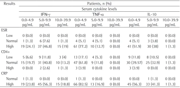
As can be seen in Table 2, there was a trend (p = 0.08) toward lower CRP production in the
patients in whom serum IFN-γ levels were low
(0.0-4.9 pg/mL) when compared with those in
whom serum IFN-γ levels were higher (> 5.0 pg/
mL). In the patients in whom serum TNF-α levels
were low (0.0-4.9 pg/mL), there was a trend toward a higher ESR (p = 0.04). Low serum levels of IL-10 (i.e., serum IL-10 levels of 0.0-4.9 pg/
8 (9.9%) were co-infected with HIV, 52 (64.2%) reported regular alcohol use, 55 (67.9%) were
smokers or former smokers, 20 (24.7%) reported illicit drug use, and 64 (79.0%) had normal CD4+ counts. All patients had smear-positive pulmonary tuberculosis.
In the analysis of the prevalence of IFN-γ
gene polymorphisms at position +874, of TNF-α
gene polymorphisms at position −238, and of IL-10 gene polymorphisms at positions −592, −819, and −1082, the mutant allele frequency was found to be 0.56, 0.56, 0.29, 0.43, and
0.68, respectively.
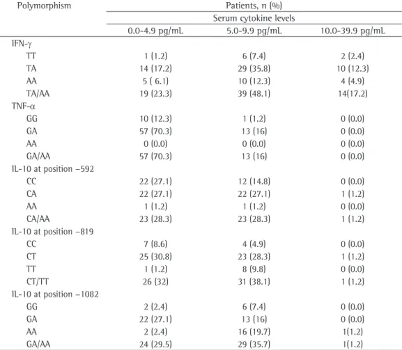
Table 1 shows the distribution of polymorphisms in the patients under study, by serum cytokine
levels. Serum IFN-γ levels were found to range
from 0 (zero) pg/mL to 20.5 pg/ml, and there was
Table 1 - Distribution of the polymorphisms found in the patients under study, by serum cytokine levels.
Polymorphism Patients, n (%)
Serum cytokine levels
0.0-4.9 pg/mL 5.0-9.9 pg/mL 10.0-39.9 pg/mL IFN-γ
TT 1 (1.2) 6 (7.4) 2 (2.4)
TA 14 (17.2) 29 (35.8) 10 (12.3)
AA 5 ( 6.1) 10 (12.3) 4 (4.9)
TA/AA 19 (23.3) 39 (48.1) 14(17.2)
TNF-α
GG 10 (12.3) 1 (1.2) 0 (0.0)
GA 57 (70.3) 13 (16) 0 (0.0)
AA 0 (0.0) 0 (0.0) 0 (0.0)
GA/AA 57 (70.3) 13 (16) 0 (0.0)
IL-10 at position −592
CC 22 (27.1) 12 (14.8) 0 (0.0)
CA 22 (27.1) 22 (27.1) 1 (1.2)
AA 1 (1.2) 1 (1.2) 0 (0.0)
CA/AA 23 (28.3) 23 (28.3) 1 (1.2)
IL-10 at position −819
CC 7 (8.6) 4 (4.9) 0 (0.0)
CT 25 (30.8) 23 (28.3) 1 (1.2)
TT 1 (1.2) 8 (9.8) 0 (0.0)
CT/TT 26 (32) 31 (38.1) 1 (1.2)
IL-10 at position −1082
GG 2 (2.4) 6 (7.4) 0 (0.0)
GA 22 (27.1) 13 (16) 0 (0.0)
AA 2 (2.4) 16 (19.7) 1(1.2)
GA/AA 24 (29.5) 29 (35.7) 1(1.2)
(IFN-γ): TT: wild-type homozygous genotype; TA: heterozygous genotype; and AA: mutant homozygous genotype. (TNF-α): GG: wild-type homozygous genotype; GA: heterozygous genotype; and AA: mutant homozygous genotype. (IL-10 at position −592): CC: wild-type homozygous genotype; CA: heterozygous genotype; and AA: mutant homozygous genotype. (IL-10 at position −819): CC: wild-type homozygous genotype; CT: heterozygous genotype; and TT: mutant homozygous genotype. (IL-10 at position −1082): GG: wild-type homozygous genotype; GA: heterozygous genotype;
markers, inflammatory markers, and immunogenetic markers in pulmonary tuberculosis patients in Brazil. The clinical features of the patients in our sample were similar to those of patients admitted to tuberculosis hospitals in developing countries.(13)
The genetic component of the host response to infection with M. tuberculosis in restricted
ethnic groups is evident in the literature.(5) In
the present study, the frequency of the mutant
allele for IFN-γ gene polymorphisms at position
+874 was 0.56, which is similar to that reported
by other authors in various countries(14-16) but
different from that reported by Fitness et al.
in Africa.(17) In addition, the frequency of the
mutant allele for TNF-α gene polymorphisms
at position +238 was 0.56, which is similar
mL) were not associated with a higher ESR or with higher CRP levels. However, the ESR was negatively correlated with serum IL-10 levels (p = 0.03) and was positively correlated with serum
IFN-γ levels (p = 0.008; Table 3).
Table 4 shows that lower bacteriological conversion was associated only with high serum levels of CRP. However, by applying the bacteriological conversion coefficient, we found
a negative correlation between serum TNF-α
levels and bacteriological conversion (r = −0.43;
p < 0.001).
Discussion
To our knowledge, this is the first study to examine the relationships among biochemical
0.08 0.07 0.06 0.05 0.04 0.03 0.02 0.01 0.00
IL-10 gene polymorphism at position −592
15.00
12.00
9.00
6.00
3.00
0.00
0.25 0.20 0.15 0.10 0.05 0.00
IL-10 gene polymorphism at position −819
15.00
12.00
9.00
6.00
3.00
0.00
p< 0.001 (-0.76) p< 0.001 (-0.80)
IL-10
IL-10
A B
Figure 1 - Regression coefficient for IL-10 gene polymorphisms at positions −592 (in A) and −819 (in B).
Table 2 - Distribution of serum levels of IFN-γ, TNF-α, and IL-10 in the patients under study, by laboratory test results.
Results Patients, n (%)
Serum cytokine levels
IFN-γ TNF-α IL-10
0.0-4.9 pg/mL
5.0-9.9
pg/mL
10.0-39.9 pg/mL
0.0-4.9 pg/mL
5.0-9.9
pg/mL
10.0-39.9 pg/mL
0.0-4.9 pg/mL
5.0-9.9
pg/mL
10.0-39.9 pg/mL ESR
Low 0 (0.0) 0 (0.0) 0 (0.0) 0 (0.0) 0 (0.0) 0 (0.0) 0 (0.0) 0 (0.0) 0 (0.0) Normal 1 (1.3) 6 (7.6) 1 (1.3) 4 (5.1) 4 (5.1) 0 (0.0) 4 (5.1) 3 (3.8) 0 (0.0) High 19 (24.1) 37 (46.8) 15 (19) 61 (77.2) 10 (12.7) 0 (0.0) 41 (51.9) 30 (38) 1 (1.3) CD4+
Low 5 (6.6) 9 (11.8) 3 (4) 13 (17.1) 4 (5.3) 0 (0.0) 9 (11.8) 8 (10.5) 0 (0.0) Normal 15 (19.7) 31 (40.8) 10 (13.2) 47 (61.8) 9 (11.8) 0 (0.0) 30 (39.57) 25 (32.9) 1 (1.3) High 0 (0.0) 2 (2.6) 1 (1.3) 3 (3.9) 0 (0.0) 0 (0.0) 3 (3.9) 0 (0.0) 0 (0.0) CRP
The functional role of allele −238A (TNF-α)
in the regulation of TNF-α gene expression was
described by Kaluza et al.,(21) whose in vitro
studies showed an association between allele
−238A and a downregulation of the TNF-α
gene (and, consequently, a reduction in TNF-α
protein production). Although we found no homozygous mutations, we observed a trend
toward low serum levels of TNF-α in patients
with a heterozygous genotype, as did Abhimanyu et al.(10) in a population of individuals from India
whose ethnic characteristics were quite different from those of our study population. However,
Haroon et al.(22) found no association between
mutation and cytokine expression in a population of White individuals.
The presence of single-nucleotide
polymorphisms in the first intron of the IFN-γ
gene (at position +874) has been associated with tuberculosis(8,14) and severe tuberculosis.(23) The
to that reported in other studies.(18-20) In our
sample, the allele frequencies for IL-10 gene
polymorphisms at positions −592, −819, and −1082 were, respectively, 0.29, 0.43, and 0.68,
being similar to those reported in most of the
studies included in a meta-analysis.(6) Although
our results are consistent with those of various studies, any differences regarding the frequency of these polymorphisms can be explained by ethnic differences among the study populations.
Table 4 - Serum levels of C-reactive protein, ESR, CD4+, and cytokines, as well as frequency of genotypes, by bacteriological conversion.a
Variable Bacteriological conversion p
Yes No
CRP, mg/dL 5.37 ± 3.96 8.6 ± 4.7 0.004
ESR, mm/h 60.3 ± 43.3 53.8 ± 37.6 0.57
CD4+, cells/mm3 784.09 ± 699.44 657.5 ± 311.49 0.46
IL-10, pg/mL 4.6 ± 2.8 4.8 ± 1.21 0.73
TNF-α, pg/mL 3.6 ± 1.93 3.99 ± 1.36 0.42
IFN-γ, pg/mL 7.17 ± 4.84 7.69 ± 3.99 0.68
IL-10 at position −592b
AA 2 (3.4) 0 (0.0) 0.42
CA/CC 57 (96.6) 18 (100.0)
IL-10 at position −819b
TT 8 (13.6) 2 (11.1) 0.78
CT/CC 51 (86.4) 16 (88.9)
IL-10 at position −1082b
AA 27 (45.8) 9 (50.0) 0.75
GA/GG 32 (54.2) 9 (50.0)
TNF-αb
AA 0 (0.0) 0 (0.0)
--GA/GG 59 (100.0) 18 (100.0)
IFN-γb
AA 11 (18.6) 6 (33.3) 0.18
TA/TT 48 (81.4) 12 (66.7)
CRP: C-reactive protein; (IFN- γ): TT: wild-type homozygous genotype; TA: heterozygous genotype; and AA: mutant homozygous genotype. (TNF-α): GG: wild-type homozygous genotype; GA: heterozygous genotype; and AA: mutant homozygous genotype. (IL-10 at position −592): CC: wild-type homozygous genotype; CA: heterozygous genotype; and AA: mutant homozygous genotype. (IL-10 at position −819): CC: wild-type homozygous genotype; CT: heterozygous genotype; and TT: mutant homozygous genotype. (IL-10 at position −1082): GG: wild-type homozygous genotype; GA: heterozygous genotype; and AA: mutant homozygous genotype. aValues expressed as mean ± SD, except where
otherwise indicated. bValues expressed as n (%).
Table 3 - Correlation between serum cytokine levels and laboratory test results.
Biochemical variable
p
IFN-γ TNF-α IL-10
CD4+ 0.59 0.11 0.47
CRP 0.43 0.32 0.33
ESR 0.008 (+) 0.31 0.03 (−)
C-reactive protein; (+): positive correlation; and (−):
with an increased risk of recurrence, whereas low serum levels of IL-10 usually occur in mild forms of tuberculosis. The results obtained in the present study do not allow us make inferences regarding this issue, given that serum IL-10 levels were assessed only at time point zero and not during clinical follow-up (after completion of antituberculosis treatment).
In the present study, acute inflammatory response markers (CRP levels) were found to be
higher in the patients in whom serum TNF-α
levels were low (0.0-4.9 pg/mL) than in those
in whom serum TNF-α levels were above 5.0
pg/mL. These data suggest that the presence
of low concentrations of TNF-α at the time
of the initial response against the disease is associated with a worse prognosis and clinical course; however, studies involving larger samples, as well as correlation studies, should be conducted in order to test these hypotheses in the Brazilian population, as mentioned in a review article by Wallis et al.(8) The role of TNF-α
in the pathophysiology of tuberculosis has been associated with defense via macrophage activation
and the subsequent inflammatory reaction.(3)
Our findings reinforce the importance of this cytokine in the host response to M. tuberculosis. In the present study, an association was found between elevated CRP levels and lower bacteriological conversion at the 8th week of antituberculosis treatment, showing the potential role of this mediator as a marker for monitoring the clinical course of the disease. Some authors have reported that ESR normalization is a marker of good response to treatment in subacute and chronic diseases, such as tuberculosis.(27,28) Various
studies have shown increased levels of immune response markers, CRP, and ESR in the initial phase, all of which decrease during treatment.
(29,30) Similar results were reported in a study
by Peresi et al.,(4) in which CRP levels were
significantly decreased only in the 3rd and 6th months of treatment. These findings suggest that CRP can be used in order to evaluate the APR in tuberculosis patients and as a marker of response to antituberculosis treatment, together with the clinical and epidemiological history of such patients.
In our study, there was no association between
serum IFN-γ levels and bacteriological conversion,
a finding that is similar to those of another
study.(8) However, there was an association
gene encoding IFN-γ is highly conserved, and
few polymorphisms are found in the intragenic region. In our sample, we found no association
between IFN-γ gene polymorphisms at position
+874 and serum IFN-γ levels, a finding that
is similar to those of Abhimanyu et al.(10) and
Vidyarani et al.(24) but different from those of
Vallinoto et al.,(11) who found low serum levels
of IFN-γ in patients with a homozygous mutant
genotype at position +874A/A.
We found a significant relationship between
high IFN-γ levels and a high ESR, a finding that
is consistent with those of Peresi et al.(4) This is
possibly due to the fact that the presence of this mutation has been associated with decreased
production of IFN-γ (a cytokine that plays an
important role in controlling the defense against the pathogen) and, therefore, a diminished acute inflammatory response.
In our study, low serum levels of IL-10 were found to be associated with IL-10 gene
polymorphisms at positions −592 and −819 (but not −1082). This finding is similar to those of
Abhimanyu et al.(10) and Edwards-Smith et al.,(25)
who investigated IL-10 gene polymorphisms at
position −1082 and showed that individuals
carrying the AA genotype are low IL-10 producers,
those carrying the GA genotype are intermediate IL-10 producers, and those carrying the GG
genotype are high IL-10 producers; however, the ATA haplotype is associated with low IL-10 production. These discrepant results can be partly explained by the distinct and heterogeneous ethnic characteristics of the study populations. We observed a trend toward a higher ESR among carriers of IL-10 gene polymorphisms at
positions −592 (CA/AA) and −819 (CT/TT). The
authors of a recent meta-analysis including 18 studies (none of which included patients from Latin America) were unable to confirm a higher risk of tuberculosis among patients with IL-10
gene polymorphisms at positions −592, −819, or −1082 but found a higher risk of tuberculosis
among Europeans with IL-10 gene polymorphisms
at position −1082.(6) In that meta- analysis, one
of the studies assessing serum IL-10 levels also
assessed serum levels of IFN-γ and IL-10. The
authors demonstrated that a stronger relationship translated to less severe tuberculosis.
Jamil et al.(26) and Lago et al.(9) suggested that
7. Wang Q, Zhan P, Qiu LX, Qian Q, Yu LK. TNF-308
gene polymorphism and tuberculosis susceptibility: a meta-analysis involving 18 studies. Mol Biol Rep. 2012;39(4):3393-400.
http://dx.doi.org/10.1007/s11033-011-1110-x PMid:21735105
8. Wallis RS, Kim P, Cole S, Hanna D, Andrade BB, Maeurer
M, et al. Tuberculosis biomarkers discovery: developments, needs, and challenges. Lancet Infect Dis. 2013;13(4):362-72. http://dx.doi.org/10.1016/S1473-3099(13)70034-3 9. Lago PM, Boéchat N, Migueis DP, Almeida AS, Lazzarini
LC, Saldanha MM, et al. Interleukin-10 and interferon-gamma patterns during tuberculosis treatment: possible association with recurrence. Int J Tuberc Lung Dis.
2012;16(5):656-9. PMid:22410761 PMCid:PMC3729476
10. Abhimanyu, Mangangcha IR, Jha P, Arora K, Mukerji
M, Banavaliker JN, et al. Differential serum cytokine levels are associated with cytokine gene polymorphisms in north Indians with active pulmonary tuberculosis.
Infect Genet Evol. 2011;11(5):1015-22. http://dx.doi.
org/10.1016/j.meegid.2011.03.017 PMid:21463712 11. Vallinoto AC, Graça ES, Araújo MS, Azevedo VN,
Cayres-Vallinoto I, Machado LF, et al. IFNG +874T/A
polymorphism and cytokine plasma levels are associated with susceptibility to Mycobacterium tuberculosis infection and clinical manifestation of tuberculosis. Hum Immunol. 2010;71(7):692-6. http://dx.doi.org/10.1016/j.
humimm.2010.03.008 PMid:20353805
12. BD™Cytometric Bead Array (CBA). Human Th1/Th2 Cytokine
Kit II - Instruction Manual. San Jose: Becton, Dickinson
and Company BD Biosciences; 2008.
13. Oliveira HM, Brito RC, Kritski AL, Ruffino-Netto A.
Epidemiological profile of hospitalized patients with TB at a referral hospital in the city of Rio de Janeiro,
Brazil. J Bras Pneumol. 2009;35(8):780-7. http://dx.doi. org/10.1590/S1806-37132009000800010 PMid:19750331
14. Amim LH, Pacheco AG, Fonseca-Costa J, Loredo CS,
Rabahi MF, Melo MH, et al. Role of IFN-gamma +874 T/A single nucleotide polymorphism in the tuberculosis outcome among Brazilians subjects. Mol Biol Rep.
2008;35(4):563-6.
http://dx.doi.org/10.1007/s11033-007-9123-1 PMid:17682837
15. Sallakci N, Coskun M, Berber Z, Gürkan F, Kocamaz H, Uysal G, et al. Interferon-gamma gene+874T-A polymorphism
is associated with tuberculosis and gamma interferon
response. Tuberculosis (Edinb). 2007;87(3):225-30. http://
dx.doi.org/10.1016/j.tube.2006.10.002 PMid:17276141 16. Lio D, Marino V, Serauto A, Gioia V, Scola L, Crivello
A, et al. Genotype frequencies of the +874T-->A
single nucleotide polymorphism in the first intron of the interferon-gamma gene in a sample of Sicilian patients affected by tuberculosis. Eur J Immunogenet.
2002;29(5):371-4. http://dx.doi.org/10.1046/j.1365-2370.2002.00327.x PMid:12358843
17. Fitness J, Floyd S, Warndorff DK, Sichali L, Malema S,
Crampin AC, et al. Large-scale candidate gene study
of tuberculosis susceptibility in the Karonga district of
northern Malawi. Am J Trop Med Hyg. 2004;71(3):341-9.
PMid:15381817
18. Oh JH, Yang CS, Noh YK, Kweon YM, Jung SS, Son JW, et
al. Polymorphisms of interleukin-10 and tumour necrosis factor-alpha genes are associated with newly diagnosed and recurrent pulmonary tuberculosis. Respirology.
2007;12(4):594-8. http://dx.doi.org/10.1111/j.1440-1843.2007.01108.x PMid:17587429
between initially low serum levels of TNF-α
and higher bacteriological conversion. These
data are similar to those reported by Su et al.(30)
Regarding bacteriological conversion (or lack thereof) and immunological and biochemical variables, we found a positive correlation between the inflammatory marker CRP and the absence of conversion. However, no such correlation was found for the remaining inflammatory markers (ESR and CD4+).
The limitations of the present study include the fact that we did not analyze IL-10 haplotypes, the fact that we did not include other cytokines that play a relevant role in the immune response to active tuberculosis, and the fact that we did not monitor the clinical and bacteriological response of the patients throughout the antituberculosis treatment period.
It is of note that, to our knowledge, this is the first study in Brazil to investigate the presence of proinflammatory and anti-inflammatory cytokines, acute inflammatory response mediators (by measuring serum CRP levels and the ESR), and the genetic background of patients in an attempt to elucidate certain mechanisms of the
immunopathogenesis of tuberculosis. Given that
this was a descriptive study, there was no control group, which is why we were careful to present the statistical associations without referring to the variables as “risk factors” for any given event.
References
1. World Health Organization. Global tuberculosis control - epidemiology, strategy, financing - WHO Report 2009.
Geneva: World Health Organization; 2009.
2. Ministério da Saúde. PNCT – Tuberculose - Situação no Brasil. Brasília: Ministério da Saúde; 2012.
3. Ottenhoff TH. New pathways of protective and pathological host defense to mycobacteria. Trends Microbiol. 2012;20(9):419-28. http://dx.doi.org/10.1016/j.
tim.2012.06.002 PMid:22784857
4. Peresi E, Silva SM, Calvi SA, Marcondes-Machado J. Cytokines and acute phase serum proteins as markers of inflammatory regression during the treatment of pulmonary tuberculosis. J Bras Pneumol. 2008;34(11):942-9.
http://dx.doi.org/10.1590/S1806-37132008001100009
PMid:19099101
5. Takiff HE. Host Genetics and Susceptibility. In: Palomino
JC, Leao SC, Ritacco V, editors. Tuberculosis 2007 - From Basic Science to Patient Care. Amedeo Challenge; 2007. p. 207-62.
6. Zhang J, Chen Y, Nie XB, Wu WH, Zhang H, Zhang M, et al. Interleukin-10 polymorphisms and tuberculosis susceptibility: a meta-analysis. Int J Tuberc Lung
polymorphism predicts initial response of chronic hepatitis
C to interferon alfa. Hepatology. 1999;30(2):526-30. http://dx.doi.org/10.1002/hep.510300207 PMid:10421663
26. Jamil B, Shahid F, Hasan Z, Nasir N, Razzaki T, Dawood
G, et al. Interferon gamma/IL10 ratio defines the disease
severity in pulmonary and extra pulmonary tuberculosis. Tuberculosis (Edinb). 2007;87(4):279-87. http://dx.doi.
org/10.1016/j.tube.2007.03.004 PMid:17532265
27. Dubost JJ, Soubrier M, Meunier MN, Sauvezie B. From sedimentation rate to inflammation profile [Article in
French]. Rev Med Interne. 1994;15(11):727-33. http:// dx.doi.org/10.1016/S0248-8663(05)81398-0
28. Sahiratmadja E, Alisjahbana B, de Boer T, Adnan I, Maya A, Danusantoso H, et al. Dynamic changes in pro- and anti-inflammatory cytokine profiles and gamma interferon receptor signaling integrity correlate with tuberculosis disease activity and response to curative treatment. Infect
Immun. 2007;75(2):820-9. http://dx.doi.org/10.1128/ IAI.00602-06 PMid:17145950 PMCid:PMC1828524
29. Suzuki K, Takashima Y, Yamada T, Akiyama J, Yagi K, Kawashima M, et al. The sequential changes of serum
acute phase reactants in response to antituberculous
chemotherapy [Article in Japanese]. Kekkaku.
1992;67(4):303-11. PMid:1376373
30. Su WL, Perng WC, Huang CH, Yang CY, Wu CP, Chen JH. Association of reduced tumor necrosis factor alpha, gamma interferon, and interleukin-1beta (IL-1beta) but increased IL-10 expression with improved chest radiography in patients with pulmonary tuberculosis. Clin Vaccine Immunol. 2010;17(2):223-31. http://dx.doi.org/10.1128/
CVI.00381-09 PMid:20007364 PMCid:PMC2815523
19. Oliveira MM, Da Silva JC, Costa JF, Amim LH, Loredo CC, et al. Single Nucleotide Polymorphisms (SNPs) of the TNF-α(-238/-308) gene among TB and nom TB patients: Susceptibility markers of TB occurrence? J Bras Pneumol. 2004;30(4):461-7.
20. Ates O, Musellim B, Ongen G, Topal-Sarikaya A.
Interleukin-10 and tumor necrosis factor-alpha gene polymorphisms in tuberculosis. J Clin Immunol.
2008;28(3):232-6. http://dx.doi.org/10.1007/s10875-007-9155-2 PMid:18071881
21. Kaluza W, Reuss E, Grossmann S, Hug R, Schopf RE, Galle PR, et al. Different transcriptional activity and
in vitro TNF-alpha production in psoriasis patients carrying the TNF-alpha 238A promoter polymorphism. J Invest Dermatol. 2000;114(6):1180-3. http://dx.doi.
org/10.1046/j.1523-1747.2000.00001.x PMid:10844563
22. Haroon N, Tsui FW, Chiu B, Tsui HW, Inman RD. Serum cytokine receptors in ankylosing spondylitis: relationship to inflammatory markers and endoplasmic reticulum aminopeptidase polymorphisms. J Rheumatol. 2010;37(9):1907-10. http://dx.doi.org/10.3899/
jrheum.100019 PMid:20595269
23. Paletta RM. Fatores de risco associados a ocorrência de tuberculose. [dissertation]. Rio de Janeiro: Universidade Federal do Rio de Janeiro; 2010.
24. Vidyarani M, Selvaraj P, Prabhu Anand S, Jawahar MS, Adhilakshmi AR, Narayanan PR. Interferon gamma (IFNgamma) & interleukin-4 (IL-4) gene variants & cytokine levels in pulmonary tuberculosis. Indian J Med
Res. 2006;124(4):403-10. PMid:17159260
25. Edwards-Smith CJ, Jonsson JR, Purdie DM, Bansal A, Shorthouse C, Powell EE. Interleukin-10 promoter
About the authors
Beatriz Lima Alezio Muller
Doctoral Student. Graduate Program in Clinical Medicine, Academic Program in Tuberculosis, Federal University of Rio de Janeiro School of Medicine Clementino Fraga Filho University Hospital, Rio de Janeiro, Brazil.
Daniela Maria de Paula Ramalho
Doctoral Student. Graduate Program in Clinical Medicine, Academic Program in Tuberculosis, Federal University of Rio de Janeiro School of Medicine Clementino Fraga Filho University Hospital, Rio de Janeiro, Brazil.
Paula Fernanda Gonçalves dos Santos
Doctoral Student. Graduate Program in Clinical Medicine, Academic Program in Tuberculosis, Federal University of Rio de Janeiro School of Medicine Clementino Fraga Filho University Hospital, Rio de Janeiro, Brazil.
Eliene Denites Duarte Mesquita,
Director. Instituto Estadual de Doenças do Tórax Ary Parreiras, Rio de Janeiro State Department of Health; and Master’s Student. Graduate Program in Clinical Medicine, Academic Program in Tuberculosis, Federal University of Rio de Janeiro School of Medicine Clementino Fraga Filho University Hospital, Rio de Janeiro, Brazil.
Afranio Lineu Kritski
Full Professor. Graduate Program in Clinical Medicine, Academic Program in Tuberculosis, Federal University of Rio de Janeiro School of Medicine Clementino Fraga Filho University Hospital, Rio de Janeiro, Brazil.
Martha Maria Oliveira