ABSTRACT
Objective: The objective of this study was to evaluate the reha-bilitation results among baseball players who presented pain and medial rotation deficit in their shoulders. Methods: Out of 55 baseball players assessed between April and June 2009, it was observed that 20 presented pain at some instant during throwing movements. They were advised to undergo a rehabi-litation program with exercises to stretch the posterior capsule and reinforce the muscles of the scapular belt, especially the lateral rotators. Eighteen patients followed the advice, while two were lost from the follow-up. The parameters evaluated were: pain, range of motion, strength before the program and strength after the end of the program. Results: Comparing the initial and final assessments, we observed mean increases as
REASSESSMENT OF PAINFUL SHOULDERS AMONG BASEBALL
PLAYERS AFTER CONSERVATIVE TREATMENT
Alberto Naoki Miyazaki1, Marcelo Fregoneze2, Pedro Doneux Santos3, Luciana Andrade da Silva³, Guilherme do Val Sella3,
Fábio Eduardo Ishioka4, João Roberto Polydoro Rosa4, José Renato Depari Estelles5, Sérgio Luiz Checchia6
follows: 10° of elevation (p = 0.001); three vertebral levels of medial rotation (p < 0.001); 20° of medial rotation at 90° abduction (p < 0.001); and 26° of range of motion (p < 0.001). Regarding strength, elevation force increased by 3 kgf (p = 0.002) and lateral rotation force increased by 1 kgf (p = 0.020). Out of the 18 baseball players studied, the pain level improved in 16, while two continued to present pain and underwent mag-netic resonance imaging, which showed lesions for surgical treatment. Conclusion: The rehabilitation program conducted among the baseball players was effective and enabled increases in medial rotation, elevation, range of motion and strength of elevation and lateral rotation, consequently producing pain improvements in most of the players.
Keywords – Baseball; Shoulder; Joint Range of Motion
1 – Assistant Professor and Head of the Shoulder and Elbow Surgery Group, Department of Orthopedics and Traumatology, School of Medical Sciences, Santa Casa de São Paulo, São Paulo, SP, Brazil.
2 – Assistant Professor and Attending Physician in the Shoulder and Elbow Surgery Group, Department of Orthopedics and Traumatology, School of Medical Sciences, Santa Casa de São Paulo, São Paulo, SP, Brazil.
3 – Attending Physician in the Shoulder and Elbow Surgery Group, Department of Orthopedics and Traumatology, School of Medical Sciences, Santa Casa de São Paulo, São Paulo, SP, Brazil.
4 – Trainee in the Shoulder and Elbow Surgery Group, Department of Orthopedics and Traumatology, School of Medical Sciences, Santa Casa de São Paulo, São Paulo, SP, Brazil. 5 – Resident Physician in the Department of Orthopedics and Traumatology, School of Medical Sciences, Santa Casa de São Paulo, São Paulo, SP, Brazil.
6 – Adjunct Professor; Academic Consultant and Member of the Shoulder and Elbow Surgery Group, Department of Orthopedics and Traumatology, School of Medical Sci-ences, Santa Casa de São Paulo, São Paulo, SP, Brazil.
Work performed in the Department of Orthopedics and Traumatology, School of Medical Sciences, Santa Casa de São Paulo (DOT-FCMSCSP), “Fernandinho Simonsen” Wing (Director: Prof. Dr. Osmar Avanzi), São Paulo, SP, Brazil.
Correspondence: Rua Dr. Cesário Mota Jr. 112, Vila Buarque, 01221-020 São Paulo, SP. E-mail: [email protected]. Work received for publication: July 12, 2011; accepted for publication: August 15, 2011.
INTRODUCTION
Baseball players’ throwing shoulders are subjec-ted to overloads that may cause anatomical and/or functional alterations. Among these, decreased medial rotation (MR) of the shoulder and increased lateral
rotation (LR) are observed(1-7).
The decrease in shoulder MR among these sports players, which occurs because of contraction of the posterior capsule, may have posterointernal and suba-cromial impacts and may cause muscle imbalance. It has the consequence that lesions appear in the rotator
The authors declare that there was no conflict of interest in conducting this work
This article is available online in Portuguese and English at the websites: www.rbo.org.br and www.scielo.br/rbort
cuff and glenoid labrum, thereby causing pain and
functional impotence(1,8-11).
Studies have shown that, to prevent and treat these alterations, baseball players should do exercises to stretch the posterior capsule, which leads to a gain in range of motion, particularly regarding MR, and should undergo a strengthening program to rebalance
the muscle forces of the scapular belt(7,12-18).
Wilk et al(7) and Lintner et al(18) applied a
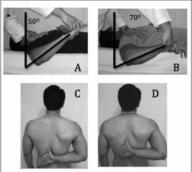
Figure 1 – Baseball player performing stretching of the posterior cap-sule, standing up (A) and in lateral decubitus (B): twice a day, with five repetitions of 30 seconds each.
Figure 2 – Baseball player performing one of the strengthening exercises, with protraction (A) and retraction (B) of the scapulas: once a day, with three series of 15 repetitions.
of achieving complete recovery of the shoulder. They also developed other programs aimed at dealing with specific lesions.
Miyazaki et al(19) conducted a study involving
ba-seball players aged 15 years or over, in which they demonstrated statistically significant correlations be-tween pain and altered range of motion; and bebe-tween length of time practicing the sport and situations of “shoulder at risk”. Such situations were described
by Burkhart and Morgan(20) as the ratio between the
deficit in MR deficit (DMR) and the gain in LR gain (GLR), which shows the imbalance in shoulder adap-tations that lead to the appearance of pain and lesions. The aim of the present study was to assess the reha-bilitation results among the baseball players previously
studied by Miyazaki et al(19), who presented diminished
MR of the dominant shoulder, associated with pain.
MATERIALS AND METHODS
In the first study, the Shoulder and Elbow Group of the Department of Orthopedics and Traumatology, School of Medical Sciences, Santa Casa de São Paulo, evaluated 55 baseball players between April and June 2009. In that study, it was observed that 20 of them presented pain at some moment during the throwing action. They were advised to undergo a rehabilita-tion program with exercises to stretch the posterior capsule and reinforce the muscles of the scapular belt (assessment 1) (Figures 1A, 1B, 2A and 2B). Among these 20 players, it was seen that two stopped playing the sport due to personal problems. Thus, these two were excluded from the study, and 18 players participated in the rehabilitation program.
Baseball players aged 15 years or over were in-cluded. They needed to have a minimum training frequency of twice a week without any interruptions greater than one month over the preceding six months, along with not presenting any type of lesion diag-nosed in the shoulders.
The 18 players who were followed up were male, with a mean age of 21 years (range: 15 to 29 years). Regarding the dominant arm, 17 (94%) were right-handed and one (6%) was left-right-handed. The mean length of time for which they had been playing base-ball was nine years (range: 2 to 23 years), with a mean of three training sessions per week. Among these 18 players, four (22%) were pitchers and 14 (78%) played in other positions (Table 1).
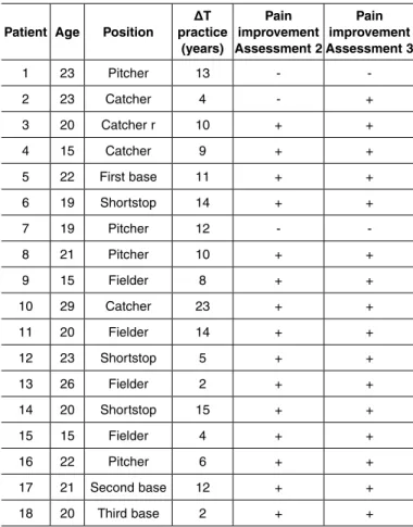
Table 1 – Results relating to pain improvement.
Patient Age Position
ΔT practice
(years)
Pain improvement Assessment 2
Pain improvement Assessment 3
1 23 Pitcher 13 - -2 23 Catcher 4 - + 3 20 Catcher r 10 + + 4 15 Catcher 9 + + 5 22 First base 11 + + 6 19 Shortstop 14 + + 7 19 Pitcher 12 - -8 21 Pitcher 10 + + 9 15 Fielder 8 + + 10 29 Catcher 23 + + 11 20 Fielder 14 + + 12 23 Shortstop 5 + + 13 26 Fielder 2 + + 14 20 Shortstop 15 + + 15 15 Fielder 4 + + 16 22 Pitcher 6 + + 17 21 Second base 12 + + 18 20 Third base 2 + +
Source: Medical archives of Irmandade Santa Casa de Misericórdia de São Paulo. Legend:
Regarding joint mobility, elevation, LR and MR were evaluated in accordance with the guidance of the American Shoulder and Elbow Surgeons (ASES). Following this, LR and MR were also measured with the patient in a supine position with the shoulder ab-ducted at 90°, the elbow flexed at 90° and the forearm in neutral rotation, in accordance with the parameters
described by Hawkins and Bokos(21) and Donatelli et
al(22) (LR90 and MR90).
All the measurements were made with a graduated goniometer, and the non-dominant shoulder was used as a parameter to calculate possible gains or losses of range of motion, and differences in elevation strength and LR.
The arc of rotation (AR) was calculated by sum-ming the LR90 and MR90 values. The GLR was mea-sured as the difference in LR90 between the dominant and non-dominant shoulders. The DMR was calcu-lated as the different in MR90 between the dominant and non-dominant shoulders. The ratio between the DMR and GLR was calculated to ascertain which baseball players presented a “shoulder at risk”, i.e. those for whom the value of this ratio was less than
one, as described by Burkhart and Morgan(20).
The isometric contraction force was measured
using a manual dynamometer (KERN®CH 50K50),
which was calibrated in accordance with the manufac-turer’s specifications. The measurements were made in the movement planes recommended by ASES and
by Hawkins and Bokos(21) and Donatelli et al(22).
To prevent compensatory muscle action in the measurements, a vertical resistance force was applied to the arm evaluated, and the joint was kept at an ap-propriate angle. Three maximum-force measurements were made along each axis evaluated, and the value of the maximum force was noted in kilograms force (kgf), for each of them. The mean for the three repeti-tions was determined for each axis. The contralateral shoulder was evaluated in the same way.
After nine months of guidance through the rehabi-litation program, a new assessment was made on each of the 18 baseball players (assessment 2). They were also asked about their adherence (or non-adherence) to this program, and a check was made on how they were doing the exercises, so that errors in implemen-ting the rehabilitation could be avoided.
From this time onwards, the players were asked every month about their physiotherapy and state of
pain. Three months later, the final assessment was made (assessment 3).
All the data relating to pain, joint mobility, muscle strength, “shoulder at risk” and rehabilitation were compiled and statistically analyzed using the SPSS software (Statistical Package for the Social Sciences), version 17.0; p-values ≤ 0.050 (5%) were taken to be significant. This study was approved by the Ethics Committee for Research on Human Beings of Santa Casa de Misericórdia de São Paulo.
RESULTS
Nine months after assessment 1 (i.e. at assessment 2), we observed that out of the 18 baseball players, 15 had achieved an improvement in their pain. Four players had done the complete rehabilitation pro-gram, 13 only did stretching exercises and one did not follow the program.
At the final assessment, one year after assessment 1 (i.e. at assessment 3), only two of the players continued to present pain. Fourteen had been doing all the exercises and four had only been doing the stretching (Table 1).
At assessment 2, it was found that there was a deficit of joint mobility on the dominant side, in relation to the contralateral limb, with increased LR90 (p = 0.001) and deficits in MR (p = 0.001) and MR90 (p = 0.018), which were considered to be statistically significant.
At assessment 3, the results were similar to those at assessment 2, but with statistically significant differences in relation to increased LR90 (p < 0.001) and diminished MR (p = 0.003) and MR90 (p = 0.009). There was an increase in AR (p = 0.001). All 18 patients showed improvements in mobility and strength (Table 2).
From analysis on muscle strength on the dominant and contralateral sides at assessment 2, statistically significant differences in elevation (p = 0.021) and LR90 (p = 0.031) could be seen, with gains in muscle strength in the dominant limb.
Also in relation to muscle strength, in assessment 2 alone there was a statistically significant gain in LR, in comparing the baseball players who had done all the exercises with those who had only done stretching (p = 0.020).
Table 2 – Mobility results
N
Assessment 1 Assessment 2 Assessment 3
Elev LR LR90° MR MR90° AR GLR DMR SR Elev LR LR90° MR MR90° AR GLR DRM SR Elev LR LR90° MR MR90° AR GLRDRM SR
1 155 70 90 T11 50 140 10 30 + 160 90 122 T8 70 192 32 20 - 170 80 125 T7 70 195 35 20 -2 155 80 100 T12 80 180 0 10 Ø 160 85 120 T8 80 200 20 10 - 170 80 120 T7 90 210 20 0 -3 170 80 140 T7 70 210 5 20 + 170 90 120 T6 80 200 10 0 - 170 80 135 T5 90 225 15 0 -4 1-40 75 110 T10 50 160 20 25 + 140 75 110 T8 75 185 0 15 Ø 170 90 120 T6 90 210 10 0 -5 170 80 110 T7 70 180 -20 10 - 170 90 130 T6 90 220 20 0 - 170 80 130 T5 90 220 20 0 -6 1-60 80 120 T8 70 190 -10 10 - 170 90 140 T7 90 230 10 0 - 170 70 120 T6 90 210 30 0 -7 1-70 90 120 T10 60 180 0 10 Ø 170 90 120 T7 80 200 0 10 Ø 170 90 100 T7 90 190 0 0 Ø 8 160 80 130 T10 70 200 10 10 - 160 90 130 T8 70 200 20 10 - 170 90 130 T8 80 210 40 10 -9 160 -90 130 T8 80 210 20 10 - 160 90 130 T7 70 200 30 5 - 170 90 130 T6 90 220 40 0 -10 160 90 100 T10 60 160 -10 20 - 160 80 100 T8 70 170 10 15 + 160 90 100 T7 80 180 10 0 -11 160 70 120 T11 60 180 0 20 Ø 160 80 120 T10 60 180 0 20 Ø 170 70 100 T8 70 170 10 20 + 12 170 90 130 T6 50 180 10 30 + 160 90 120 T5 75 195 10 5 - 170 90 125 T4 80 205 35 10 -13 160 90 110 T11 70 180 0 10 Ø 160 90 100 T10 80 180 10 -20 - 170 90 120 T8 90 210 0 10 Ø 14 150 80 120 T10 50 170 -10 0 - 160 75 120 T10 60 180 20 0 - 170 80 120 T8 70 190 20 20 -15 140 90 100 T4 70 170 10 0 - 150 90 120 T3 75 195 30 15 - 170 90 140 T3 80 220 40 10 -16 155 80 90 T7 70 160 0 20 Ø 160 70 130 T7 80 210 20 10 - 170 80 130 T6 80 210 40 10 -17 160 90 140 T8 40 180 20 30 + 170 90 130 T7 80 210 10 10 - 170 90 130 T6 90 220 30 0 -18 160 80 110 T7 80 190 -10 10 - 170 90 120 T6 90 210 0 0 - 170 90 120 T5 90 210 10 0
-Source: Medical archives of Irmandade Santa Casa de Misericórdia de São Paulo. Legend: Elev = elevation; LR = lateral rotation; MR = medial rotation; T = thoracic vertebra; L = lumbar vertebra; AR = arc of rotation; GLR = gain in lateral rotation; DMR = deficit in medial rotation; SR = shoulder at risk; Ø = impossible to calculate.
Figure 3 – Case 2: baseball player in supine position with right shoulder
abduction of 90° and elbow flexion of 90°: (A) with Mr90 of 50° at as
-sessment 1; (B) gain of 20° at as-sessment 3; (C) as-sessment 1 on the same player, with the limb in shoulder abduction of 20° and vertebral
level T11; (D) gain of four vertebral levels at assessment 3, reaching vertebral level T7.
was an increase of 10° (p = 0.001); regarding MR, an increase of three vertebral levels (p < 0.001); regarding MR90, an increase of 20° (p < 0.001); and regarding AR, an increase of 26° (p < 0.001) (Figures 3 A-D, 4 and 5).
In relation to muscle strength, an improvement in elevation strength was evident, with an increase of 3 kgf (p = 0.002). There was also an increase in LR strength, of 1 kgf (p = 0.020) (Figure 6) (Table 3).
The relationships of joint mobility, muscle strength and situation of “shoulder at risk” between the base-ball players with and without pain did not show sta-tistical correlations in any of the measurement planes at any of the assessments.
DISCUSSION
Baseball players’ shoulders are subjected to extre-me forces during the throwing action. Through this, this joint may develop a gain in LR and diminished MR(18,23). These alterations were also found in the
Degrees 250.00 200.00 150.00 100.00 50.00 0.00 Ass. 1 Elevation
(p = 0.001) Lateral rotation 0°(p = 0.157) Lateral rotation 90°(p = 0.140) Medial rotation 90°(p = 0.000)
Range of motion (p = 0.000) Ass. 3 Ass. 1 Ass. 3 Ass. 1 Ass. 3 Ass. 1 Ass. 3 Ass. 1 Ass. 3
158.6 ±8.8º 169.4 ±2.3º 82.5 ±6.9º 84.4 ±7.0º 115.0 ±15.4º 121.9 ±11.6º 63.8 ±11.9º 83.8 ±7.7º 178.8 ±17.7º 205.8 ±15.0º
Figure 4 – Comparison of mobility of the affected shoulder between assessments 1 and 3, in degrees. It was seen that there were statistically significant differences in relation to elevation, AR and MR90.
Source: Medical archives of the hospital. Legend: Ass. 1 = assessment 1; Ass. 3 = assessment 3; * = standard deviation.
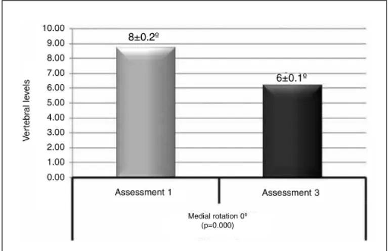
10.00 9.00 8.00 7.00 6.00 5.00 4.00 3.00 2.00 1.00 0.00 Ve rte br al le ve ls 8±0.2º 6±0.1º
Assessment 1 Assessment 3
Medial rotation 0º (p=0.000)
Figure 5 – Comparison of mean mobility of medial rotation (MR) of the affected shoulder between assessments 1 and 3, measured in vertebral levels. A statistically significant difference was seen.
Source: Medical archives of the hospital.Legend: * = standard deviation
Figure 6 – Comparison of the strength of the affected shoulder between assessments 1 and 3, in kgf. It was seen that there were statistically significant differences in elevation strength and lateral rotation (LR).
Source: Medical archives of the hospital. Legend: Ass. 1 = assessment 1; Ass. 3 = assessment 3; * = standard deviation.
18.00 16.00 14.00 12.00 10.00 8.00 6.00 4.00 2.00 0.00
Ass. 1 Ass. 3 Ass. 1 Ass. 3 Ass. 1 Ass. 3 Ass. 1 Ass. 3 Ass. 1 Ass. 3
8.8 ±2.7º 11.7 ±3.7º 10.5 ±2.4º 12.0 ±2.7º 14.8
±4.3º 14.7 ±5.9º
15.9
±4.3º 15.6 ±4.4º
14.1
±4.5º
14.7
±3.9º
Elevation
(p = 0.002) Lateral rotation 0°(p = 0.020)
Lateral rotation 90°
(p = 0.879)
Medial rotation 0°
(p = 0.396)
Medial rotation 90°
(p = 0.396)
According to Burkhart et al(8), it was observed
that shoulder lesions may occur if the AR values are less than 180° and the MR deficit is greater than 25°. In assessment 1, we observed that the 18 baseball players with pain during throwing actions had deficits of elevation and MR and a mean AR of 179°. For this reason, they were referred for physiotherapy, with the aim of recovering their MR.
If contracture of the posture capsule is conside-red to be the cause of the deficit in MR, and the latter as one of the factors responsible for shoulder lesions, appropriate stretching of the posterior cap-sule and correction of this deficit should prevent
the lesions(1,6,7). Our rehabilitation program led to a
significant improvement in pain, probably because we achieved gains in MR, MR90 and AR of 26° on average, thus reaching 205°. However, despite the improvement in AR, we noted that a difference in AR between the dominant and contralateral arms was maintained, even after the rehabilitation program. We believe that the gain was sufficient to diminish the risk of shoulder lesion, because the players experien-ced reductions in pain.
Several studies relating to exercise programs for professional baseball players have demonstrated gains in MR and MR90. In some of them, this gain was im-mediate, after the stretching exercise, but did not last.
In others, like the study by Lintner et al(18), the gain
was progressive and became more significant after
two years of exercises(13-15,24). Our program showed
progressive and long-lasting gains, i.e. the players maintained or increased the gain in movement up to the end of the rehabilitation.
In relation to muscle strength, Wilk et al(24)
de-monstrated through isokinetic tests that the LR streng-th of streng-the dominant shoulder of streng-throwers is significan-tly lower than that of the contralateral shoulder. In compensation, the MR strength is greater in the domi-nant shoulder than in the contralateral shoulder. They also demonstrated that when the LR muscle strength reached at least 65% of the MR muscle strength of the same limb, there was a state of balance between the agonist and antagonist muscles of the shoulder.
Byram et al(25) evaluated the LR muscle strength of
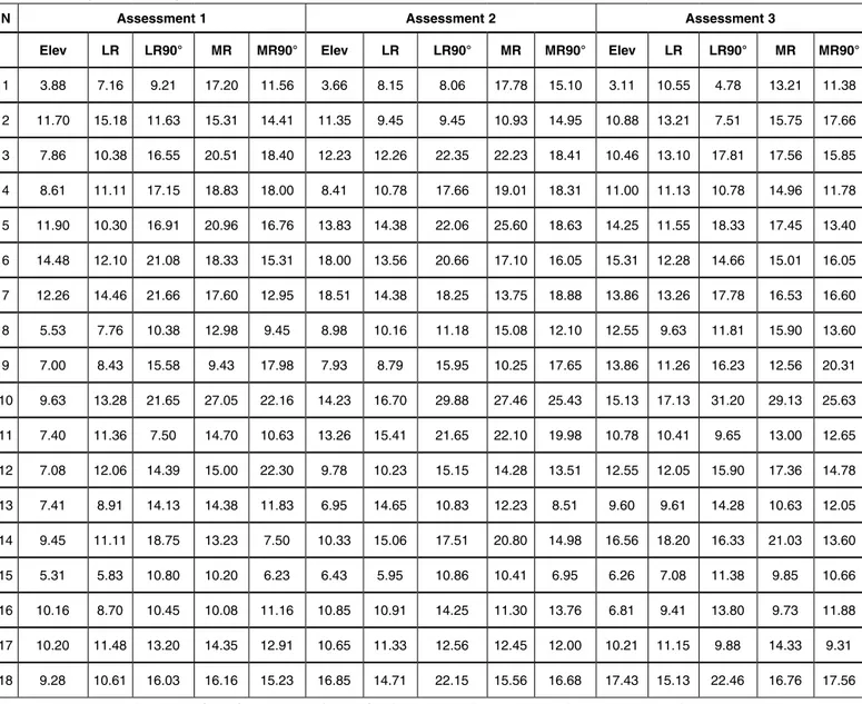
N Assessment 1 Assessment 2 Assessment 3
Elev LR LR90° MR MR90° Elev LR LR90° MR MR90° Elev LR LR90° MR MR90°
1 3.88 7.16 9.21 17.20 11.56 3.66 8.15 8.06 17.78 15.10 3.11 10.55 4.78 13.21 11.38
2 11.70 15.18 11.63 15.31 14.41 11.35 9.45 9.45 10.93 14.95 10.88 13.21 7.51 15.75 17.66
3 7.86 10.38 16.55 20.51 18.40 12.23 12.26 22.35 22.23 18.41 10.46 13.10 17.81 17.56 15.85
4 8.61 11.11 17.15 18.83 18.00 8.41 10.78 17.66 19.01 18.31 11.00 11.13 10.78 14.96 11.78
5 11.90 10.30 16.91 20.96 16.76 13.83 14.38 22.06 25.60 18.63 14.25 11.55 18.33 17.45 13.40
6 14.48 12.10 21.08 18.33 15.31 18.00 13.56 20.66 17.10 16.05 15.31 12.28 14.66 15.01 16.05
7 12.26 14.46 21.66 17.60 12.95 18.51 14.38 18.25 13.75 18.88 13.86 13.26 17.78 16.53 16.60
8 5.53 7.76 10.38 12.98 9.45 8.98 10.16 11.18 15.08 12.10 12.55 9.63 11.81 15.90 13.60
9 7.00 8.43 15.58 9.43 17.98 7.93 8.79 15.95 10.25 17.65 13.86 11.26 16.23 12.56 20.31
10 9.63 13.28 21.65 27.05 22.16 14.23 16.70 29.88 27.46 25.43 15.13 17.13 31.20 29.13 25.63
11 7.40 11.36 7.50 14.70 10.63 13.26 15.41 21.65 22.10 19.98 10.78 10.41 9.65 13.00 12.65
12 7.08 12.06 14.39 15.00 22.30 9.78 10.23 15.15 14.28 13.51 12.55 12.05 15.90 17.36 14.78
13 7.41 8.91 14.13 14.38 11.83 6.95 14.65 10.83 12.23 8.51 9.60 9.61 14.28 10.63 12.05
14 9.45 11.11 18.75 13.23 7.50 10.33 15.06 17.51 20.80 14.98 16.56 18.20 16.33 21.03 13.60
15 5.31 5.83 10.80 10.20 6.23 6.43 5.95 10.86 10.41 6.95 6.26 7.08 11.38 9.85 10.66
16 10.16 8.70 10.45 10.08 11.16 10.85 10.91 14.25 11.30 13.76 6.81 9.41 13.80 9.73 11.88
17 10.20 11.48 13.20 14.35 12.91 10.65 11.33 12.56 12.45 12.00 10.21 11.15 9.88 14.33 9.31
18 9.28 10.61 16.03 16.16 15.23 16.85 14.71 22.15 15.56 16.68 17.43 15.13 22.46 16.76 17.56
Source: Medical archives of Irmandade Santa Casa de Misericórdia de São Paulo. Legend: Elev = elevation; LR = lateral rotation; MR = medial rotation.
Table 3 – Strength results in kgf.
had predominantly been doing stretching exercises and not strengthening exercises. We advised our ba-seball players regarding the importance of doing the entire rehabilitation program. At assessment 3, gre-ater adherence to the exercises was noted, and there were gains in AR, LR and elevation muscle strength.
Our study showed at assessment 1 that the LR strength of the dominant shoulder reached 67% of the MR strength. However, at assessment 2, we saw that there had been an improvement in the balance betwe-en the muscle strbetwe-engths, with 72%, and at assessmbetwe-ent 3, with 76%. These gains were analyzed and were found to present statistical significance, comparing evaluations 1 and 3, thus showing that the baseball players’ shoulders presented an adequate response to the physiotherapy.
Two of the players did not achieve improvements in pain, which led us to investigate the possibility of anatomical injuries developed during sports
practice. Burkhart and Morgan(20) also reported that
CONCLUSION
The rehabilitation program applied to the baseball players was effective and enabled increases in MR, elevation, AR, elevation muscle strength and LR, with
consequent improvement of pain. A mean gain in AR of 26° was enough to improve the shoulder pain of these baseball players.
REFERENCES
1. Muraki T, Yamamoto N, Zhao KD, Sperling JW, Steinmann SP, Cofield RH, et al. Effect of posteroinferior capsule tightness on contact pressure and area beneath the coracoacromial arch during pitching motion. Am J Sports Med. 2010;38(3):600-7.
2. Bigliani LU, Codd TP, Connor PM, Levine WN, Littlefield MA, Hershon SJ.Shoulder motion and laxity in the professional baseball player. Am J Sports Med. 1997;25(5):609-13.
3. Brown LP, Niehues SL, Harrah A, Yavorsky P, Hirshman HP. Upper extremity range of motion and isokinetic strength of the internal and external shoulder rotators in major league baseball players. Am J Sports Med. 1988;16(6):577-85.
4. Downar JM, Sauers EL. Clinical Measures of Shoulder Mobility in the Professional Baseball Player. J Athl Train. 2005;40(1):23-29.
5. Johnson L. Patterns of shoulder flexibility among college baseball players. J Athl Train. 1992;27(1):44-9.
6. Myers JB, Laudner KG, Pasquale MR, Bradley JP, Lephart SM. Glenohumeral range of motion deficits and posterior shoulder tightness in throwers with patho-logic internal impingement. Am J Sports Med. 2006;34(3):385-91.
7. Wilk KE, Meister K, Andrews JR. Current concepts in the rehabilitation of the overhead throwing athlete. Am J Sports Med. 2002;30(1):136-51.
8. Burkhart SS, Morgan CD, Kibler WB. The disabled throwing shoulder: spectrum of pathology Part I: pathoanatomy and biomechanics. Arthroscopy. 2003;19(4):404-20.
9. Grossman MG, Tibone JE, McGarry MH, Schneider DJ, Veneziani S, Lee TQ. A cadaveric model of the throwing shoulder: a possible etiology of superior labrum anterior-to-posterior lesions. J Bone Joint Surg Am. 2005;87(4):824-31.
10. Bach HG, Goldberg BA. Posterior capsular contracture of the shoulder. J Am Acad Orthop Surg. 2006;14(5):265-77.
11. Ejnisman B, Andreoli CV, Carrera EF, Abdalla RJ, Cohen M. Lesões músculo--esqueléticas no ombro do atleta: mecanismo de lesão e retorno a prática espor-tiva. Rev Bras Ortop. 2001;36(10):389-93.
12. Sauers E, August A, Snyder A. Fauls stretching routine produces acute gains in throwing shoulder mobility in collegiate baseball players. J Sport Reha-bil.2007;16(1):28-40.
13. Goldman B, Sauers EL. The acute effectiveness of PNF stretching and joint mo-bilizations for increasing posterior shoulder mobility of the professional baseball player. J Athl Train. 2004; 39(Suppl 2):S-56–80.
14. McClure P, Balaicuis J, Heiland D, Broersma ME, Thorndike CK, Wood A. A randomized controlled comparison of stretching procedures for posterior shoulder tightness. J Orthop Sports Phys Ther. 2007;37(3):108-14.
15. Decicco PV, Fisher MM. The effects of proprioceptive neuromuscular facili-tation stretching on shoulder range of motion in overhand athletes. J Sports Med Phys Fitness. 2005;45(2):183-7.
16. Johansen RL, Callis M, Potts J, Shall LM. A modified internal rotation stre-tching technique for overhand and throwing athletes. J Orthop Sports Phys Ther. 1995;21(4):216-9.
17. Burkhart SS, Morgan CD, Kibler WB. The disabled throwing shoulder: spec-trum of pathology Part III: The SICK scapula, scapular dyskinesis, the kinetic chain, and rehabilitation. Arthroscopy. 2003;19(6):641-61.
18. Lintner D, Mayol M, Uzodinma O, Jones R, Labossiere D. Glenohumeral internal rotation deficits in professional pitchers enrolled in an internal rotation stretching program. Am J Sports Med. 2007;35(4):617-21.
19. Miyazaki AN, Santos PD, Fregoneze M, Silva LA, Sella GV, Checchia SL, et al. Avaliação clínica do ombro doloroso nos jogadores de beisebol. Revis Bras Ortop. 2011;46(2):165-71.
20. Burkhart SS, Morgan CD. The peel-back mechanism: its role in producing and extending posterior type II SLAP lesions and its effect on SLAP repair rehabilitation. Arthroscopy. 1998;14(6):637-40.
21. Hawkins RJ, Bokos DJ. Clinical evaluation of shoulder problems. In: Ro-ckwood CA Jr, Matsen FA 3rd. The Shoulder. 2nd ed. Philadelphia: Saunders; 1998. p. 175-80
22. Donatelli R, Ellenbecker TS, Ekedahl SR, Wilkes JS, Kocher K, Adam J. Assessment of shoulder strength in professional baseball pitchers. J Orthop Sports Phys Ther. 2000;30(9):544-51.
23. Murachovsky J, Ikemoto RY, Nascimento LGP, Bueno RS, Coelho JA, Kome-çu MT, et al. Avaliação da retroversão do úmero em jogadores de handebol. Acta Ortop Bras. 2007;15(5):258-61.
24. Wilk KE, Obma P, Simpson CD, Cain EL, Dugas JR, Andrews JR. Shoulder injuries in the overhead athlete. J Orthop Sports Phys Ther. 2009;39(2):38-54.
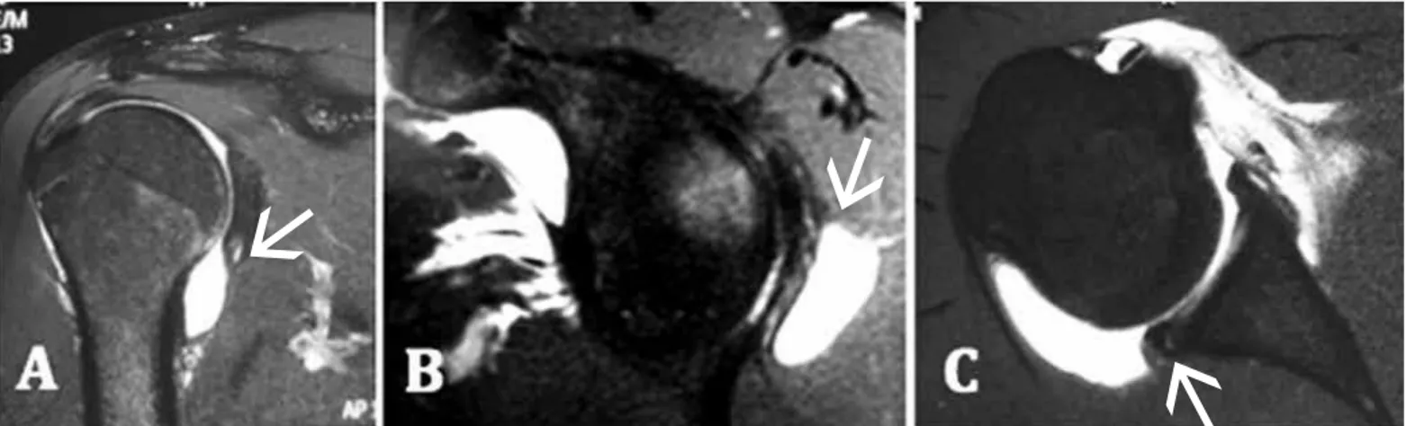
25. Byram IR, Bushnell BD, Dugger K, Charron K, Harrell FE Jr, Noonan TJ. Preseason shoulder strength measurements in professional baseball pitchers: identifying players at risk for injury. Am J Sports Med. 2010;38(7):1375-82. Figure 7 – Case 1: Magnetic arthro-resonance images of the right shoulder of a baseball player who did not achieve improvement of his pain through the rehabilitation program: (A) T2 coronal slice showing lesion of the posteroinferior labrum (arrow); (B) T2 sagittal slice showing posteroinferior paralabral cyst associated with lesion of the posteroinferior labrum (arrow); (C) T2 axial slice showing posteroinferior detachment of labrum (arrow).