Alzheimer Tg2576 Mice Following Modulation of Brain
Amyloid-
b
Levels
Anna M. Lilja1, Jennie Ro¨jdner1,2, Tamanna Mustafiz1, Carina M. Thome´1, Elisa Storelli1,
Daniel Gonzalez1, Christina Unger-Lithner1, Nigel H. Greig3, Agneta Nordberg1,2, Amelia Marutle1*
1Alzheimer Neurobiology Center, Department of Neurobiology, Care Sciences and Society, Karolinska Institutet, Stockholm, Sweden,2Department of Geriatric Medicine, Karolinska University Hospital Huddinge, Stockholm, Sweden,3Drug Design & Development Section, Laboratory of Neurosciences, Intramural Research Program, National Institute on Aging, National Institutes of Health, Baltimore, Maryland, United States of America
Abstract
The objective of this study was to investigate the effects of modulating brain amyloid-b(Ab) levels at different stages of amyloid pathology on synaptic function, inflammatory cell changes and hippocampal neurogenesis, i.e. processes perturbed in Alzheimer’s disease (AD). Young (4- to 6-month-old) and older (15- to 18-month-old) APPSWE transgenic (Tg2576) mice were treated with the AD candidate drug (+)-phenserine for 16 consecutive days. We found significant
reductions in insoluble Ab1-42 levels in the cortices of both young and older transgenic mice, while significant reductions in soluble Ab1-42 levels and insoluble Ab1-40 levels were only found in animals aged 15–18 months. Autoradiography binding with the amyloid ligand Pittsburgh Compound B (3H-PIB) revealed a trend for reduced fibrillar Abdeposition in the brains of older phenserine-treated Tg2576 mice. Phenserine treatment increased cortical synaptophysin levels in younger mice, while decreased interleukin-1band increased monocyte chemoattractant protein-1 and tumor necrosis factor-alpha levels were detected in the cortices of older mice. The reduction in Ab1-42 levels was associated with an increased number of bromodeoxyuridine-positive proliferating cells in the hippocampi of both young and older Tg2576 mice. To determine whether the increased cell proliferation was accompanied by increased neuronal production, the endogenous early neuronal marker doublecortin (DCX) was examined in the dentate gyrus (DG) using immunohistochemical detection. Although no changes in the total number of DCX+-expressing neurons were detected in the DG in Tg2576 mice at either age following (+)-phenserine treatment, dendritic arborization was increased in differentiating neurons in young Tg2576
mice. Collectively, these findings indicate that reducing Ab1-42 levels in Tg2576 mice at an early pathological stage affects synaptic function by modulating the maturation and plasticity of newborn neurons in the brain. In contrast, lowering Ab
levels in Tg2576 mice when Abplaque pathology is prominent mainly alters the levels of proinflammatory cytokines and chemokines.
Citation:Lilja AM, Ro¨jdner J, Mustafiz T, Thome´ CM, Storelli E, et al. (2013) Age-Dependent Neuroplasticity Mechanisms in Alzheimer Tg2576 Mice Following Modulation of Brain Amyloid-bLevels. PLoS ONE 8(3): e58752. doi:10.1371/journal.pone.0058752
Editor:Hemachandra Reddy, Oregon Health & Science University, United States of America
ReceivedDecember 10, 2012;AcceptedFebruary 5, 2013;PublishedMarch 15, 2013
Copyright:ß2013 Lilja et al. This is an open-access article distributed under the terms of the Creative Commons Attribution License, which permits unrestricted use, distribution, and reproduction in any medium, provided the original author and source are credited.
Funding:This work was supported by grants from the Swedish Medical Research Council (Project no. 05817), the Stockholm County Council-Karolinska Institutet (ALF grant), the Karolinska Institutet Strategic Neuroscience Program, Swedish Brain Power, the Brain Foundation, the INMiND consortium (Imaging of Neuroinflammation in Neurodegenerative Diseases) under the Seventh Research Framework Program of the European Union, Gun and Bertil Stohne’s Foundation, the Foundation for Old Servants, Magnus Bergvall’s Foundation, The Dementia Association, The Lars Hierta Memorial Foundation, the Olle Engkvist Byggma¨stare Foundation, Karolinska Institutet Foundations, an academic grant from Johnson & Johnson administered through the Karolinska Institutet, and the Alzheimer Association Sweden. AML was supported by a long-term scholarship from the Karolinska Institutet for Funding for new doctoral students at Karolinska Institutet (KID), a grant from Erik and Edith Fernstro¨m’s Foundation, and a stipend from Gun and Bertil Stohne’s Foundation. NHG was supported by the Intramural Research Program, National Institute on Aging, National Institutes of Health. The funders had no role in study design, data collection and analysis, decision to publish, or preparation of the manuscript.
Competing Interests:Agneta Nordberg is PI for clinical trials sponsored by Torrey pines Therapeutics, GSK, Wyeth, and has been a consultant for AstraZeneca, served on the advisory board for Elan, Pfizer, GSK, Novartis, Lundbeck AB, GE Health Care, and received honorarium for lectures from Novartis, Jansen-Cilag, Pfizer, Merck and received research grants from Novartis, Pfizer, GE Healthcare, Johnson & Johnson. The experimental Alzheimer drugs, (2)-phenserine and (+
)-phenserine, both derive from Nigel H. Greig’s research and are covered in patents that have been assigned to the National Institutes of Health and Dr. Greig has no ownership of these patents. None of the other authors have any conflicts of interest and potential competing interests. This does not alter the authors’ adherence to all the PLOS ONE policies on sharing data and materials.
* E-mail: [email protected]
Introduction
The accumulation of amyloid-b(Ab) aggregates in the brain is a pathological hallmark of Alzheimer’s disease (AD). Abis thought to play a central role in the disease pathogenesis, triggering a cascade of neurodegenerative processes including the activation of inflammatory mediators, altered protein kinase and neurotrophic
signaling, oxidative stress, and neuronal and synaptic dysfunction, ultimately resulting in the impairment of cognitive functions in AD patients [1], [2], [3], [4], [5].
Pittsburgh Compound B (11C-PIB), has demonstrated that increased fibrillar Abdeposition in the brain precedes functional changes and cognitive decline in AD patients [6], [7], [8].11C-PIB PET imaging has also been used to measure changes in brain Ab
load in patients with mild AD treated with the potential therapeutical drug (2)-phenserine [9], a non-competitive acetyl-cholinesterase inhibitor with reported modulatory effects on Ab
production [10], [11]. A reduction in 11C-PIB retention was observed in some patients, and cerebrospinal fluid (CSF) Ab40 levels correlated positively with improvement in brain metabolic function and cognition in AD patients [9].
(+)-Phenserine (also known as posiphen) is also under consid-eration for AD treatment. Unlike its enantiomer (2)-phenserine, this molecule provides little acetylcholinesterase inhibitory action; it lowers the generation of Abby suppressing amyloid precursor protein (APP) synthesis [12]. A recent pharmacokinetic study undertaken in a small group of patients with mild cognitive impairment who were treated with (+)-phenserine reported significantly lower levels of sAPPa, sAPPb and proinflammatory markers in the CSF, and alterations in CSF Ab42 levels [13].
Clinical and biomarker changes assessed in patients with a genetic predisposition for familial AD (FAD) suggest that pathological changes start approximately two decades before cognitive symptoms appear [14].
Transgenic mice harboring similar mutations in human APP and presenilin-1 (PS1) genes exhibit early and progressive accumulation of Ab, associated with compromised neocortical synaptic plasticity and synaptic dysfunction, traits similar to those observed in FAD patients [15], [16], [17]. The complex composition of pathological alterations in the AD brain microen-vironment is thought to lead to impairment of neurotrophic signaling and inadequate synaptic repair [18], [19], [20].
Neurogenesis, the birth of new neurons, has been shown to persist in the adult brain, although it is largely restricted to two regions: the subventricular zone of the lateral ventricles and the subgranular zone of the dentate gyrus (DG) in the hippocampus.
However, the ability of new neurons to incorporate into the brain circuitry and form functional synaptic connections declines with age [21].
In the few existing neurogenesis studies performed in the autopsied brains of AD patients, an increase in the number of neural progenitors in the DG has been demonstrated, compared to age-matched controls [22], [23], [24]. Whether this rise in neural progenitors is a consequence of triggered compensatory mecha-nisms related to the neurodegenerative changes in the brain or is induced by medications the patients received before death remains unclear. Nevertheless, this interesting finding implies that stimu-lation of the neuroprotective and regenerative mechanisms in the AD brain is feasible and, hence, this area holds promise as a novel treatment approach for slowing AD progression.
Studies in various AD animal models have tested the potential of stimulating neurogenesis with different treatment approaches. We have previously demonstrated improved survival andin vivo
neuronal differentiation of transplanted neural progenitors in the brains of APP23 transgenic mice receiving (+)-phenserine treat-ment before the onset of Ab plaque deposition, compared with APP23 mice that did not receive treatment [25]. Other pharmacological strategies, including Ab immunization or treat-ment with neurotrophic peptides, have also been associated with enhanced hippocampal neurogenesis in other AD transgenic mice strains [26], [27], [28].
A paradigm shift in recent years has resulted in the suggestion that anti-Ab therapies, in combination with disease-modifying therapies, could be highly effective in preventing or delaying the development of the disease in asymptomatic patients with very early signs of AD pathology [29]. In line with this view, clinical studies focusing on early presymptomatic treatment and preven-tion are being initiated, as presented at the most recent meeting for clinical trials on AD (CTAD) (see http://www.alzforum.org/new/ detail.asp?id = 3312).
This preclinical study was designed to examine the effects on synaptic function and neuroinflammatory and regenerative
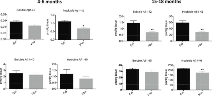
Figure 1. Effects of (+)-phenserine on brain amyloid levels in Tg2576 mice.Levels of tris-soluble (soluble) and guanidine-soluble (insoluble) Ab1-42 and Ab1-40 in the cerebral cortices of 4- to 6-month-old (A) and 15- to 18-month-old (B) Tg2576 transgenic mice treated with saline (Sal) or (+)-phenserine (Phe) for 16 days.*P,0.05 and **P,0.01 compared to saline-treated mice (Mann-Whitney test). Values are expressed as means6SEM throughout the manuscript.
processes of an Ab-lowering therapeutic intervention at different stages of amyloid pathology in an AD transgenic mouse model. We treated transgenic mice overexpressing human APP with the Swedish double mutation (Tg2576), at 4–6 and 15–18 months of age, with (+)-phenserine for 16 days. These mice exhibit impaired cognitive functioning and high soluble Ablevels in the brain when very young (,6 months old); insoluble Ablevels are increased and Abplaques are formed later [30], [17].
We measured a selective decrease in Ab42 levels in the cortices of young Tg2576 mice and demonstrated attenuation of both Ab40 and Ab42 levels after the onset of Abplaque pathology in the brains of older mice. These changes were assessed in relation to levels of brain-derived neurotrophic factor (BDNF), the presynaptic vesicle protein synaptophysin, pro-inflammatory cytokines [interleukin-1b (IL-1b), tumor necrosis factor-alpha (TNF-a) and chemokine monocyte chemoattractant protein-1 (MCP-1)], and hippocampal neurogenesis. Concurrent with the reduction in amyloid levels, phenserine treatment primarily elevated cortical synaptophysin levels and increased the matura-tion of newborn neurons in the DG of 4- to 6-month-old Tg2576
mice, while significant alterations in IL-1b and MCP-1 were detected in the cortices of 15- to 18-month-old mice.
Materials and Methods
Animals
Male mice expressing the APP Swedish mutation (APPS-WE2576Kha; Tg2576) were bred in the Karolinska Institutet animal care facility by backcrossing with B6SJL (F1) females (Taconic). Their genotype was determined using polymerase chain reaction technology [31]. Wild type (wt) littermates served as control animals throughout the study. All mice were housed in enriched cages with a 12-hr light-dark cycle andad libitumaccess to food and water.
Ethics Statement
All animal experimental procedures were carried out in strict accordance with the guidelines for the Swedish National Board for Laboratory Animals, and the protocol was approved by the Regional Ethics and Animal Research Committee at the Karolinska Institute, Stockholm, Sweden (Permit Numbers: S43/
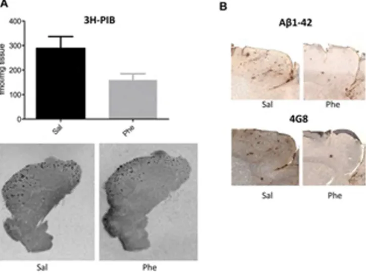
Figure 2. Modulation of amyloid-b(Ab) burden in 15- to 18-month-old Tg2576 mice treated with (+)-phenserine.(A)topBinding of the amyloid ligand3H-PIB (1.5 nM) to fibrillar Abin the brains of 15- to 18-month-old Tg2576 mice treated with saline or (+)-phenserine (Phe). A decrease in 3H-PIB binding was observed following (+)-phenserine treatment compared to saline treated mice (p= 0.05; Mann-Whitney test). Bottom,
representative autoradiography distributions of 3H-PIB in sagittal hemibrain sections of saline- and (+)-phenserine-treated Tg2576 mice. (B) Representative sections illustrating immunohistochemical staining of amyloid plaques with antibodies Ab1-42 and 4G8 in the cerebral cortices of 15-to 18-month-old Tg2576 mice treated with (+)-phenserine and saline.
07 and S53/10). All surgery was performed under ketamine/ xylazine anesthesia, and all efforts were made to minimize animal suffering.
Drug Treatment
Tg2576 mice at 4–6 months (n = 16) and 15–18 months (n = 11–13) of age and age-matched wt controls (n = 13–16) were divided into groups of 6–8 mice and injected once daily with intraperitoneal (i.p.) (+)-phenserine tartrate (25 mg/kg) or physi-ological saline solution for 16 consecutive days. On the final three days, all animals were co-administered the thymidine analog 5-bromo-29-deoxyuridine (BrdU; 50 mg/kg per day i.p.) to measure ongoing cell proliferation. Within 12 hr of the final administration, the mice were anesthetized with a 1:1 mixture of ketamine (100 mg/kg) and xylazine (20 mg/kg) and euthanized by trans-cardial perfusion with phosphate buffered saline (PBS). The brains were collected and divided into hemispheres. One hemisphere was stored at 280uC and used later in the biochemical assays. The other hemisphere was post-fixed with 4% paraformaldehyde (PFA; pH 7.4) and transferred to a sucrose cryoprotectant for 24 h at 4uC, after which it was frozen at 280uC. Sagittal 20mm brain sections were subsequently cut and processed for autoradiography binding and immunohistochemical staining.
Tissue Extractions
Dissected cortical brain tissue was processed individually and sequential extraction steps were performed to obtain membranous and soluble fractions for Western blot and sandwich enzyme-linked immunosorbent assay (ELISA) analyses. Briefly, tissues were homogenized in ice-cold tris-buffered saline (50 mM Tris HCl, 150 mM NaCl, 1 mM EDTA, 1 mM DDT) containing protease inhibitors (Sigma). The homogenates were centrifuged (4206g, 10 min, 4uC) and the supernatant collected and re-centrifuged (15,0006g, 60 min, 4uC). The supernatant (cytosolic fraction) was
saved, and the remaining pellet was resuspended in five volumes RIPA buffer containing detergent (2% Triton X-100, 0.2% SDS),
centrifuged (15,0006g, 20 min, 4uC) and the supernatant (mem-branous fraction) collected. Both the cytosolic and mem(mem-branous fractions were stored at280uC until use.
AbMeasurements
Tris-soluble and -insoluble (guanidine-extracted) Ab1-40 and Ab1-42 levels were quantified in cortical homogenates using commercial colorimetric ELISA kits (Signal SelectTM Human Ab1–40 and 1–42, BioSource International Inc., Camarillo, CA, USA) as previously described [32]. Levels of Abwere expressed as pg/mg tissue.
3
H-PIB autoradiography binding was carried out by pre-incubating triplicate sections from each animal for 15 min in PBS buffer (pH 7.4) containing 1% bovine serum albumin (BSA) and then incubating them with 1.5 nM 3H-PIB (specific activity 68 Ci/mmol, custom synthesis, GE Healthcare, Ger-many) in PBS containing 0.1% BSA for 1 hr, as described earlier [33]. Non-specific binding was determined by incubating adjacent sections with 1mM unlabeled PIB. After washing in PBS, the sections were exposed along with calibrated tritium standards (American Radiolabeled Chemicals, Saint Louis, MO, USA) on Fuji BAS-TR2040 phosphor imaging plates (Science Imaging Scandinavia AB, Nacka, Sweden) for 3 days. The plates were processed with a Fujifilm BAS5000 phosphorimager (Fuji, Tokyo, Japan), and binding densities were analyzed using Multigauge software V3.0 (Fuji). The relative optical density calculated from the tritium standards was used to calculate binding values in fmol/mg tissue.
Ab1-42 and 4G8 immunohistochemistry analysis was carried out on 4% PFA sagittal brain sections treated with formic acid and 0.3% H2O2to deplete endogenous peroxidase activity. Following washing and blocking steps, the sections were incubated with antibodies Ab1-42 and 4G8 (reactive to amino acid residue 17–24; 1:500, Covance, Denver, PA) overnight at 4uC. The following day, the sections were rinsed and incubated for 1 hr with a biotinylated anti-mouse antibody IgG (1:200; Vector laboratories Inc.,
Figure 3. Synaptophysin protein levels in the cerebral cortices of 4- to 6- and 15- to 18-month-old wild-type (Wt, white bars) and Tg2576 mice (black bars) after treatment with saline (Sal) or (+)-phenserine (Phe).The signals corresponding to the synaptophysin levels were normalized tob-actin in each gel, and a pooled sample (P) was used to control the intergel variability. **P,0.01 compared to saline-treated 4- to 6-month-old Tg2576 mice (Mann-Whitney test). Values are expressed as means6SEM.
Burlingame, CA). Immunoreactivity was visualized using the avidin biotinylated peroxidase complex (ABC) method (Vector Laboratories Inc., Burlingame, CA) with the sections counter-stained with hematoxylin.
Growth Factor and Cytokine Measurements
The levels of BDNF and the proinflammatory cytokines IL-1b, TNF-aand chemokine MCP-1 were measured in cortical cytosolic fractions using commercial ELISA kits, the mouse BDNF ELISA kit (Abnova, Immunkemi F&D AB, Stockholm, Sweden) and the MSD Multi-arrayH mouse cytokine ultra-sensitive assay (Meso Scale Discovery, Gaithersburg, MD, USA), according to the manufacturer’s instructions.
Western Blot Detection of Synaptophysin Protein
Samples from the membranous fractions (15mg protein) were subjected to electrophoresis in 12% Tris-Bis gels (Invitrogen) and transferred to polyvinylidene difluoride (PVDF) membranes (GE Healthcare). The membranes were blocked with 5% non-fat dry milk and incubated overnight at 4uC with rabbit anti-synapto-physin (1:2000, DAKO), with rabbit anti-b-actin as the loading control (1:2000, Abcam) and, thereafter, with a horseradish peroxidase (HRP)-conjugated donkey rabbit secondary anti-body (1:2000, Santa Cruz Biotechnology) for 2 hr at 21uC. Protein bands were visualized using an enhanced chemiluminescence detection reagent (GE HealthCare) and the density of each band was normalized withb-actin and compared with a pooled sample using the National Institutes of Health Image J analysis software program.
BrdU and Doublecortin (DCX) Immunohistochemistry
A one-in-six series of sections throughout the entire rostral-caudal extent of the hippocampus was used to assess the number of BrdU-positive (BrdU+
) cells. BrdU fluorescent immunohistochem-istry analysis was carried out by incubating the free-floating sections with sheep anti-BrdU (1:100, Abcam, Cambridge, UK) overnight at 4uC and then with a Texas Red conjugated donkey anti-sheep IgG antibody (1:500, Santa Cruz Biotechnology, Inc., Santa Cruz, CA) for 2 hr at 21uC. The sections were mounted with 49, 6-diamidino-2-phenylindole (DAPI) for nuclear counter-staining (Vector Laboratories). The total number of BrdU+cells in the hippocampus was quantified using a Nikon E800 Eclipse fluorescence microscope (Nikon, Tokyo, Japan) at 20X magnifi-cation. An average of three sections was counted for each animal and quantification was blinded.
For the detection of DCX-expressing cells, sections were incubated overnight with goat anti-DCX (1:500, Santa Cruz) and the immunoreactivity was visualized with rabbit anti-goat streptavidin-horseradish peroxidase conjugate (1:500, Vector Laboratories) using the ABC method with nickel-enhanced diaminobenzidine as substrate. Images were captured with a light microscope (Leica Microsystems, Wetzlar, Germany) with an attached ProgResH video camera and the ProgResHCapture Pro2.8.8 image analysis system (Jenoptik AG, Jena, Germany) was used at 20X magnification. The total number of DCX+
cells and the number of DCX+
cells with clearly defined dendrites
Figure 4. (+)-Phenserine-induced effects on proinflammatory cytokines (IL-1b, TNF-a) and a chemokine (MCP-1) in Tg2576 and wt mice brains. Effects of (+)-phenserine treatment on IL-1b
levels (A), MCP-1 levels (B), and TNF-alevels (C) in the cerebral cortices of 4- to 6- and 15- to 18-month-old wild-type (Wt, white bars) and Tg2576 mice (black bars) treated with saline (Sal) or (+)-phenserine (Phe). *P,0.05 compared to saline-treated 15- to 18-month-old Tg2576 mice (Mann-Whitney test), **p,0.01 compared to saline-treated 4- to 6-month-old wildtype and Tg2576 mice (Mann-Whitney test). Values are expressed as means6SEM.
extending from the cell body were counted in the DG in 4- to 6-and 15- to 18-month-old Tg2576 6-and wt mice (triplicate sections per animal; blinded analysis).
Statistical Analyses
Data are expressed as mean values 6 SEM. Statistical differences between groups were determined with the non-parametric Mann-Whitney test or the unpaired t-test using a statistical software package (GraphPad Prism, version 4.00; GraphPad Software, San Diego, CA). The non-parametric Spearman’s correlation was used to assess the relationships between the different parameters. The significance level was set at p,0.05.
Results
(+)-Phenserine Decreases Brain AbLevels in 4- to 6- and 15- to 18-month-old Tg2576 Mice
A significant decline (38%) in insoluble (guanidine-soluble) Ab 1-42 levels was measured in the cortices of 4- to 6-month-old (+ )-phenserine-treated Tg2576 mice (p,0.05 vs saline-treated con-trols; Fig. 1A). In 15- to 18-month-old mice, both soluble and insoluble Ab1-42 levels were significantly reduced (by 53% and 52%, respectively). A 20% decline in insoluble Ab1-40 levels was also observed in (+)-phenserine-treated versus saline-treated older Tg2576 mice (p,0.01, Fig. 1B).
Autoradiography with the PET ligand 3H-PIB was used to quantify the effects of (+)-phenserine on fibrillar Abdeposition in brain sections from 15- to 18-month-old Tg2576 mice. At this age, these mice normally have pronounced fibrillar Ab deposition in the brain. Reduced binding to fibrillar Ab was observed in the cortical regions of (+)-phenserine-treated versus saline-treated mice
Figure 5. The effects of (+)-phenserine treatment on cell proliferation in the hippocampi of Tg2576 mice. The number of bromodeoxyuridine-positive (BrdU+
) cells was increased in hippocampal regions of 4- to 6-month-old Tg2576 mice treated with saline (Sal) or (+)-phenserine (Phe). The number of BrdU+cells was counted in the dentate gyrus (DG) (A), and in the CA1 region (B) in the hippocampi of wild-type (Wt) and Tg2576 mice. Representative images of the DG and CA1 in Tg2576 mice treated with saline (Sal) or (+)-phenserine (Phe) are shown in (C). The number of BrdU+
(p = 0.05, Fig. 2A). Moreover, as illustrated in Figure 2B, Ab1-42 and 4G8 immunoreactivity appeared weaker in the cerebral cortices of 15- to 18-month-old Tg2576 mice treated with (+ )-phenserine versus saline treatment.
Synaptophysin and BDNF Levels in the Cortices of 4- to 6- and 15- to 18-month-old Tg2576 Mice Treated with (+)-phenserine
To elucidate the effects of treatment on processes associated with synaptic plasticity, the expression of the presynaptic vesicle
protein synaptophysin and neurotrophic BDNF protein was quantified in the cerebral cortices of Tg2576 mice at 4–6 and 15–18 months of age. Synaptophysin levels were significantly higher in the cerebral cortices of 4- to 6-month-old Tg2576 mice treated with (+)-phenserine than in those administered saline (p,0.01; Fig. 3). There were no significant differences in synaptophysin levels between the older Tg2576 mice and wt controls, or between (+)-phenserine and saline treatment in the older mice.
A significant elevation in BDNF levels was evident in older versus young Tg2576 mice (3626681 versus 8676129 pg/mg
Figure 6. Increased dendritic arborization of newborn neurons in the dentate gyrus (DG) following treatment with (+)-phenserine. The number of doublecortin (DCX)-positive cells in the DG of 4- to 6- and 15- to 18-month-old mice after saline (Sal) or (+)-phenserine (Phe) treatedment (A), and the number of DCX+
cells with dendrites in 4- to 6-month-old mice (B). Illustrative images (20X magnification) of DCX immunoreactivity in 4- to 6-month-old Tg2576 mice that had been given saline or (+)-phenserine, and a 40X magnification of DCX+
cells with (black arrow) and without (white arrow) dendrites (C). Illustrative images (20X magnification) of DCX immunoreactivity in 15- to 18-month-old saline-treated and (+)-phenserine-treated Tg2576 mice (D). *P,0.05 compared to saline-treated 4- to 6-month-old Tg2576 mice, **p,0.01 compared to 4- to 6-month-old mice (Mann-Whitney test). Values are expressed as means6SEM.
protein; p,0.01). A trend towards increased BDNF levels was observed in the drug-treated young and older Tg2576 and wt mice versus saline treatment, but this did not reach statistical significance (data not shown).
(+)-Phenserine Treatment Alters the Levels of
Proinflammatory Cytokines IL-1b, TNF-a and Chemokine MCP-1 in the Brains of 15- to 18-month-old Tg2576 Mice
A highly sensitive sandwich ELISA assay was used to examine whether the reduced cortical Ablevels in the brains of young and older Tg2576 mice altered levels of the proinflammatory cytokines IL-1b, TNF-a and chemokine MCP-1. A significant decrease in IL-1blevels was observed in (+)-phenserine-treated Tg2576 mice at 15–18 months, but not at 4–6 months of age (Fig. 4A). Irrespective of treatment, MCP-1 levels were much lower in young Tg2576 and wt mice than in older animals (p,0.01; Fig. 4B). In the 15- to 18-month-old group of Tg2576 mice, MCP-1 levels increased after treatment but the difference did not reach statistical significance (Fig. 4B). In 4- to 6-month-old mice, there were no differences in TNF-a levels between drug- and saline-treated Tg2576 or wt mice, and the levels of TNF-awere lower than in older animals (Fig. 4C). At 15–18 months of age, we observed a
trend for decreased TNF-alevels in (+)-phenserine-treated wt mice and increased TNF-alevels in (+)-phenserine-treated Tg2576 mice versus saline-treated mice (Fig. 4C).
(+)-Phenserine Induces Age-dependent Increases in the
Number of Proliferating Cells and Stimulates Dendritic Arborization of Newborn Neurons in the Hippocampi of Tg2576 Mice
We then assessed both BrdU+
immunoreactivity (a marker for cell proliferation) and the expression of DCX (an endogenous neurogenesis marker for the microtubule-associated phosphopro-tein that is expressed by migrating and differentiating neurons) in hippocampal sub-regions to see whether lowered Ab levels stimulated regenerative mechanisms in the brains of Tg2576 mice at 4–6 and 15–18 months of age. At 4–6 months, the trend toward a greater number of BrdU+
cells observed in the DG (69% increase; Fig. 5A) reached significance in the CA1 region (119% increase,p,0.05; Fig. 5B and 5D) in Tg2576 mice treated with (+)-phenserine versus saline-treated mice. At age 15–18 months, the BrdU+
cell number was significantly elevated in the DG of Tg2576 (+)-phenserine-treated mice versus saline treatment (76% increase,p,0.05; Fig. 5C, E).
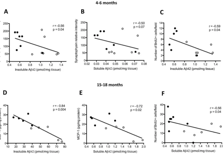
Figure 7. Correlations of amyloid-b(Ab) levels with synaptophysin levels, cell proliferation and proinflammatory markers in the brains of 4- to 6- and 15- to 18-month-old Tg2576 mice.Correlation of synaptophysin levels with cortical guanidine-soluble (insoluble) Ab1-42 levels (A) and tris-soluble (soluble) Ab1-42 levels (B). Correlation of insoluble Ab1-42 levels with the number of bromodeoxyuridine-positive (BrdU+
) cells (C) in the dentate gyri of 4- to 6-month-old Tg2576 mice. In Tg2576 mice aged 15–18 months, the relationships between MCP-1 levels and insoluble and soluble Ab1-42 levels in the cortex are shown in (D)and (E), respectively, and the relationship between the number of proliferating BrdU+cells in the dentate gyrus and cortical soluble Ab1-42 levels is shown in (F) (Spearman rank test). Open circles indicate saline-treated animals (n = 5–7) and closed circles indicate (+)-phenserine-treated animals (n = 5–8).
Immunohistochemical staining revealed the presence of DCX+ neurons in the granule cell layer and subgranular zone of the DG in both Tg2576 and wt mice. DCX+
cells were not detected in other hippocampal sub-regions. A marked reduction in DCX immunoreactivity was evident in the DG of Tg2576 mice at 15–18 months of age compared to those aged 4–6 months (p,0.05, Fig. 6A, C and D). However, the total number of DCX+neurons in the DG of mice in both age groups was unaffected by (+ )-phenserine treatment. After further quantification of DCX+ neurons, those possessing dendrites were determined to be significantly elevated in number in the DG of 4- to 6-month-old Tg2576 (+)-phenserine-treated mice versus those receiving saline (p,0.05, Fig. 6B).
Ab Reduction is Associated with Age-related Effects on Cell Proliferation, Synaptogenesis and Inflammatory Cytokines and Chemokines in Tg2576 Mice
The relationship between Ab levels and the expression of synaptophysin, BDNF, and proinflammatory markers was exam-ined in the brains of both young and older Tg2576 mice. At 4–6 months of age, a significant inverse correlation was observed between insoluble Ab1-42 levels and synaptophysin protein levels (p,0.05; Fig. 7A), and a weaker inverse correlation was seen between soluble Ab1-42 levels and synaptophysin levels (p = 0.07; Fig. 7B). The levels of insoluble Ab1-42 correlated negatively with the number of BrdU+
cells in the DG (p,0.05; Fig. 7C). No correlation, however, was apparent between Ab1-42 levels and DCX+-expressing cells (data not shown). In 15- to 18-month-old Tg2576 mice, levels of MCP-1 but not of the cytokines IL-1band TNF-ademonstrated a significant negative correlation with both insoluble (p,0.01; Fig. 7D) and soluble (p,0.05; Fig. 7E) Ab1-42 levels. Similarly, the number of BrdU+
cells in the DG was inversely correlated with cortical soluble Ab1-42 levels (p,0.05) but not insoluble Ab1-42 levels (Fig. 7F).
Discussion
The aim of this study was to elucidate the extent to which pharmacological modulation of brain Ablevels at different stages of amyloid pathology influences synaptic, neuroinflammatory and regenerative processes in the brains of AD Tg2576 mice.
These mice accumulate Ab in an age-related manner, particularly in the cortex and the hippocampus, and although overt neuronal loss is not observed with ageing, synaptic aberrations and progressive learning and memory deficits arise. These are attributed to the presence of soluble Ab assemblies at younger ages and, at later stages, with heavy Abplaque deposits [34], [30]. We report here a pronounced reduction in Ab, primarily Ab1-42, levels in the cortices of 4- to 6- and 15- to 18-month-old Tg2576 mice following treatment with (+)-phenserine. This drug is currently undergoing clinical optimization and evaluation for AD therapy [13]. Decreases in insoluble Ab1-42 levels detected by ELISA were more prominent in 15- to 18-month-old Tg2576 mice than in younger mice, in accord with the higher rate of Ab plaque deposition with age. These decreases were supported by reduced Ab42 immunohistochemistry and autoradiographic distribution of 3H-PIB, which binds to fibrillar Ab. The finding that (+)-phenserine has a greater impact on Ab 1-42 than on Ab1-40 levels is in agreement with a prior preclinical study [12].
The neurodegenerative process in AD is initially characterized by synaptic damage accompanied by neuronal loss. Synaptic dysfunction is one of the strongest correlates to cognitive impairment in patients with AD [35]. Repair mechanisms in the
brain can be associated with neuroplasticity at multiple levels, including structural remodeling of degenerating neuronal circuits, synaptic changes and strengthening of the dendritic branches that connect synapses. In this study, elevated levels of the synaptic protein synaptophysin were seen in the cortices of 4- to 6-month-old (+)-phenserine-treated Tg2576 mice, supporting the premise that lowering brain Ab levels early in the disease could have beneficial effects on synaptogenesis.
The neurotrophic factor BDNF plays an important role in promoting neuronal survival and synaptic plasticity [36], [37]. The levels of BDNF are decreased in both the cerebral cortex and the hippocampus in the AD brain, and it is thought that the early impairments in synaptic function could result in part from neurotrophin signaling deficits [18], [19].
A previous study in AD Tg2576 mice demonstrated that Ab
reduces BDNF signaling by impairing axonal transport of BDNF [38] while another study reported that engrafted neural stem cells expressing high levels of BDNF could improve the spatial learning and memory deficits observed in aged 36Tg AD mice [39]. In our study, only slight increases in BDNF brain levels were measured in young and older Tg2576 mice treated with (+)-phenserine. Interestingly, we found that BDNF levels were significantly higher in the older versus younger saline treated Tg2576 cohorts, suggesting an age-associated compensatory up-regulation of brain BDNF due to increasing Abin the brain. Thus, the regulation of BDNF expression as well as signaling in AD brain appears to be complex, and further studies are warranted to fully characterize (+)-phenserine’s actions on BDNF signaling and also that of other neurotrophins during the course of AD neurodegeneration.
An inflammatory response invariably accompanies elevated Ab
levels in the brain, with a hallmark of increased presence of activated microglia and reactive astrocytes in the brains of AD patients [40], [41]. Compelling evidence supports a key role for microglia and astrocytes in regulating and maintaining neuronal activity, which can be adversely influenced by elevated Ablevels [42]. Ab increases the synthesis of microglia and reactive astrocytes and the release of pro-inflammatory cytokines [43], [44], [45], as well as inhibiting adult neurogenesis [46], [47], [48]. Our study showed elevated brain IL-1blevels in both 4- to 6- and 15- to 18-month-old Tg2576 mice, signifying early and persistent activation of inflammatory processes. (+)-Phenserine reduced IL-1blevels in the older cohort in comparison to those measured in wt littermates. IL-1b has been shown to decrease synaptophysin levels in cortical neuronal cultures, and to inhibit cell proliferation in the DG of rodents [49]. However, the changes in IL-1blevels that we measured did not correlate with either synaptophysin levels or with increased cell proliferation.
An age-related rise in MCP-1 levels was also detected in the cortices of aged wt and Tg2576 mice. MCP-1 induces astrocyte chemotaxis and contributes to the recruitment of astrocytes around Ab plaques [50]. It has recently been suggested that astrocytes play an additional role as key integrators of neurogenic permissiveness [51]. In neurogenic brain regions, new astrocytes are produced alongside new neurons [52], [53].
In the present study, we can not dismiss the possibility that the increase in cell proliferation observed in the hippocampi following Ab reduction may have contributed to a rise in astroglia precursors. Since BrdU was administered to the mice only during the last three days following (+)-phenserine treatment, co-staining BrdU+
MCP-1 levels only in older mice, which suggests induction of astrocyte-mediated neuroprotective effects. A similar elevation in TNF-a levels was observed in older wt and Tg2576 mice compared to the younger group. In addition, 15- to 18-month-old Tg2576 mice treated with (+)-phenserine demonstrated a tendency for increased TNF-alevels versus saline treatment but, because of variations among the mice in this group, this difference did not reach statistical significance. Both experimental and clinical evidence implicates the involvement of TNF-a in the pathogenesis of AD [43], [54], [55]. However, there are also a few studies that report a neuroprotective function for TNF-a and a role in modulating neuronal cell function through an indirect mechanism by which TNF-a stimulates the production of neurotrophic factors [56], [57]. In support of the latter scenario, our results suggest the possibility that (+)-phenserine potentiates the neuroprotective function of TNF-a, as elevated levels of this cytokine were measured in the drug-treated 15- to 18-month-old mice, concomitant with high BDNF levels.
Neurogenesis plays an important role in structural neuronal plasticity and network maintenance in the adult brain. It is a complex developmental process characterized by five stages, in which neural stem/progenitor cells in the subgranular layer of the DG develop through proliferation, differentiation, migration, axonal and dendritic targeting, and finally functional synaptic integration into neuronal circuits [36], [23]. Previous studies in nonhuman primates and rodents have shown that immature neurons in the adult DG have high synaptic plasticity, and that this declines with age [58]. Critical for learning and memory, the hippocampus is one of the earliest regions to be affected in AD [59] and dysfunctional neurogenesis consequent to subtle early disease manifestations in the brain could in turn render neurons more vulnerable to AD and contribute to memory impairment [58], whereas enhanced neurogenesis could provide a compensa-tory, endogenous repair mechanism. Prior investigations into hippocampal neurogenesis in mouse models of AD have provided conflicting findings. The majority of these studies report compro-mised neurogenesis [60], [61], [62], but others have described increased neurogenesis [63], [64]. These contradictory findings may stem from the different transgenic models studied, the age of the mice, or the detection methods used to label proliferating cells. In our study, (+)-phenserine treatment of young adult Tg2576 mice resulted in significantly increased numbers of BrdU+
proliferating cells in the CA1 region, an area particularly vulnerable to Ab deposition [65], and a trend for increased numbers in the DG. A similar increase in BrdU+
proliferating cells was observed in the DG of older Tg2576 mice following treatment. DCX is expressed in transiently amplifying neuroblasts during the migration and early differentiation phase of neurogen-esis. DCX expression was therefore quantified to determine whether the increase in the number of BrdU+
proliferating cells in the DG was accompanied by an increase in neuronal production. No net rise in DCX+
cells was apparent in the DG of 4- to 6-month-old Tg2576 mice, but increased dendritic arborization was seen in differentiating neurons in this region following (+ )-phenserine treatment. Markedly fewer DCX+
cells were identified in the DG of both saline- and drug-treated 15- to 18-month-old Tg2576 mice, indicating that neural stem/progenitor cells are more vulnerable in older animals.
In conclusion, the findings reported in this study suggest that early modulation of Ablevels with (+)-phenserine could influence the maturation and plasticity of newborn neurons in the brain. In addition, reducing the Abload in the AD brain when Abplaque pathology is prominent could support neuroprotective functions by altering the levels of proinflammatory cytokines and chemokines. In light of current discussions regarding anti-Ab treatment in clinical trials aiming to promote effective Ab reduction or clearance in presymptomatic patients and patients with mild AD, it is important to continue carrying out translational studies in preclinical AD transgenic animal models to test the long-term effects of Ab-lowering drugs and to elucidate whether targeting Ab
pathways early in the disease will lead to positive effects on brain function, including the stimulation of brain repair.
Acknowledgments
The authors sincerely thank Pasi Miettinen for contributing methodolog-ical expertise in the doublecortin immunohistochemistry work and Ida Niss Blomqvist for contributing to the synaptophysin assays.
Author Contributions
Conceived and designed the experiments: AML AM. Performed the experiments: AML JR TM CMT ES DG CUL AM. Analyzed the data: AML JR TM ES CUL AM. Contributed reagents/materials/analysis tools: NHG. Wrote the paper: AML JR NHG AN AM.
References
1. Hardy J (2009) The amyloid hypothesis for Alzheimer’s disease: a critical reappraisal. J Neurochem 110(4): 1129–34.
2. Hyman BT (2011) Amyloid-dependent and amyloid-independent stages of Alzheimer disease. Arch Neurol 68(8): 1062–4.
3. Mattson MP (2004) Pathways towards and away from Alzheimer’s disease. Nature 430(7000): 631–9.
4. Querfurth HW, LaFerla FM (2010) Alzheimer’s disease. N Engl J Med 362(4): 329–44.
5. Walsh DM, Teplow DB (2012) Alzheimer’s Disease and the Amyloid beta-Protein. Prog Mol Biol Transl Sci 107: 101–24.
6. Forsberg A, Engler H, Almkvist O, Blomquist G, Hagman G, et al. (2008) PET imaging of amyloid deposition in patients with mild cognitive impairment. Neurobiol Aging 29(10): 1456–65.
7. Kadir A, Almkvist O, Forsberg A, Wall A, Engler H, et al. (2012) Dynamic changes in PET amyloid and FDG imaging at different stages of Alzheimer’s disease. Neurobiol Aging 33(1): 198.e1–14.
8. Nordberg A, Rinne JO, Kadir A, Langstrom B (2010) The use of PET in Alzheimer disease. Nat Rev Neurol 6(2): 78–87.
9. Kadir A, Andreasen N, Almkvist O, Wall A, Forsberg A, et al. (2008) Effect of phenserine treatment on brain functional activity and amyloid in Alzheimer’s disease. Ann Neurol 63(5): 621–31.
10. Greig NH, Sambamurti K, Yu QS, Brossi A, Bruinsma GB, et al. (2005) An overview of phenserine tartrate, a novel acetylcholinesterase inhibitor for the treatment of Alzheimer’s disease. Curr Alzheimer Res 2(3): 281–90.
11. Shaw KT, Utsuki T, Rogers J, Yu QS, Sambamurti K, et al. (2001) Phenserine regulates translation of beta -amyloid precursor protein mRNA by a putative interleukin-1 responsive element, a target for drug development. Proc Natl Acad Sci U S A 98(13): 7605–10.
12. Lahiri DK, Chen D, Maloney B, Holloway HW, Yu QS, et al. (2007) The experimental Alzheimer’s disease drug posiphen [(+)-phenserine] lowers amyloid-beta peptide levels in cell culture and mice. J Pharmacol Exp Ther 320(1): 386–96.
13. Maccecchini ML, Chang MY, Pan C, John V, Zetterberg H, et al. (2012) Posiphen as a candidate drug to lower CSF amyloid precursor protein, amyloid-beta peptide and tau levels: target engagement, tolerability and pharmacokinet-ics in humans. J Neurol Neurosurg Psychiatry 83(9): 894–902.
14. Bateman RJ, Xiong C, Benzinger TL, Fagan AM, Goate A, et al. (2012) Clinical and biomarker changes in dominantly inherited Alzheimer’s disease. N Engl J Med 367(9): 795–804.
15. Battaglia F, Wang HY, Ghilardi MF, Gashi E, Quartarone A, et al. (2007) Cortical plasticity in Alzheimer’s disease in humans and rodents. Biol Psychiatry 62(12): 1405–12.
16. Crews L, Adame A, Patrick C, Delaney A, Pham E, et al. (2010) Increased BMP6 levels in the brains of Alzheimer’s disease patients and APP transgenic mice are accompanied by impaired neurogenesis. J Neurosci 30(37): 12252–62. 17. Lithner CU, Hedberg MM, Nordberg A (2011) Transgenic mice as a model for
18. Connor B, Young D, Yan Q, Faull RL, Synek B, et al. (1997) Brain-derived neurotrophic factor is reduced in Alzheimer’s disease. Brain Res Mol Brain Res 49(1–2): 71–81.
19. Hock C, Heese K, Hulette C, Rosenberg C, Otten U (2000) Region-specific neurotrophin imbalances in Alzheimer disease: decreased levels of brain-derived neurotrophic factor and increased levels of nerve growth factor in hippocampus and cortical areas. Arch Neurol 57(6): 846–51.
20. Rothman SM, Mattson MP (2010) Adverse stress, hippocampal networks, and Alzheimer’s disease. Neuromolecular Med 12(1): 56–70.
21. Gage FH (2000) Mammalian neural stem cells. Science 287(5457): 1433–8. 22. Jin K, Peel AL, Mao XO, Xie L, Cottrell BA, et al. (2004) Increased
hippocampal neurogenesis in Alzheimer’s disease. Proc Natl Acad Sci U S A 101(1): 343–7.
23. Perry EK, Johnson M, Ekonomou A, Perry RH, Ballard C, et al. (2012) Neurogenic abnormalities in Alzheimer’s disease differ between stages of neurogenesis and are partly related to cholinergic pathology. Neurobiol Dis. 47(2): 155–62.
24. Ziabreva I, Perry E, Perry R, Minger SL, Ekonomou A, et al. (2006) Altered neurogenesis in Alzheimer’s disease. J Psychosom Res 61(3): 311–6. 25. Marutle A, Ohmitsu M, Nilbratt M, Greig NH, Nordberg A, et al. (2007)
Modulation of human neural stem cell differentiation in Alzheimer (APP23) transgenic mice by phenserine. Proc Natl Acad Sci U S A 104(30): 12506–11. 26. Biscaro B, Lindvall O, Hock C, Ekdahl CT, Nitsch RM (2009) Abeta
immunotherapy protects morphology and survival of adult-born neurons in doubly transgenic APP/PS1 mice. J Neurosci 29(45): 14108–19.
27. Chohan MO, Li B, Blanchard J, Tung YC, Heaney AT, et al. (2011) Enhancement of dentate gyrus neurogenesis, dendritic and synaptic plasticity and memory by a neurotrophic peptide. Neurobiol Aging 32(8): 1420–34. 28. Rockenstein E, Ubhi K, Doppler E, Novak P, Moessler H, et al. (2011) Regional
comparison of the neurogenic effects of CNTF-derived peptides and cerebrolysin in AbetaPP transgenic mice. J Alzheimers Dis 27(4): 743–52.
29. Golde TE, Schneider LS, Koo EH (2011) Anti-abeta therapeutics in Alzheimer’s disease: the need for a paradigm shift. Neuron 69(2): 203–13.
30. Lesne S, Koh MT, Kotilinek L, Kayed R, Glabe CG, et al. (2006) A specific amyloid-beta protein assembly in the brain impairs memory. Nature 440(7082): 352–7.
31. Hsiao K, Chapman P, Nilsen S, Eckman C, Harigaya Y, et al. (1996) Correlative memory deficits, Abeta elevation, and amyloid plaques in transgenic mice. Science 274(5284): 99–102.
32. Hellstrom-Lindahl E, Court J, Keverne J, Svedberg M, Lee M, et al. (2004) Nicotine reduces A beta in the brain and cerebral vessels of APPsw mice. Eur J Neurosci 19(10): 2703–10.
33. Johnson AE, Jeppsson F, Sandell J, Wensbo D, Neelissen JA, et al. (2009) AZD2184: a radioligand for sensitive detection of beta-amyloid deposits. J Neurochem 108(5): 1177–86.
34. Jacobsen JS, Wu CC, Redwine JM, Comery TA, Arias R, et al. (2006) Early-onset behavioral and synaptic deficits in a mouse model of Alzheimer’s disease. Proc Natl Acad Sci U S A 103(13): 5161–6.
35. Shankar GM, Walsh DM (2009) Alzheimer’s disease: synaptic dysfunction and Abeta. Mol Neurodegener 4: 48.
36. Kempermann G, Gage FH (2000) Neurogenesis in the adult hippocampus. Novartis Found Symp 231, 220–35: discussion 35–41, 302–6.
37. Ming GL, Song H (2005) Adult neurogenesis in the mammalian central nervous system. Annu Rev Neurosci 28: 223–50.
38. Poon WW, Blurton-Jones M, Tu CH, Feinberg LM, Chabrier MA, et al. (2011) beta-Amyloid impairs axonal BDNF retrograde trafficking. Neurobiol Aging 32(5): 821–33.
39. Blurton-Jones M, Kitazawa M, Martinez-Coria H, Castello NA, Muller FJ, et al. (2009) Neural stem cells improve cognition via BDNF in a transgenic model of Alzheimer disease. Proc Natl Acad Sci U S A 106(32): 13594–9.
40. Carter SF, Scholl M, Almkvist O, Wall A, Engler H, et al. (2012) Evidence for astrocytosis in prodromal Alzheimer disease provided by 11C-deuterium-L-deprenyl: a multitracer PET paradigm combining 11C-Pittsburgh compound B and 18F-FDG. J Nucl Med 53(1): 37–46.
41. Edison P, Archer HA, Gerhard A, Hinz R, Pavese N, et al. (2008) Microglia, amyloid, and cognition in Alzheimer’s disease: An [11C](R)PK11195-PET and [11C]PIB-PET study. Neurobiol Dis 32(3): 412–9.
42. Graeber MB (2010) Changing face of microglia. Science 330(6005): 783–8. 43. Combs CK, Karlo JC, Kao SC, Landreth GE, (2001) beta-Amyloid stimulation
of microglia and monocytes results in TNFalpha-dependent expression of inducible nitric oxide synthase and neuronal apoptosis. J Neurosci 21(4): 1179– 88.
44. Lindberg C, Selenica ML, Westlind-Danielsson A, Schultzberg M (2005) Beta-amyloid protein structure determines the nature of cytokine release from rat microglia. J Mol Neurosci 27(1): 1–12.
45. Meda L, Cassatella MA, Szendrei GI, Otvos L Jr, Baron P, et al. (1995) Activation of microglial cells by beta-amyloid protein and interferon-gamma. Nature 374(6523): 647–50.
46. Das S, Basu A (2008) Inflammation: a new candidate in modulating adult neurogenesis. J Neurosci Res 86(6): 1199–208.
47. Ekdahl CT, Claasen JH, Bonde S, Kokaia Z, Lindvall O (2003) Inflammation is detrimental for neurogenesis in adult brain. Proc Natl Acad Sci U S A 100(23): 13632–7.
48. Russo I, Barlati S, Bosetti F (2011) Effects of neuroinflammation on the regenerative capacity of brain stem cells. J Neurochem 116(6): 947–56. 49. Kaneko N, Kudo K, Mabuchi T, Takemoto K, Fujimaki K, et al. (2006)
Suppression of cell proliferation by interferon-alpha through interleukin-1 production in adult rat dentate gyrus. Neuropsychopharmacology 31(12): 2619– 26.
50. Wyss-Coray T, Loike JD, Brionne TC, Lu E, Anankov R, et al. (2003) Adult mouse astrocytes degrade amyloid-beta in vitro and in situ. Nat Med 9(4): 453– 7.
51. Morrens J, Van Den Broeck W, Kempermann G (2012) Glial cells in adult neurogenesis. Glia 60(2): 159–74.
52. Russo I, Barlati S, Bosetti F (2011) Effects of neuroinflammation on the regenerative capacity of brain stem cells. J Neurochem 116(6): 947–56. 53. Kuhn HG, Cooper-Kuhn CM, Boekhoorn K, Lucassen PJ (2007) Changes in
neurogenesis in dementia and Alzheimer mouse models: are they functionally relevant? Eur Arch Psychiatry Clin Neurosci 257(5): 281–9.
54. Suh H, Consiglio A, Ray J, Sawai T, D’Amour KA, et al. (2007) In vivo fate analysis reveals the multipotent and self-renewal capacities of Sox2+neural stem cells in the adult hippocampus. Cell Stem Cell 1(5): 515–28.
55. Tarkowski E, Andreasen N, Tarkowski A, Blennow K (2003) Intrathecal inflammation precedes development of Alzheimer’s disease. J Neurol Neurosurg Psychiatry 74(9): 1200–5.
56. Tarkowski E, Liljeroth AM, Minthon L, Tarkowski A, Wallin A, et al. (2003) Cerebral pattern of pro- and anti-inflammatory cytokines in dementias. Brain Res Bull 61(3): 255–60.
57. Hattori A, Tanaka E, Murase K, Ishida N, Chatani Y (1993) Tumor necrosis factor stimulates the synthesis and secretion of biologically active nerve growth factor in non-neuronal cells. J Biol Chem 268(4): 2577–82.
58. Sriram K, O’Callaghan JP (2007) Divergent roles for tumor necrosis factor-alpha in the brain. J Neuroimmune Pharmacol 2(2): 140–53.
59. Mu Y, Gage FH (2011) Adult hippocampal neurogenesis and its role in Alzheimer’s disease. Mol Neurodegener 6: 85.
60. Braak H, Braak E, Bohl J (1993) Staging of Alzheimer-related cortical destruction. Eur Neurol 33(6): 403–8.
61. Demars M, Hu YS, Gadadhar A, Lazarov O (2010) Impaired neurogenesis is an early event in the etiology of familial Alzheimer’s disease in transgenic mice. J Neurosci Res 88(10): 2103–17.
62. Haughey NJ, Nath A, Chan SL, Borchard AC, Rao MS, et al. (2002) Disruption of neurogenesis by amyloid beta-peptide, and perturbed neural progenitor cell homeostasis, in models of Alzheimer’s disease. J Neurochem 83(6): 1509–24. 63. Zhang C, McNeil E, Dressler L, Siman R (2007) Long-lasting impairment in
hippocampal neurogenesis associated with amyloid deposition in a knock-in mouse model of familial Alzheimer’s disease. Exp Neurol 204(1): 77–87. 64. Jin K, Galvan V, Xie L, Mao XO, Gorostiza OF (2004) Enhanced neurogenesis
in Alzheimer’s disease transgenic (PDGF-APPSw,Ind) mice. Proc Natl Acad Sci U S A 101(36): 13363–7.
65. Lopez-Toledano MA, Shelanski ML (2007) Increased neurogenesis in young transgenic mice overexpressing human APP(Sw, Ind). J Alzheimers Dis 12(3): 229–40.