Assessment of the Relationship between Monocyte to High-Density
Lipoprotein Ratio and Myocardial Bridge
Asim Enhos,
1Kahraman Cosansu,
2Mustafa Ahmet Huyut,
3Fahrettin Turna,
2Erdem Karacop,
1Nijad Bakshaliyev,
1Aydin Nadir,
1Ramazan Ozdemir,
1Mahmut Uluganyan
1Bezmialem University,1 Istanbul – Turkey
Sakarya Educational and Research Hospital,2 Istanbul – Turkey Bezmialem University,3 Istanbul – Turkey
Mailing Address: Mahmut Uluganyan •
Bezmialem Vakif University - Vatan Caddesi. Fatif. 34093, Istanbul – Turkey E-mail: [email protected], [email protected]
Manuscript received January 22, 2018, revised manuscript June 19, 2018, accepted August 02, 2018
DOI: 10.5935/abc.20180253
Abstract
Background: Assessing the monocyte to high-density lipoprotein ratio (MHR) is a new tool for predicting inflamation, which plays a major role in atherosclerosis. Myocardial bridge (MB) is thought to be a benign condition with development of atherosclerosis, particularly at the proximal segment of the brigde.
Objective: To evaluate the relationhip between MHR and the presence of MB.
Methods: We consecutively scanned patients referred for coronary angiography between January 2013- December 2016, and a total of 160 patients who had a MB and normal coronary artery were enrolled in the study.The patients’ angiographic, demographic and clinic characteristics of the patients were reviewed from medical records. Monocytes and HDL-cholesterols were measured via complete blood count. MHR was calculated as the ratio of the absolute monocyte count to the HDL-cholesterol value. MHR values were divided into three tertiles as follows: lower (8.25 ± 1.61), moderate (13.11 ± 1.46), and higher (21.21 ± 4.30) tertile. A p-value of < 0.05 was considered significant.
Results: MHR was significantly higher in the MB group compared to the control group with normal coronary arteries. We found the frequency of MB (p = 0.002) to increase as the MHR tertiles rose. The Monocyte-HDL ratio with a cut-point of 13.35 had 59% sensitivity and 65.0% specificity (ROC area under curve: 0.687, 95% CI: 0.606–0.769, p < 0.001) in accurately predicting a MB diagnosis. In the multivariate analysis, MHR (p = 0.013) was found to be a significant independent predictor of the presence of MB, after adjusting for other risk factors.
Conclusion: The present study revealed a significant correlation between MHR and MB. (Arq Bras Cardiol. 2019; 112(1):12-17)
Keywords: Biomarkers/blood; Cholesterol, HDL/blood; Monocytes/citology; Myocardial Bridging; Atherosclerosis; Inflammation.
Introduction
Myocardial bridge (MB), which was described early in the cardivascular literature, is an anatomical variation characterized by the narrowing of some of the epicardial coronary arterial segments during systole. MB, also known as muscular bridge, is a rare congenital disease with a relatively good prognosis.1-3 It has an estimated frequency of 0.5-2.5% in angiographic series, and it frequently involves the left anterior descending artery.1 Although it is considered a benign anomaly, it may lead to complications such as angina pectoris, acute myocardial infarction, coronary spasm, arrhythmias, syncope, and sudden cardiac death.4,5 Systolic compression of the epicardial artery is visible on angiographic imaging. Diagnosis can be made using quantitative angiography, intracoronary ultrasound, or Doppler flow measurement.6-8
Monocyte activation has been known to play an important role in chronic inflammation and cardiovascular disease, in which monocytes and differentiated macrophages can modulate inflammatory cytokines.9HDL is highly effective at inhibiting the
endothelial expression of adhesion molecules and preventing monocyte recruitment to the artery wall.9 Therefore, while monocytes exert a proinflammatory effect, HDL functions as a reversal factor during this process. Monocyte to HDL-cholesterol ratio (MHR) is a simple assessment method for inflammatory status.10 MHR has also been reported as a new prognostic marker in cardiovascular diseases.
It is known that atherosclerosis is an inflammatory process and that MHR is a simple tool for assessing proinflamatory status.9,10 Atherosclerosis has been shown to develop especially at the proximal and distal segments of MB in most patients.11-13 In the present study, we evaluate the association between MHR and MB.
Methods
Study Population
artery were enrolled in the study. The patients’ angiographic, demographic and clinic characteristics of the patients were reviewed from medical records. Patients with acute coronary syndrome, previous cardiac surgery, known coronary artery disease, concomitant valvular disease, cardiomyopathy, heart failure, atrial fibrillation, congenital heart defects, renal or hepatic disease, malignancy, hematological disorders, and acute or chronic inflammatory disorders were excluded from this study. The study was approved by the local ethics committee.
Angiographic analysis
Coronary angiography was performed using the standard Judkins’ technique with a biplane cineangiography system. Coronary arteries in the left and right oblique planes and in the cranial and caudal angles were demonstrated. Iopromide (Ultravist-370; Schering AG, Berlin, Germany) was used as the contrast agent, and it was manually injected (4–6 ml of contrast agent in each position) during the coronary arteriography. All of the angiograms were evaluated by two experienced physicians. The presence of MB was defined according to the following criteria: narrowing of coronary vessel lumen during systole and dilation during diastole; no evidence of coronary vasospasm. Based on the findings of coronary angiography, the patients were divided in two subgroups: group A (n = 84) with normal coronary arteries; and group B (n = 76) with MB.
Laboratory measurements
Blood sample was collected from the antecubital vein using a 21-gauge sterile syringe in laboratory. Monocytes and HDL-cholesterols were measured via complete blood count. MHR was calculated as the ratio of the absolute monocyte count to the HDL-cholesterol value.
Statistical analysis
All the statistical data were analyzed using SPSS 15.0 for Windows (SPSS Inc., Chicago, IL, USA). Continuous data were expressed as mean ± standard deviation, and the categorical data were expressed as percentages. Continuous variables were tested for normal distribution using Kolmogorov-Smirnov test. Both groups were compared using chi-square test or Fisher’s exact test for qualitative variables when appropriate, and independent t-test for normally distributed continuous variables. The non-normally distributed continuous variables are presented as median and interquantile range. Pearson test was used in the correlation analysis between parametric variables. Receiver-operating characteristic (ROC) analysis was performed for MHR in order to determine optimal cut-off values and to obtain the sensitivity and specificity for each variable to predict the presence of MB. A multivariate logistic regression model was performed by including the parameters that differed significantly between the groups in order to identify the independent predictor of patients with MB. A p-value of < 0.05 was considered significant.
Results
Seventy-six MB (mean age: 52.3 ± 11.7 years, 82.0% male) and 84 age- and gender-matched control participants with
normal coronary arteries (mean age: 53.8 ± 12.2 years, 75.0% male) were enrolled in this study.
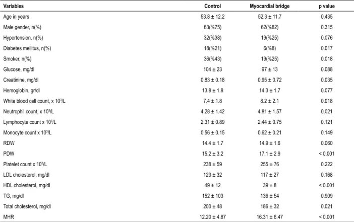
Both groups’ baseline demographics, as well as their clinic and laboratory characteristics, are summarized in Table 1. Diabetes mellitus and smoking were found to be lower in the MB group compared to the control group. There was no difference between two groups in terms of other demographic or clinic findings. When laboratory parameters were compared, creatinine, white blood cell and neutrophil were significantly higher in the MB group compared to the control group. However, HDL and total cholesterol were found to be significantly lower in the MB patients. Moreover, the monocyte/ HDL ratio was found to be significantly higher in the MB group compared to the control group. The remaining laboratory parameters did not differ between both groups.
MHR values were divided into three tertiles as follows: lower (8.25 ± 1.61); moderate (13.11 ± 1.46); and higher (21.21 ± 4.30) tertile (Table 2). We found the frequency of MB (p = 0.002), male gender (p = 0.04) and the WBC count (p < 0.001) to increase as the MHR tertiles rose.
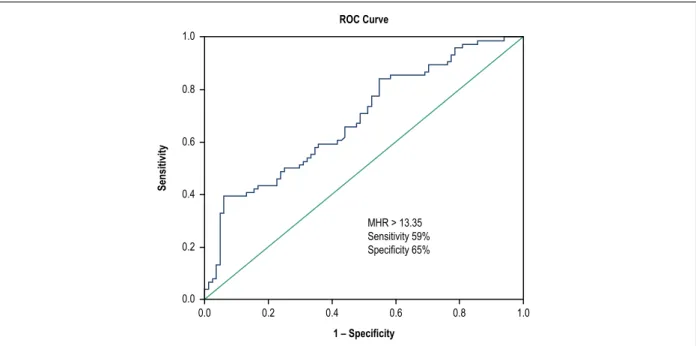
A receiver operating curve (ROC) was generated for sensitivity and specificity, with the respective areas under the curve (AUC), to investigate the predictive value of monocyte/ HDL ratio for the presence of MB (Figure 1). The Monocyte/ HDL ratio with a cut-point of 13.35 had 59.0% sensitivity and 65.0% specificity (ROC area under curve: 0.687, 95% CI: 0.606–0.769, p < 0.001) in accurately predicting MB diagnosis.
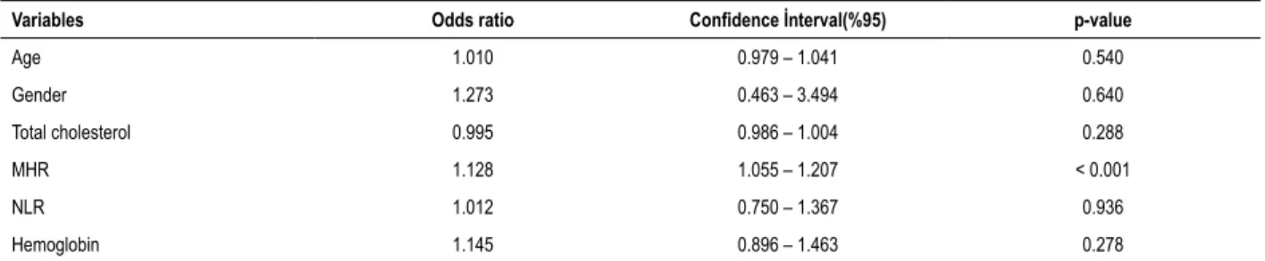
In a univariate regression analysis, age, gender, total cholesterol, neutrophil to lymphocyte ratio (NLR), and hemoglobin were significantly related with MB. In the multivariate analysis, MHR (p = 0.013) was found to be significant as the independent predictor of MB, after adjusting for other risk factors (Table 3).
Discussion
The main findings of the present study were as follows: 1) A raised monocyte/HDL ratio was found to be significantly higher in patients with MB; 2) The monocyte/HDL ratio with a cut-point of 13.35 had moderate sensitivity and specifity to diagnose MB; and 3) MHR was found to be a significant independent predictor for presence of MB, after adjusting for other risk factors in multivariate analysis.
Table 1 – Demographic, clinic and laboratory characteristics of the groups studied
Variables Control Myocardial bridge p value
Age in years 53.8 ± 12.2 52.3 ± 11.7 0.435
Male gender, n(%) 63(%75) 62(%82) 0.315
Hypertension, n(%) 32(%38) 19(%25) 0.076
Diabetes mellitus, n(%) 18(%21) 6(%8) 0.017
Smoker, n(%) 36(%43) 19(%25) 0.018
Glucose, mg/dl 104 ± 23 97 ± 13 0.088
Creatinine, mg/dl 0.83 ± 0.18 0.95 ± 0.72 0.035
Hemoglobin, gr/dl 13.8 ± 1.8 14.3 ± 1.7 0.077
White blood cell count, x 103/L 7.4 ± 1.8 8.2 ± 2.1 0.018
Neutrophil count, x 103/L 4.28 ± 1.42 4.81 ± 1.57 0.021
Lymphocyte count x 103/L 2.31 ± 0.89 2.44 ± 0.75 0.121
Monocyte count x 103/L 0.56 ± 0.15 0.62 ± 0.21 0.149
RDW 14.4 ± 1.7 14.9 ± 1.6 0.060
PDW 15.2 ± 3.2 17.1 ± 2.9 < 0.001
Platelet count x 103/L 238 ± 59 255 ± 76 0.222
LDL cholesterol, mg/dl 123 ± 32 117 ± 27 0.168
HDL cholesterol, mg/dl 49 ± 12 39 ± 8 < 0.001
TG, mg/dl 152 ± 103 136 ± 54 0.909
Total cholesterol, mg/dl 200 ± 48 186 ± 32 0.021
MHR 12.20 ± 4.87 16.31 ± 6.47 < 0.001
RDW: red cell distribution width; PDW: platelet distribution width; HDL: high density lipoprotein; LDL: low density lipoprotein; TG: triglyceride; MHR: mononcyte count/ HDL cholesterol ratio.
convert enzyme in the proximal segment of the MB artery, are the main pathophysiological mechanisms for increased atherosclerotic plaque formation.13,17 Coronary angiography, intracoronary doppler ultrasonography, intravascular ultrasound, fractional flow reserve and cardiac computed tomography angiography are main tools for diagnosing coronary MB.18
Monocytes are a source of various cytokines and molecules that interact with endothelial cells, which leads to an aggravation of inflammatory pathways.19 Inflamation play a major role in atherosclerosis development and progression.10 HDL cholesterol, which has antiinflammatory, antioxidant, and antithrombotic properties, strongly decreases the endothelial expression of adhesion molecules and prevents monocyte recruitment to the artery wall.20 Furthermore, HDL decrease pro-inflammatory and pro-oxidant effects of monocytes by inhibiting the migration of macrophages and the oxidation of the low-density lipoprotein (LDL) molecules, as well as by promoting the efflux of cholesterol from these cells.21 Therefore, it seems logical to combine these two parameters into a single ratio as an MHR, which can reflect the underlying inflammation process. A prognostic value of MHR has been reported in various cardiovascular diseases.22-24 MHR was found to be related with major cardiovascular adverse events (MACE) including stent thrombosis and mortality after primary percutaneous coronary intervention (PCI) in ST-segment elevation myocardial infarction (STEMI) patients.25 Moreover, it has been demonstrated to be a new potential marker for predicting bare metal stent restenosis.26
An important association between pre-procedural MHR levels and atrial fibrillation recurrence after ablation procedures was demonstrated by the study of Canbolat et al.24 MHR is alwo well demontrated to be associated with coronary slow flow and coronary actesia, which are different forms of inflammation and atherosclerosis.10,27 Our study has reported, for the first time, an important relationship between admission MHR and the presence of MB. Moreover, and concordant with previous studies on various cardiovascular diseases, MHR was found to be a significant independent marker associated with MB, with moderate sensitivity and specifity.
The main pathophysiological links between MHR and MB can be endothelial dysfunction and inflammation. Inflammation not only leads to monocyte secretion and aggregation, but it also reduces HDL blood levels and its anti-oxidative feature.10 Increased MHR was associated with systemic inflammation and endothelial dysfunction, and it was defined as a novel inflammation-based prognostic marker in cardiovascular diseases.22-24 In our study, concordant with previous studies on cardiovascular disease, increased MHR was found to be related with the presence of MB, in whose pathophysiology inflammation plays a significant role.
Figure 1 – The receiver operative characteristic curve analysis of monocyte to high density lipoprotein cholesterol rate for predicting the presence of myocardial bridge.
1.0
1.0 0.8
0.8 0.6
0.6 0.4
0.4 0.2
0.2 0.0
0.0
ROC Curve
Sensitivity
1 – Specificity
MHR > 13.35 Sensitivity 59% Specificity 65%
Table 2 – Demographic, clinic and laboratory characteristics of the MHR tertiles
Variables 1st tertile (n:54) 2nd tertile (n:53) 3rd tertile (n:53) p-value
MHR 8.25 ± 1.61 13.11 ± 1.46 21.21 ± 4.30 < 0.001
NLR 2.10 ± 1.35 1.98 ± 0.96 2.31 ± 1.16 0.332
Myocardial bridge, n(%) 16(%30) 26(%49) 34(%64) 0.002
Male gender, n(%) 37(%69) 41(%77) 47(%88) 0.041
Hypertension, n(%) 20(%37) 16(%30) 15(%28) 0.593
Diabetes mellitus, n(%) 8(%15) 10(%19) 6(%11) 0.553
Smoker, n(%) 13(%24) 19(%36) 23(%43) 0.105
Age 56 ± 11 55 ± 10 49 ± 14 0.006
White blood cell count, x 103/L 6.80 ± 1.63 7.80 ± 1.99 8.72 ± 1.88 < 0.001
Hemoglobin, gr/dl 13.5 ± 1.8 14.1 ± 1.5 14.5 ± 1.8 0.011
RDW 14.6 ± 1.9 14.6 ± 1.4 14.7 ± 1.6 0.973
Platelet count x 103/L 250 ± 65 240 ± 76 248 ± 63 0.739
PDW 9.2 ± 1.6 9.1 ± 1.5 9.1 ± 1.6 0.940
Glucose, mg/dl 100 ± 15 101 ± 21 101 ± 21 0.964
Creatinine, mg/dl 0.84 ± 0.17 0.85 ± 0.18 0.86 ± 0.16 0.703
LDL cholesterol, mg/dl 127 ± 31 121 ± 29 111 ± 27 0.020
HDL cholesterol, mg/dl 53 ± 11 43 ± 8 37 ± 8 < 0.001
TG, mg/dl 123 ± 47 153 ± 88 159 ± 104 0.060
Total cholesterol, mg/dl 204 ± 40 197 ± 46 179 ± 34 0.004
RDW: red cell distribution width; PDW: platelet distribution width; MHR: Mononcyte count/HDL cholesterol ratio; NLR: neutrophil / lymphocyte ratio; TG: triglyceride.
the MB. We suppposed that MHR could demonstrate not just systemic artheriosclerosis, but also local artheriosclerosis. With the addition of the local changes at the near of the MB atherosclerosis could be started earlier.
1. Bourassa MG, Butnaru A, Lespérance J, Tardif JC. Symptomatic myocardial bridges: Overview of ischemic mechanisms and current diagnostic and treatment strategies. J Am Coll Cardiol. 2003;41(3):351-9.
2. Reyman HC. Disertatio de vasis cordis propriis [dissertation]. Gottingen: Med Diss Univ; 1737.
3. Portmann W, Iwig J. Die intramurale koronarie im angiogramm. Fortschr Rontgenstr 1960;92(2):129-133.
4. Bauters C, Chmait A, Tricot O, Lamblin N, Van Belle E, Lablanche JM. Coronary thrombosis and myocardial bridging. Circulation. 2002;105(1):130.
5. Rossi L, Dander B, Nidasio GP, Arbustini E, Paris B, Vassanelli C, et al. Myocardial bridges and ischemic heart disease. Eur Heart J. 1980;1(4):239-45.
6. Schwarz ER, Klues HG, Vom Dahl J, Klein I, Krebs W, Hanrath P. Functional, angiographic and intracoronary Doppler flow characteristicsin symptomatic patients with myocardial bridging: effect ofshort-term intravenous beta-blocker medication. J Am Coll Cardiol. 1996;27(7):1637-45.
7. Ge J, Erbel R, Rupprecht HJ, Koch L, Kearney P, Gorge G, et al. Comparison of intravascular ultrasound and angiography in the assessment of myocardial bridging. Circulation. 1994;89(4):1725-32.
8. Kneale BJ, Stewart AJ, Coltart DJ. A case of myocardial bridging: evaluation using intracoronary ultrasound, Doppler flow measurement, and quantitative coronary angiography. Heart. 1996;76(4):374-6.
9. Libby P. Inflammation in atherosclerosis. Arterioscler Thromb Vasc Biol. 2012;32(9):2045-51.
10. Canpolat U, Çetin EH, Cetin S, Aydin S, Akboga MK, Yayla C, et al. Association of Monocyte-to-HDL cholesterol ratio with slow coronary flow is linked to systemic inflammation. Clin Appl Thromb Hemost. 2016;22(5):476-82.
11. Cockerill GW, Rye KA, Gamble JR, Vadas MA, Barter PJ. High-density lipoproteins inhibit cytokine-induced expression of endothelial, cell adhesion molecules. Arterioscler Thromb Vasc Biol. 1995;15(11):1987-94.
12. Mohlenkamp S, Hort W, Ge J, Erbel R. Update on myocardial bridging. Circulation. 2002;106(20):2616-22.
13. Ishikawa Y, Ishii T, Asuwa N, Masuda S. Absence of atherosclerosis evolution in the coronary arterial segment covered by myocardial tissue in cholesterol-fed rabbits. Virchows Arch. 1997;430(2):163-71.
14. Ishii T, Hosoda Y, Osaka T, Imai T, Shimada H, Takami A, et al. The significance of myocardial bridge upon atherosclerosis in the left anterior descending coronary artery. J Pathol. 1986;148(4):279-91.
References
Table 3 – Multivariate analysis to detect independent variables for the diagnosis of myocardial bridge
Variables Odds ratio Confidence İnterval(%95) p-value
Age 1.010 0.979 – 1.041 0.540
Gender 1.273 0.463 – 3.494 0.640
Total cholesterol 0.995 0.986 – 1.004 0.288
MHR 1.128 1.055 – 1.207 < 0.001
NLR 1.012 0.750 – 1.367 0.936
Hemoglobin 1.145 0.896 – 1.463 0.278
MHR: Mononcyte count/HDL cholesterol ratio; NLR: neutrophil / lymphocyte ratio.
determined due to a lack of follow-up of the study patients. Moreover, the effect of other inflamatory markers, like C-reactive protein, was not assesed due to a lack of records.
Conclusions
In conclusion, since increased MHR is a marker of inflammation and atheroclerosis, MB could be one of the factors associated with increased MHR.
Author contributions
Conception and design of the research: Enhos A, Bakshaliyev N; acquisition of data: Enhos A, Cosansu K, Huyut MA, Bakshaliyev N, Nadir A; analysis and interpretation of the data: Enhos A, Cosansu K, Huyut MA; statistical analysis: Turna F; obtaining funding: Enhos A, Cosansu K, Turna F, Karacop E, Nadir A; writing of the manuscript and critical revision of the manuscript for intellectual content: Enhos A, Karacop E, Ozdemir R, Uluganyan M.
Potential Conflict of Interest
No potential conflict of interest relevant to this article was reported.
Sources of Funding
There were no external funding sources for this study.
Study Association
This study is not associated with any thesis or dissertation work.
Ethics approval and consent to participate
This is an open-access article distributed under the terms of the Creative Commons Attribution License
15. Utuk O, Bilge A, Bayturan O, Tikiz H, Tavli T, Tezcan U. Thrombosis of a coronary artery related to the myocardial bridging. Heart Lung Circ. 2010;19(8):481-2.
16. Kramer JR, Kitazume H, Proudfit WL, Sones FM Jr. Clinical significance of isolated coronary bridges: benign and frequent condition involving the left anterior descending artery. Am Heart J. 1982;103(2):283-8.
17. Masuda T, Ishikawa Y, Akasaka Y, Itoh K, Kiguchi H, Ishii T. The effect of myocardial bridging of the coronary artery on vasoactive agents and atherosclerosis localization. J Pathol. 2001;193(3):408-14.
18. Lee MS, Chen CH. Myocardial Bridging: an up-to-date review. J Invasive Cardiol. 2015;27(11):521-8.
19. Ancuta P, Wang J, Gabuzda D. CD16+ monocytes produce IL-6, CCL2, and matrix metalloproteinase-9 upon interaction with CX3CL1-expressing endothelial cells. J Leukoc Biol. 2006;80(5):1156-64.
20. Cockerill GW, Rye KA, Gamble JR, Vadas MA, Barter PJ. High-density lipoproteins inhibit cytokine-induced expression of endothelial cell adhesion molecules. Arterioscler Thromb Vasc Biol. 1995;15(11):1987-94.
21. Farb A, Sangiorgi G, Carter AJ, Walley VM, Edwards WD, Schwartz RS, et al. Pathology of acute and chronic coronary stenting in humans. Circulation. 1999;99(1):44-52.
22. Akboga MK, Balci KG, Maden O, Ertem AG, Kirbas O, Yayla C, et al. Usefulness of monocyte to HDLcholesterol ratio to predict high SYNTAX score in patients with stable coronary artery disease. Biomark Med. 2016;10(4):375-83.
23. Akboga MK, Yayla C, Balci KG, Ozeke O, Maden O, Kisacik H, et al. Relationship between serum albumin level and monocyte-to-high-density lipoprotein cholesterol ratio with saphenous vein graft disease in coronary bypass. Thorac Cardiovasc Surg. 2017;65(4):315-21.
24. Canpolat U, Aytemir K, Yorgun H, Şahiner L, Kaya EB, Çay S, et al. The role of preprocedural monocyte-to high-density lipoprotein ratio in prediction of atrial fibrillation recurrence after cryoballoon-based catheter ablation. Europace. 2015;17(12):1807-15.
25. Cetin EH, Cetin MS, Canpolat U, Aydin S, Topaloglu S, Aras D, et al. Monocyte/HDL-cholesterol ratio predicts the definite stent thrombosis after primary percutaneous coronary intervention for ST-segment elevation myocardial infarction. Biomark Med. 2015;9(10):967-77.
26. Ucar FM. A potential marker of bare metal stent restenosis: monocyte count - to- HDL cholesterol ratio. BMC Cardiovasc Disord. 2016;16(1):186.