Comparison between refraction measured by Spot Vision
Screening
TM
and subjective clinical refractometry
Daniela Lima de Jesus,I,* Fla´vio Fernandes Villela,ILuis Fernando Orlandin,IFernando Naves Eiji,I Daniel Oliveira Dantas,II Milton Ruiz AlvesI
IHospital das Clı´nicas da Faculdade de Medicina da Universidade de Sa˜o Paulo, Disciplina de Oftalmologia e Otorrinolaringologia, Sa˜o Paulo/SP, Brazil. IIUniversidade Federal de Sergipe, Departamento de Computac
¸a˜o, Sa˜o Cristo´va˜o/SE, Brazil.
OBJECTIVE:The purpose of this study was to evaluate the accuracy of Spot Vision ScreeningTMas an autorefractor by comparing refraction measurements to subjective clinical refractometry results in children and adult patients.
METHODS:One-hundred and thirty-four eyes of 134 patients were submitted to refractometry by Spot and clinical refractometry under cycloplegia. Patients, students, physicians, staff and children of staff from the Hospital das Clı´nicas (School of Medicine, University of Sa˜o Paulo) aged 7-50 years without signs of ocular disease were examined. Only right-eye refraction data were analyzed. The findings were converted in magnitude vectors for analysis.
RESULTS:The difference between Spot Vision ScreeningTM and subjective clinical refractometry expressed in spherical equivalents was+0.66±0.56 diopters (D),+0.16±0.27 D for the vector projected on the 90 axis and
+0.02±0.15 D for the oblique vector.
CONCLUSIONS:Despite the statistical significance of the difference between the two methods, we consider the difference non-relevant in a clinical setting, supporting the use of Spot Vision ScreeningTM as an ancillary method for estimating refraction.
KEYWORDS: Refraction; Ocular; Equipment Design; Comparative Study.
Jesus DL, Villela FF, Orlandin LF, Eiji FN, Dantas DO, Alves MR. Comparison between refraction measured by Spot Vision ScreeningTMand
subjective clinical refractometry. Clinics. 2016;71(2):69-72
Received for publication onNovember 13, 2015;First review completed onDecember 1, 2015;Accepted for publication onDecember 1, 2015
E-mail: [email protected]
*Corresponding author
’ INTRODUCTION
Uncorrected refractive errors are the main cause of visual impairment and the second cause of blindness worldwide (1). In 2011, the World Health Organization estimated that visual impairment affects 284 million people worldwide, among whom 43% have uncorrected refractive errors (2).
In preschool and school-aged children, refractive errors pose serious limitations (3,4) that can compromise intellectual and psychological development as well as academic performance (5-8), resulting in potentially life-long negative effects (9). Amblyopia and amblyopic factors, such as strabismus and refractive errors, are most commonly observed in children. Visual disorders should be detected as early as possible to increase the chances of effective treatment and minimize the burden on public health. In adults, screening for refractive
errors provides an opportunity to identify conditions that may lead to blindness, including cataracts, glaucoma and diabetic retinopathy.
Currently, refractive error may also be detected using photo-screeners. The method is an attractive alternative for examining children with a risk for amblyopia or severe refractive errors, especially preschool, preverbal and mentally challenged children (10-15). In adults, photoscreeners may be used in refractive error screening campaigns, improving access to ocular health care services for socioeconomically disadvantaged populations (16).
Developed and marketed in October 2011 by PediaVision (Lake Mary, FL), Spot Vision ScreeningTM(SVS) technology was recently acquired by Welch Allyn (Skaneateles Falls, NY) (17). The SVS is a non-invasive, handheld, portable laser device that automatically captures vision data for both eyes concomitantly in children of all ages. The built-in software displays readings in a one-page summary and indicates whether the child should be referred to an ophthalmologist. Wi-Fi-enabled for easy data transfer and printing, the device is held at a distance of one meter from the face of the patient, much like a camera. The measuring range extends up to
±7.50 diopters (D) for spherical errors and ±3.50 D for
cylindrical errors. The interpupillary distance, pupil diameter and ocular alignment may also be evaluated.
DOI:10.6061/clinics/2016(02)03
Copyright&2016CLINICS–This is an Open Access article distributed under the
terms of the Creative Commons License (http://creativecommons.org/licenses/by/ 4.0/) which permits unrestricted use, distribution, and reproduction in any medium or format, provided the original work is properly cited.
No potential conflict of interest was reported.
69
The purpose of this study was to evaluate the accuracy of SVS refraction measurements by comparing SVS readings to subjective clinical refractometry (SCR) results in volunteers consisting of children and adults.
’ METHODS
Following approval of the study protocol by the National Research Ethics Committee (CONEP), 134 healthy adult volunteers with no signs of ocular disease were submitted to refractometry at the ophthalmology out-patient service of Hospital das Clínicas (School of Medicine, University of São Paulo/USP).
The participants were aged 7-50 years and included patients, medical students (USP), physicians from the Hospital das Clínicas, staff and children of staff. All participants/caretakers were informed about the study objectives and procedures and provided their written consent.
Information was collected regarding age, date of birth, gender and ophthalmological findings. Subjects presenting a visual acuity o20/20 with correction in one eye and/or
ocular disease were not eligible for inclusion in the study. Three patients were excluded because readings could not be obtained by SVS due to small pupil (n=1), pterygium (n=1) and signs of pigmentary glaucoma in one eye (n=1).
The ophthalmological measurements were obtained in the following sequence: i) visual acuity (Snellen chart at 5 m) with-out optical correction, ii) three static SVS refraction measure-ments under cycloplegia, iii) subjective clinical refractometry (SCR) under cycloplegia using a Greens refractor, iv) visual acuity with correction, and v) slit lamp biomicroscopy and fundoscopy.
The Spot Vision Screener used in this study was provided free of charge by Loktal Medical Electronics (São Paulo, Brazil).
Statistical analysis
Data were collected from the right eye only to avoid challenges associated with the interdependence of observa-tions of eyes from the same individual.
The refraction measures obtained using the two methods were averaged and compared. For the purpose of the refractive error analysis, the spherical component was expressed in spherical diopters, the cylindrical component in cylindrical diopters and the main axis of the cylinder in degrees.
To calculate average values and perform statistical analyses, readings were converted into a spherical equivalent (SE), which corresponded to the spherical value plus half the astigmatism value. In addition, the spherical and cylindrical components were converted into power vectors according to the Naeser equation (18): MV 90=m(sen2a-cos2a), where MV 90 is the
magnitude vector on the 90o
axis, m is astigmatism in diopters, and a is the meridian of astigmatism in degrees
(vertical and horizontal refraction components). The equa-tion MV 135=m(sen2(a-45)-cos2(a-45) allows the calculation
of the difference between diopter components projected on the 135o
axis and the 45o
axis. To maintain the spherical equivalent format, the MV components were divided in half. The statistical analysis was performed using the software R-project (19). The univariate analysis was conducted as follows. The difference in spherical equivalent (SE) values was calculated as the SVS spherical equivalent value minus the SCR spherical equivalent value. The same procedure was applied to MV90 and MV135, respectively. A positive difference indicated that SVS overestimated the corresponding value.
Comparisons between measurements were performed using a paired two-tailed Student’s t-test. The level of statistical signi-ficance was set at 5% (po0.05).
’ RESULTS
The final sample consisted of 134 eyes of 134 subjects, among whom 54 (40.3%) were male and 80 (59.7%) were female. The average age was 29.7 years.
Table 1 shows the differences between the refraction values obtained by SVS and SCR, both under cycloplegia. The SVS produced the following mean±SD: SE was+0.66 ±0.56, MV90 was +0.16±0.27 and MV135 was +0.02 ±0.15. The data showed that SVS tended to overestimate
both the Spherical and Cylindrical powers, although this difference was not large. The greatest difference was obtained for the spherical equivalent, which was slightly greater than half dioptry.
A bivariate analysis was performed to evaluate the influence of astigmatism on the differences between the refraction values obtained by SVS and SCR, both under cycloplegia (Figure 1).
In the trivariate analysis, a 3D plot was used to assess the relationship between the parameters SE, MV 90 and MV 135 and their influence on differences between the right-eye refraction values obtained under cycloplegia using SVS and SCR (Figure 2).
The conversion of refraction expressed in magnitude vec-tors to the conventional form revealed an average difference between SVS and SCR of+0.63 DE, with -0.33 DC on the 4 axis, for the right eye of each patient.
’ DISCUSSION
Photoscreening technology is gaining increasing popular-ity because of its range of advantages. Photoscreeners collect swift and binocular measurements, require minimal training, detect refractive errors, strabismus and risk factors for amblyopia and constitute a cost-efficient screening tool for populations with limited access to ocular health care.
In a recent study of 151 children submitted for photo-screening with SVS, Silbert and Matta (20) reported a sensitivity and specificity of 87% and 74%, respectively, for the detection of risk factors for amblyopia, according to the revised criteria of the American Association for Pediatric Ophthalmology and Strabismus. Using SVS, Arnold and Armitage (21) photoscreened 108 children aged 1-12 years and obtained a sensitivity and specificity of 80% and 85%, respectively; inconclusive results were collected in 4% of the
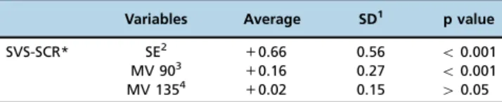
Table 1-Univariate analysis of differences between right-eye refraction values obtained under cycloplegia by Spot Vision
ScreeningTMand by subjective clinical refractometry in a sample
of 134 healthy Brazilian volunteers.
Variables Average SD1 p value
SVS-SCR* SE2
+0.66 0.56 o0.001
MV 903
+0.16 0.27 o0.001
MV 1354
+0.02 0.15 40.05
1) SD: standard deviation; 2) SE: spherical equivalent; 3) MV 90: magnitude vector on the 90o
axis; 4) MV 135: difference between diopter components projected on the 135o
axis and the 45o
axis; *) SVS-SCR: difference between refraction values obtained by Spot Vision ScreeningTM
and subjective clinical refractometry, both under cycloplegia.
70
SpotTMand clinical refractometry
children. To our knowledge, the performance of SVS in adults has not been assessed.
Before widespread adoption, new screening methods must be validated against the gold standard for diagnosis. To some authors, a difference of ±1.00 D SE between methods is
relevant. Therefore, we adopted this value as a failure criterion (22,23). In the present study, we analyzed the performance of Spot Vision ScreeningTM as a refractor against subjective clinical refractometry under cycloplegia and observed significant quantitative differences between the methods with regard to SE (+0.66) and MV 90 (+0.16). However, this difference may be considered of minor relevance in the clinical setting. The difference with regard to MV 135 was negligible (+0.02) and non-significant.
The bivariate analysis (Figure 2) revealed astigmatism had little influence (greater for MV 90 than for MV 135) on the results. Naeser obtained similar results in a study that demon-strated greater differences for the vertical and horizontal components than for the oblique component, from which the
author inferred that the vertical component was more suscep-tible to eyelid tone and the blink mechanism (19).
Demirci et al. compared photoscreening (Plusoptix S08) to retinoscopy. The methods differed with regard to average SE (0.46±0.35;p=0.007) but presented comparable astigmatism
values. The authors concluded that the device is a feasible alternative for patients in whom conventional autorefractors cannot be employed (24).
In a similar study, Lang et al. compared dynamic and static subjective refractometry readings obtained using the auto-refractor Topcon RM8800 and the Near-Eye Tool for Refractive Assessment (NETRA), a new method based on smartphones. The study subjects (n=33; 38 eyes) were aged 14-61 years (34.2±13.0) and had a visual acuity X20/40.
Under cycloplegia, the methods did not differ significantly with regard to diopters in SE (absolute average difference: 0.94±0.7 D; p=0.99), but in the absence of cycloplegia, the
difference was significant (SE 2.88±1.8 D; p=0.02). In
conclusion, the authors demonstrated a good agreement between NETRA and autorefractometry (differenceo1D in
SE) in patients under cycloplegia (23).
In this study, Spot Vision Screening (SVS) was found to be a simple and quick refractometry method. From the clinical perspective, comparable measurements were obtained by SVS and subjective clinical refractometry. This finding validated the use of SVS as an ancillary method for estima-ting refraction, especially in cases in which conventional clinical examinations are difficult to perform.
’ FUNDING INSTITUTION
Coordenac¸ão de Aperfeic¸oamento de Pessoal de Nível Superior (CAPES).
’ AUTHOR CONTRIBUTIONS
Jesus DL and Villela FF examined the patients, analyzed the results and wrote the manuscript. Orlandin LF and Eiji FN examined the patients, reviewed the literature and wrote the manuscript. Dantas DO reviewed the literature, performed the statistical analysis and wrote the article. Alves MR directed the study design and participated in manuscript writing.
’ REFERENCES
1. World Health Organization. Vision 2020 The Right to Sight: Global Initia-tive for the Elimination of Avoidable Blindness Action Plan 2006-2011. Geneva, Switzerland: 2007.
2. World Health Organization. Vision impairment and blindness. Fact sheet 282. April 2011. Available in: http://www.who.int/mediacentre/fact-sheets/fs282/en/
3. Alves MR, Kara-José N. Manual de Orientac¸ão aos professores - Veja Bem
Brasil, CBO, Imprensa Oficial [Guidance Manual for Teachers - Get Well Brazil, CBO, Press Officer], 1998.
4. Temporini ER, Carvalho RS, Kara-José N, Oliveira DF. A importância dos professores e da escolar [The importance of teachers and the school]. In: Kara-José N, Gonc¸alves ER, Carvalho RS, eds. Olho no olho: Campanha
Nacional de Prevenc¸ão à Cegueira e Reabilitac¸ão Visual do Escolar [Eye
to eye: National Campaign to Prevent Blindness and Visual Rehabilitation of School]. Rio de Janeiro Cultura Médica. 2006.
5. Moore, BD. Eye care for Infants & Young Children. Butterworth-Heinemann, Boston, 1997, p. 361.
6. Alves MR, Kara-José N. O Olho e a Visão: o que fazer pela saúde ocular das nossas crianc¸as [The Eye and Vision: what to do for the eye health of
our children]. Petrópolis, Vozes, 1996, 160p.
7. Toledo CC, Paiva APG, Camilo GB, Maior MRS, Leite ICG, Guerra MR. Detecc¸ão precoce de deficiência visual e sua relac¸ão com o rendimento
escolar [Early detection of visual impairment and its relationship to aca-demic achievement]. Rev Assoc Med Bras. 2010; 56(4):415-9.
8. Lauretti Filho A, Romão E. Estudo da acuidade visual e dos vícios de refrac¸ão em crianc¸as com baixo rendimento escolar [Study of visual
acuity and refractive errors in children with poor school performance]. Rev Bras Oftalmol. 1982;41:31-6.
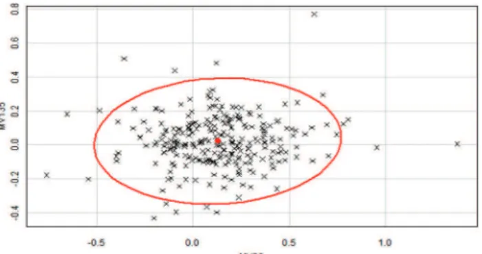
Figure 1 -Bivariate analysis of the influence of the parameters magnitude vector 90 and magnitude vector 135 on differences between right-eye refraction values obtained under cycloplegia
by Spot Vision ScreeningTM and by subjective clinical
refracto-metry in a sample of 134 healthy Brazilian volunteers.
Magni-tude vector 90: magniMagni-tude vector on the 90o
axis; MV 135:
difference between diopter components projected on the 135o
axis and the 45o
axis.
Figure 2 - Trivariate analysis showing the influence of the parameters spherical equivalent, magnitude vector 90 and magnitude vector 135 on differences between right-eye refrac-tion values obtained under cycloplegia by Spot Vision
Screen-ingTMand by subjective clinical refractometry) in a sample of 134
healthy Brazilian volunteers. SE: spherical equivalent; MV 90:
magnitude vector on the 90o
axis, magnitude vector 135:
difference between diopter components projected on the 135o
axis and the 45o
axis.
71
CLINICS 2016;71(2):69-72 SpotTMand clinical refractometry
9. Burns MJ. Building a priority for national vision heath care. Eye Ear Nose Throat Mon. 1973;52(10):353-6.
10. Alley CL. Preschool vision screening: update on guidelines and techniques. Curr Opin Ophthalmol. 2013;24(5):415-20, http://dx.doi.org/10.1097/ ICU.0b013e3283641c56.
11. Ottar WL, Scott WE, Holgado SI. Photoscreening for amblyogenic factors. J Pediatr Ophthalmol Strabismus. 1995;32(5):289-95.
12. Ugurbas SC, Alpay A, Tutar H, Sagdik HM, Ugurbas SH. Validation of plusoptiX S04 photoscreener as a vision screening tool in children with intellectual disability. J AAPOS. 2011;15(5):476-9, http://dx.doi.org/10.1016/ j.jaapos.2011.05.023.
13. Longmuir SQ, Boese EA, Pfeifer W, Zimmerman B, Short L, Scott WE. Practical community photoscreening in very young children. Pediatrics. 2013;131(3):e764-9.
14. McCurry TC, Lawrence LM, Wilson ME, Mayo L. The plusoptiX S08 photoscreener as a vision screening tool for children with autism. J AAPOS. 2013;17(4):374-7, http://dx.doi.org/10.1016/j.jaapos.2013.05.006.
15. Yanovitch T, Wallace DK, Freedman SF, Enyedi LB, Kishnani P, Worley G, Crissman B, Burner E, Young TL. The accuracy of photoscreening at detecting treatable ocular conditions in children with Down syndrome. J AAPOS. 2010;14(6):472-7, http://dx.doi.org/10.1016/j.jaapos.2010.09.016. 16. Alves MR, Jesus DL, Villela FF, Baptista GV. Métodos de rastreamento
refrativo baseados em equipamentos [Methods of refractive screening based on equipments]. In: Alves MR, Nishi M, Carvalho KM, Ventura LM, Schellini SA, Kara-José N. Refrac¸ão Ocular: Uma necessidade
social [Ocular Refraction: A social need]. Rio de Janeiro: Cultura Médica, 2014.
17. SpotTM Vision Screening by Welch Allyn. http://www.spotvision
screening.com acessed August 25, 2014.
18. Naeser K. Hjortdal J. Multivariate analysis of refractive data: mathematics and statistics of spheroclylinders. J Cataract Refract Surg. 2001;27(1)129-42, http://dx.doi.org/10.1016/S0886-3350(00)00816-6.
19. The R-Project for Statistical Computing. Available in: http://www. r-project.org
20. Silbert DI, Matta NS. Performance of the Spot vision screener for the detection of amblyopia risk factors in children. J AAPOS. 2014; 18(2):169-72, http://dx.doi.org/10.1016/j.jaapos.2013.11.019.
21. Arnold RW, Armitage D. Performance of four photoscreeners on pediatric patients with high risk amblyopia. J Ped Ophthalmol Strabismus. 2014;51:1-7, http://dx.doi.org/10.3928/01913913-20131223-02. 22. Czinder NC. A Retrospective study comparing the accuracy of the
PediaVision Assessment Solution (PAS) Photo-Screener refraction cap-abilities on spacial needs patients against other refraction methods. Doc-torate Tesis. Ferris State University Michigan College of Optometry. 2013. 23. Lang MP, Pakter HM, Ferreira LB, Mohan A, Raskar R, Pamplona VF, Oliveira MM. Comparison of a Cell Phone-Based Refraction Technique (NETRA) With Auto-Refraction ARVO 2012. Poster 3506/D1109. 24. Demirci G, Arslan B, Ozsutc¸u M, Eliac¸ik M, Gulkilik G. Comparison of
photorefraction, autorefractometry and retinoscopy in children. Int Oph-thalmol. 2014;34(4):739-46, http://dx.doi.org/10.1007/s10792-013-9864-x.
72
SpotTMand clinical refractometry