T
QUANTITATIVE ANALYSES OF MAXILLARY
SINUS USING COMPUTED TOMOGRAPHY
ANÁLISE QUANTITATIVA DOS SEIOS MAXILARES POR MEIO DA
TOMOGRAFIA COMPUTADORIZADA
Andréia PERRELLA
Aluna do Curso de Graduação da Faculdade de Odontologia da Universidade de São Paulo. Bolsista de Iniciação Científica CNPq/PIBIC.
Sara dos Santos ROCHA
Mestre em Odontologia Legal pela Faculdade de Odontologia da Universidade de São Paulo.
Marcelo de Gusmão Paraiso CAVALCANTI
Professor Livre-Docente da Disciplina de Radiologia do Departamento de Estomatologia da Faculdade de Odontologia da Universidade de São Paulo, São Paulo.
Professor Adjunto Assistente do Departamento de Radiologia da Faculdade de Medicina da Universidade de Iowa, EUA.
he aim of this study was to evaluate the precision and accuracy of linear measurements of maxillary sinus made in tomographic films, by comparing with 3D reconstructed images. Linear measurements of both maxillary sinus in computed tomography CT of 17 patients, with or without lesion by two calibrated examiners independently, on two occasions, with a single manual caliper. A third examiner has done the same measurements electronically in 3D-CT reconstruction. The statistical analysis was performed using ANOVA (analyses of variance). Intra-observer percentage error was little in both cases, with and without lesion; it ranged from 1.14% to 1.82%. The inter-observer error was a little higher reaching a 2.08% value. The accuracy presented a higher value. The perceptual accuracy error was higher in samples, which had lesion compared to that which had not. CT had provided adequate precision and accuracy for maxillary sinus analyses. The precision in cases with lesion was considered inferior when compared to that without lesion, but it can’t affect the method efficacy.
UNITERMS: Tomography; X-ray computed; Maxillary sinus; Quantitative study.
INTRODUCTION
The maxillary sinus is a pneumatic cavity of the facial skeleton within maxillary bone; it has a quadrangular pyramid form with an internal base6. The
maxillary sinus fluids drain into the nasal cavity by a narrow osteomeatal complex, with the obstruction of the outflow causing sinusitis, mucosal thickening, and polyps or retention cyst formation14. Also it can be
affected too by other lesions in the face, like tumors or fractures but its clinical access is difficult and radiographic examination is considered to be very helpful in the better clarification of those diseases.
Computed tomography (CT) proved to be the most accurate technique used in detection of simulated lesions of all surfaces of the maxillary sinus, allowing access to all the walls of the sinus and should be used when
there is definite evidence of maxillary sinus pathosis12.
The defect can be exactly located by CT scans using three-dimensional reconstructed images.
Spiral CT is a fast scanning method, permitting improved contrast enhancement and decreased motion artifacts, so it enables the radiologist to visualize lesions quickly and manipulate volumetric data in three-dimensional reconstructed images (3D-CT). Quantitative studies have validated linear measurements of bone structures in the maxillofacial region, for treatment planning using 3D reconstructed spiral CT5.
MATERIALS AND METHODS
The study population consisted of 17 patients, with or without maxillary sinus lesion (17 sinuses with lesion and 17 without), who were submitted to a CT scan (Toshiba X/Press, Toshiba Medical System, Tustin, CA, EUA) with the following protocol: 3 mm axial slices, 3 mm table feed and 3 mm reconstructed slice interval, in 1 second, at 130 kVp and 200mA, and 512X512 matrix. The original CT data were transferred to a workstation (DELL hardware Precision 420 WINDOWS NT with Vitrea® 2.3 software, Vital
Images Inc., Minneapolis, MN, USA). The data were archived in a standard computer workstation and on a CD-ROM in order to postprocess any study.
Linear measurements were done on the tomographic films in the antero-posterior and right-left largest diameters by two calibrated examiners independently, on two occasions, with a single manual caliper, which was previously calibrated. To perform these measurements it was used a scale, which vary from 5cm to 15cm, on the right side of the CT films (Figure 1). So we pointed on the caliper in a boundary of the region we wanted (each side of the large diameter A-P or R-L) and then we transferred this distance obtained to the scale to obtain the value of this distance.
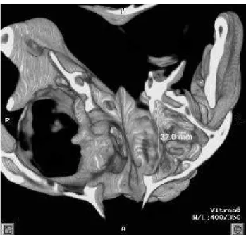
The 3D-CT reconstructed images were used as a gold standard, to evaluate the accuracy of 2D-CT in films. To obtain the measurements in 3D-CT we used the crosshair tool, which allowed selecting the antero-posterior and right-left largest measurements of the maxillary sinus, in axial view and marked it with an arrow in order to guide the measurement in the 3D. Subsequently, we displayed the 3D-CT images with
the arrows showing the exact localization of the measurements, and we have done it electronically with the ruler tool (Figures 2 and 3). In order to make easy this work, we have used other tools from the software, which allowed us to rotate and to section the skull. The statistical analysis was performed using ANOVA (analyses of variance).
FIGURE 1- 2D-CT axial view is demonstrating the largest diameter delimitation of maxillary sinuses (arrows). The scale, which was used to perform the measurements, is also shown on the right side of the figure (open arrow)
FIGURE 2- 3D-CT supero-inferior view using bone protocol is demonstrating the electronically measurement of the left maxillary sinus’ antero-posterior largest diameter (32.00 mm)
RESULTS
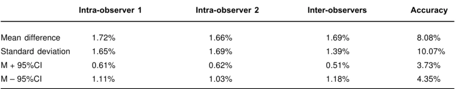
The Tables 1 and 2 showed the error percentage found in linear measurements of maxillary sinus with lesion and without lesion, respectively. The statistical evaluation displayed the data like the group means, standard deviations and 95% of confidence intervals for individual predicted values.
The intra-observer percentage error was little in both cases, with and without lesion; it ranged from 1.14% to 1.82%. The inter-observer error was higher reaching a 2.08% value. The accuracy presented a higher value.
The perceptual accuracy error was higher in samples, which had lesion compared to that which had not (8.08% and 5.19%, respectively), but this difference isn’t significant because we have a small sample”.
DISCUSSION
Inaccurate measurements from conventional radiographs occurs due to superimposition of the other paranasal sinuses and craniofacial skeleton on the upper portion of the maxillary sinus. However, CT has the advantage of plainly demonstrating the components of the maxillary sinus1. CT and
multiplanar reconstructed images (MPR) are considered standard images for detecting both bony and soft tissue lesions improving the accuracy in evaluation of the soft tissues and subtle changes in
bones and air- filled spaces 12.
In general, the films are more available to the clinicians who don’t need extra equipment to interpret them. In our paper, we used an independent workstation, where 3D-CT images were processed, becoming a more practical and faster tool to assess the images. We used the 3D-CT images as being the gold standard of this study, since many authors have already found accuracy of measurements in these images5,8,10,15. Matteson, et
al.10 (1989) found the 3D-CT images of the craniofacial
region to be very accurate, and Waitzman, et al15 in 1992
related excellent agreement between direct measurements made in dry skull and in CT images. Quantitative studies have validated linear measurements of bone structures in the maxillofacial region, for treatment planning using spiral 3D-CT5. In CT images
there is no significant enlargement or distortion of the image, overlap of structures, or tracing error, so it is an accurate and reproducible means of recording quantitative information15.Simulated 3D-CT reformats
conventional imaging data into series of images that closely resemble the original studied structure8.
Cavalcanti, Ruprecht3 in 2000, studying neoplastic
lesions associated with the mandible concluded that spiral CT allowed accurate computer graphics and film-based measurements. They showed there was no statistically significant differences between computer graphics or film-based measurements and physical measurements or between inter and intra-observer measurements. These findings are in agreement with those we found in our results regarding to maxillary sinus.
Intra-observer 1 Intra-observer 2 Inter-observers Accuracy
Mean difference 1.14% 1.82% 2.08% 5.19% Standard deviation 1.63% 1.70% 1.88% 5.091% M + 95%CI 0.54% 1.18% 1.39% 3.31% M – 95%CI 1.75% 2.45% 2.78% 7.08%
TABLE 1- Statistical analysis of measurements in maxillary sinus without lesion
Intra-observer 1 Intra-observer 2 Inter-observers Accuracy
Mean difference 1.72% 1.66% 1.69% 8.08% Standard deviation 1.65% 1.69% 1.39% 10.07% M + 95%CI 0.61% 0.62% 0.51% 3.73% M – 95%CI 1.11% 1.03% 1.18% 4.35%
Measurements of skeletal dimensions are essential for accurate diagnosis and planned reconstructive surgery10,15. An inaccurate localization of anatomical
structures can lead to undesirable complications as a perforation of the maxillary sinus13.In our research,
several factors were considered as potential errors when the measurements were made: 1. Lesions, which caused destruction or expansion on the sinus cortex, made more difficult to localize the landmarks (the internal walls of the maxillary sinuses in their largest diameter antero-posterior and right–left) to perform the measurements, especially in the 3D-CT. 2. Another point, which influenced on the results, was how the measurements on the tomographic films were done. In materials and methods section, we used the film’s scale. However those scales were not millimetricaly divided and each interval corresponds to a large value varying from 5 mm to 10 mm not in real distances (this 5 mm does not correspond to 5 mm in a ruler, but 5 mm in real image). Subsequently, our measurements presented values lower than these, so we may suppose the lower intervals and this perception varied from person to person making our measurements less precise than it could be if the scale were divide in lower intervals. This did not occur in 3D-CT images, because the measurements are made electronically with ruler tool software. The measurements in 3D-CT are made easier by the software tools like translation, rotation and segmentation, as was stated by Cavalcanti, Vannier4 in 1998. The value of
computer graphics in manipulating craniofacial images and the importance of 3D-CT images in quantitative and qualitative information about the craniofacial complex are clearly recognized. Computer graphics technology and current workstations allow better visualization and segmentation that enable assessment of volume, area, linear and angular measurements4.
3D –CT – based measurements are characterized by a number of features: spiral CT scanning, 3D-image reconstruction, image manipulation (translation, rotation, and segmentation) by computer graphics and interactive landmark identification. Ace contributes to the overall improvement in measurement accuracy4.
Our results demonstrated that reproducibility of measurements was high, and the error intra and inter-observers were less than 2.0%. Similar results were already found by Christiansen, Thompson, Kopp7,
1989 when studying accuracy in linear and angular measurements in CT done in vitro in human mandible7,
and by Cavalcanti, et al3 in 2000 in their studies with
simulated neoplastic lesions in mandible too. 2D-CT and 3D-CT images can be useful to the clinician in diagnosis and treatment planning. These methods
enhance the accuracy of diagnostic decisions and establishment of appropriate treatment plans2.
CONCLUSIONS
The mean difference of measurements were higher in maxillary sinus with lesion when compared to that without lesion
The measurements of CT-based films provided adequate precision (reproducibility) and accuracy for maxillary sinus analyses.
CT-based films can be used in study and in attendance of clinical cases in all their phases as treatment planning and a follow up of cases.
ACKNOWLEDGEMENTS
The support for this research was provided by grants from CNPq (520425/01.5) and CNPq/PIBIC (undergraduate student). The authors gratefully acknowledge the contribution of José Leopoldo F. Antunes, from the Department of Social Dentistry, College of Dentistry, University of São Paulo for the statistical analysis.
RESUMO
quanto intra-examinadores, sem alterar, porém, a eficácia do método.”
UNITERMOS: Tomografia; Raios x por computador; Seio maxilar; Estudo quantitativo.
REFERENCES
1- Ariji Y, Kuroki T, Moriguchi S, Ariji E, Kanda S. Age changes in the volume of the human maxillary sinus: a study using computed tomography. Dentomaxillofac Radiol 1994; 23:163-8.
2- Alder ME, Deahl T, Matteson SR. Clinical usefulness of two-dimensional reformatted and three-two-dimensionally rendered computerized tomographic images – literature review had a survey of surgeons’ opinions. J Oral Maxillofac Surg 1995; 53:375-86.
3- Cavalcanti MGP, Ruprecht A, Bonomie JM, Vannier MW. Accuracy and precision of spiral CT in the assessment of neoplastic lesions associated with the mandible.. Acad Radiol. 2000; 7:94-9.
4- Cavalcanti MGP, Vannier MW. Quantitative analysis of spiral computed tomography for craniofacial clinical applications. Dentomaxillofac Radiol 1998; 27:344-50.
5- Cavalcanti MGP, Vannier MW. Measurement of the volume of oral tumors by three dimensional spiral computed tomography. Dentomaxillofac Radiol2000; 29:35-40.
6-Chanavaz M. Maxillary sinus: anatomy, physiology, surgery and bone grafting related to implantology–Eleven years of surgical experience (1979-1990). J Oral Implantol 1990;16(3):199-209.
7- Christiansen EW, Thompson JR, Kopp S. Intra- and inter-observer variability and accuracy in the determination of linear and angular measurements in computed tomography. an in vitro and in situ study in human mandibles. Acta Odontol Scand 1986; 44:221-9.
8- Fishman EK, Magid D, Ney DR, Chaney EL, Pizer SM, Rosenman JG, et al. Three- dimensional imaging. Radiology 1991; 181:321-37.
9- Frederiksen NL. Specialized radiographic techniques. In: White CS, Pharoah MJ. Oral Radiology : principles and interpretation. St. Louis, Mosby; 2000.
10- Matteson SR, Bechtold W, Phillips C, Staab E. A method for three-dimensional image reformation for quantitative cephalometric analysis. J Oral Maxillofac Surg 1989; 47:1053-61.
11- Ohba T, Ogawa Y, Shinohara Y, Hiromatsu T, Uchida A, Toyoda Y. Limitations of panoramic radiography in the detection of bony defects on the posterior wall of the maxillary sinus: an experimental study. Dentomaxillofac Radiol. 1994 Aug; 23:149-53.
12- Perez CA, Farman AG. Diagnostic radiology of maxillary sinus defects Oral Surg Oral Med Oral Pathol. 1988; 66:507-12.
13- Regev E, Smith RA, Perrot DH, Pogrel MA Maxillary sinus complications related to endosseous implants. International J Oral Maxillofac Implants 1995 10: 451-61.
14- Rothman SLG Dental applications of computerized tomography : surgical planning for implant placement. Illinois, Quintessence Books;1998.
15- Waitzman AA, Posnick JC, Armstrong DC, Pron G. Craniofacial skeletal measurements based on computed tomography: Part II. Normal values and growth trends. Cleft Palate Craniofac. J. 1992; 29(2): 118-28.
Recebido para publicação em: 12/03/2003 Aceito após reformulações: 11/07/2003
Endereço para correspôndencia:
Prof. Dr. Marcelo de Gusmão Paraiso Cavalcanti Av. Prof. Lineu Prestes, 2227, FOUSP
Disciplina de Radiologia 05508-900 - São Paulo, SP e-mail: [email protected]