Glutamine treatment attenuates hyperglycemia-induced
mitochondrial stress and apoptosis in umbilical vein
endothelial cells
Sher Zaman Safi,I,*Kalaivani Batumalaie,I Marzida Mansor,IIKaruthan Chinna,IIIHamed Karimian,IV Rajes Qvist,IMuhammad Aqeel Ashraf,V Syam Mohan,VIGarcie Ong Siok YanII
IUniversity of Malaya, Faculty of Medicine, Department of Medicine, Kuala Lumpur, Malaysia.IIUniversity of Malaya, Faculty of Medicine, Department of
Anesthesiology, Kuala Lumpur, Malaysia.IIIUniversity of Malaya, Faculty of Medicine, Department of Social and Preventive Medicine, Kuala Lumpur,
Malaysia.IVUniversity of Malaya, Faculty of Medicine, Department of Pharmacy, Kuala Lumpur, Malaysia.VUniversity of Malaya, Faculty of Science,
Department of Geology, Kuala Lumpur, Malaysia.VIMedical Research Center, Jazan University, 11420, Jazan, Saudi Arabia.
OBJECTIVE:The aim of this study was to determine the in vitro effect of glutamine and insulin on apoptosis, mitochondrial membrane potential, cell permeability, and inflammatory cytokines in hyperglycemic umbilical vein endothelial cells.
MATERIALS AND METHODS:Human umbilical vein endothelial cells were grown and subjected to glutamine and insulin to examine the effects of these agents on the hyperglycemic state. Mitochondrial function and the production of inflammatory cytokines were assessed using fluorescence analysis and multiple cytotoxicity assays. Apoptosis was analyzed by the terminal deoxynucleotidyl transferase dUTP nick end-labeling assay.
RESULTS: Glutamine maintains the integrity of the mitochondria by reducing the cell permeability and cytochrome c levels and increasing the mitochondrial membrane potential. The cytochrome c level was significantly (po0.005) reduced when the cells were treated with glutamine. An apoptosis assay revealed significantly reduced apoptosis (po0.005) in the glutamine-treated cells. Moreover, glutamine alone or in combination with insulin modulated inflammatory cytokine levels. Interleukin-10, interleukin-6, and vascular endothelial growth factor were up-regulated while tumor necrosis factor-a was down-regulated after treatment with glutamine.
CONCLUSION:Glutamine, either alone or in combination with insulin, can positively modulate the mitochondrial stress and cell permeability in umbilical vein endothelial cells. Glutamine regulates the expression of inflammatory cytokines and maintains the balance of the mitochondria in a cytoprotective manner.
KEYWORDS: Hyperglycemia; Sepsis; Apoptosis; Cytokine; Glutamine.
Safi SZ, Batumalaie K, Mansor M, Chinna K, Mohan S, Karimian H, Qvist R, et al. Glutamine treatment attenuates hyperglycemia-induced mitochondrial stress and apoptosis in umbilical vein endothelial cells. Clinics. 2015;70(8):569-576
Received for publication onApril 29, 2015;First review completed onJune 9, 2015;Accepted for publication onJune 9, 2015 E-mail: [email protected]
*Corresponding author
’ INTRODUCTION
Hyperglycemia is the abnormality underlying diabetes and several other complications. Chronic hyperglycemia condition initiates a wide range of complications, including cardiovascular disease, which is the most frequent cause of death in the diabetic population (1,2). Hyperglycemia also induces the production of reactive oxygen species (ROS) and, ultimately, DNA damage and apoptosis (3). Apoptosis has
been observed in the vascular cells, myocardium, and nerves of diabetic experimental animals and human subjects, although whether it contributes to or is a marker for complications in these tissues is unclear. Vascular diseases are the major cause of morbidity and mortality in diabetic patients. Several studies suggest that the endothelium is the initial site where these vascular complications develop (4). During critical illness, hyperglycemia alone or hyperglyce-mia coupled with a relative insulin deficiency may directly or indirectly yield a predisposition to a spectrum of complica-tions, such as multi-organ failure and death (5-7). Morpho-logical correlates of these functional abnormalities were not initially identified; however, several later studies showed an increase in apoptosis in several organs affected by diabetes and sepsis, including the eye (8,9), heart, and vascular endothelium (10,11).
DOI:10.6061/clinics/2015(08)07
Vascular endothelial cells are among the first cells in the body to interact with bacterial endotoxins during sepsis. Sepsis is a very complex and heterogeneous clinical condition that is associated with hyperglycemia and insulin resistance (12). Vascular endothelial cells possess mechan-isms that recognize structural patterns of bacterial constitu-ents and initiate the expression of proinflammatory and anti-inflammatory pathways, which are tightly controlled to maintain homeostatic balance (13). However, in severe sepsis, external stimuli, such as a severe infection, can activate the immune cells and unleash a systemic inflamma-tory response that is expressed through various pathways (14). There is also increasing evidence to suggest that cell death by apoptosis plays an important role in the pathogen-esis of severe sepsis/septic shock (15).
Several studies have shown that endothelial cells can undergo apoptosis in response to sepsis-related factors such as Lipopolysaccharide (LPS) and Tumor necrosis factor alpha (TNF-a) (16-18). In addition, studies usingin vitromodels of infection have demonstrated that certain organisms are capable of inducing endothelial apoptosis (19). Along with the interaction of inflammation with apoptosis in sepsis, mitochondrial dysfunction seems to have a major impact in sepsis patients because it has been closely linked to pro-grammed cell death. Alterations in mitochondrial function have been described in the muscle and liver mitochondria from septic rats and primates. Furthermore, mitochondrial dysfunction has been suggested as a potential mechanism to explain tissue hypoxia despite normal oxygen availability during sepsis (20,21). Pro-inflammatory cytokines, such as interleukin 6 (IL-6), TNF-a, and other molecules, are released during acute inflammation and result in endothelial activation and a significant increase in the expression of endothelial leukocyte adhesion molecule 1, vascular cell adhesion mole-cule 1 (VCAM-1), intercellular adhesion molemole-cule 1 (ICAM-1), and vascular endothelial growth factor (VEGF). These proteins promote leukocyte rolling, adherence, and migration, which initiate inflammation in the endothelium and other cells (22,23). We included IL-10 as an anti-inflammatory marker. Therefore, the aim of this study was to determine the mechanism of endothelial cell apoptosis and the expression of inflammatory cytokines under hyperglycemic conditions and to examine the effects of glutamine and insulin.
’ MATERIALS AND METHODS
Cell culture
Endothelial cells were obtained from VEC Technologies (New York, USA). The cells were thawed at 37˚C and cultured in T25 flasks coated with 50 mg/ml of fibronectin. The cells were immersed in 5 ml of complete medium (MCDB-B-131), supplemented with 10% FBS, 1% penicillin-streptomycin, and epidermal growth factor (EGF, 10 ng/ml). The cells were incubated at 37 ˚C with 5% CO2. Trypsin/
EDTA (1 ml for each flask) was used to detach the cells upon confluency. All the experiments were performed at passages 2-5.
Cell treatment
The cells were seeded at 1x10⁴ cells in each well and
incubated for 24 hours. Various concentrations of glucose, ranging from a normal value (5 mM) to a hyperglycemic level (20 mM), were added to the individual wells. The hyperglycemic cells (glucose concentration 20 mM) were
divided into three groups. In the first group, 40 mM of gluta-mine was added. In the second group, 1.0 x 10⁻⁶units/ml of
insulin was added. In the third group, glutamine (40 mM) and insulin (1.0 x 10⁻⁶units/ml) were added. The cells were then incubated for the required length of time (24 hours). For the cytokine and TUNEL analyses, 0.7x10⁶cells were grown in T25 flasks using the same treatment groups. The cells were harvested and frozen until required for analysis.
Western blotting
The endothelial cells were first lysed in cold lysis buffer containing 20 mmol/l of TRIS HCl, 140 mmol/l of NaCl, 1 mmol/l of EDTA and complete miniprotease inhibitor cock-tail, 1% Triton X-100, 0.1% SDS, 1% sodium deoxycholate, 1 mmol/l NaF, and 1 mmol orthovanadate. The proteins (30mg) were then loaded on 10% SDS polyacrylamide gels and transferred to activated nitrocellulose membranes. The mem-branes were blocked with Tris-buffered saline (TBS) contain-ing 5% nonfat milk and incubated overnight with the primary antibodies to IL-10 and TNF-a, obtained from Santa Cruz, at 4˚C. Beta-actin was used as a loading control. After extensive washes in TBS, the membranes were incubated for one hour at room temperature with the appropriate horseradish perox-idase-conjugated secondary antibodies, and the proteins were visualized using a chemiluminescence substrate according to the manufacturer’s instructions (Amersham Life Sciences).
Multiple cytotoxicity assays
The Cellomics Multiparameter Cytotoxicity 3–kit was used
as previously reported in detail by Cheah et al. (24). The Multiparameter Cytotoxicity 3–kit enables parallel
measure-ments of six independent parameters that monitor cell health, namely, changes in cell permeability, cell loss and nuclear size; changes in mitochondrial membrane potential; cytochrome c release; and morphological changes. Briefly, the cells were plated at 1x10⁴cells per well for 24 hours. Glucose (5 or 20 mM), glutamine (40 mM), and insulin (1.0 x 10⁻⁶ units/ml) were added in different combinations as described in the cell treatment section, and the incubation was continued for 24 hours. The MMP dye and the cell permeability dye were added to the live cells, and the cells were incubated for 1 hour. The cells were fixed, permeabi-lized, and blocked with 1 blocking buffer before they were incubated with the primary cytochrome c antibody and conjugated secondary antibody for 1 hour. The cells were rinsed three times with wash buffer II, and the plates were analyzed using the Array Scan HCS high content system (Cellomics, PA, USA).
Measurement of transmembrane mitochondrial potential
Terminal deoxynucleotidyl transferase dUTP nick end-labeling (TUNEL) assay
DNA damage was investigated using a 96–well
colori-metric apoptosis detection kit (R&D System) according to the manufacturer’s instructions. Umbilical vein endothelial cells were cultured and transferred to a 96-well plate (1x10⁵cells/ well). The cells were then fixed with 3.7% buffered formaldehyde for 5 minutes, followed by washing with PBS. The washing was followed by permeabilization of the cells with 100% methanol for 20 minutes and another wash with PBS. Following the manufacturer’s protocol, the cells were then subjected to the labeling procedure, and the reaction was stopped with 0.2 N HCl after 30 minutes. The cells were treated with NucleaseTM to generate DNA breaks and to confirm the permeabilization and labeling reactions. An unlabeled control was included to indicate the level of background labeling associated with non-specific binding of the Strep-HRP. The absorbance at 450 nm was measured using a microplate reader.
Cytokine measurements
The cytokines TNF-a, IL-6, and IL-10 were measured in triplicate using the Protein Bio-Plex Cytokine Assay (Bio-Rad Laboratories). T25 flasks containing 0.7x10⁶ cells were cultured, and the lysate was filter-sterilized (0.22-mm pore size). The protein concentrations were determined, and the
Bio-Plex Cytokine Assay (Bio-Rad Laboratories) was con-ducted according to the manufacturer’s protocol. The calculated concentrations for each cytokine were averaged, and the standard deviations were determined. Statistical significance was determined using the t-test, wherepo0.05 designated increased/decreased cytokine production in the presence of glutamine or glutamine in combination with insulin.
Statistical analysis
Each experiment was performed at least two times. Statistical analysis was performed using one-way analysis of variance (ANOVA).
’ RESULTS
Glutamine reduces the cytochrome C levels and apoptosis in hyperglycemic human umbilical vein endothelial cells
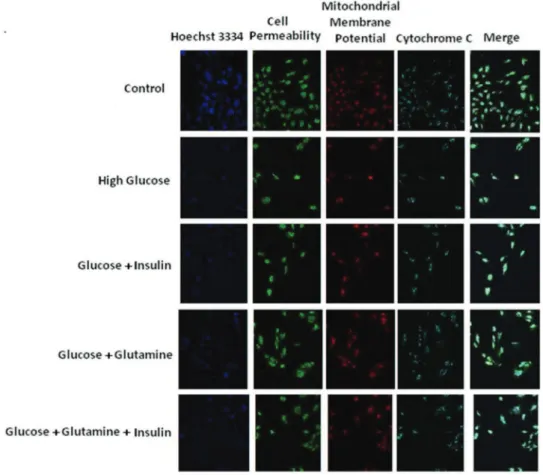
Umbilical vein endothelial cells were stained with Hoechst in the presence of glucose (20 mM) alone, 20 mM glucose+
40 mM glutamine, 20 mM glucose + 10-6 M insulin, or 20 mM glucose+40 mM glutamine+10-6M insulin, and the
staining intensity was determined. As shown in Figures 1 and 2, glucose alone (20 mM) reduced the number of cells, possibly by apoptosis, as well as reduced the level of
Figure 1 -Representative images of endothelial cells treated with medium alone (control), glucose alone (20mM), glucose (20mM)+
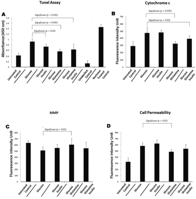
cytochrome c. There was a significant change (po0.005) in the fluorescence intensity in the cells that were treated with glucose along with glutamine. This result indicates that these cells were more viable. In addition, the cytochrome c levels in the endothelial cells treated with glucose (20 mM) +
glutamine (40 mM) were significantly (po0.005) lower than the levels in the cells treated with glucose (20 mM) alone. The cytochrome c levels in the cells treated with glucose (20 mM)
+insulin (10-6M) and glucose (20 mM)+insulin (10-6M)+
glutamine (40 mM) also changed significantly (po0.05) compared with the levels in the cells treated with glucose (20 mM) alone (Figures 1 and 2B). Similarly, the TUNEL assay revealed a significantly reduced level of apoptosis when the hyperglycemic cells were treated with insulin (po0.05), glutamine (po0.005), or glutamine + insulin (po0.005) (Figure 2A).
Glutamine restores the loss of mitochondrial membrane potential (MMP) in Human Umbilical Endothelial Cells (HUVEC)
According to previous studies, a reduction in the mitochondrial membrane potential occurs when cells undergo oxidative stress and hyperglycemia in the presence of other co-factors. The cells showed a signifi-cant change in the MMP when treated with glutamine and a considerable, but not significant, change in the MMP when they were treated with glucose+glutamine
+ insulin compared with 20 mM glucose only. Glucose (20 mM)+insulin (10-6M) and glucose (20 mM)+insulin (10-6M)+glutamine (40 mM) also yielded an increase in the MMP (DCm) compared with the control. These results show that the MMP was reduced after the cells had been treated
with 20 mM glucose alone; however, glucose (20 mM) in combination with glutamine (40 mM) was shown to be more protective against the fall in the MMP and oxidative stress-induced cell death (Figure 2C).
Effect of glutamine on cell permeability
When the MMP decline reaches a certain point, the opening of the permeability transition pore (PTP) starts to cause extensive cell damage and, consequently, cell death. The hyperglycemia-induced permeability was reversed when we treated the HUVECs with glucose + glutamine or glucose + glutamine + insulin compared with glucose (20 mM) alone. However, there was a slight increase in the cell permeability when the cells were treated with glucose (20 mM)+insulin (10-6M) (Figure 2D).
Glutamine and inflammatory cytokines
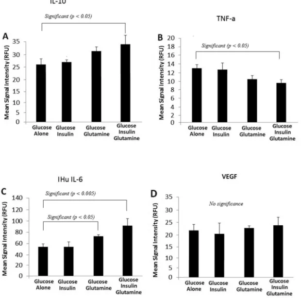
When glucose (20 mM) and insulin (10-6M) were added to the culture, there was no difference in the expression of IL-6. However, glucose + glutamine significantly increased the expression of IL-6. The combined treatment with glucose (20 mM), glutamine (40 mM), and insulin (10-6 M) also
significantly increased the expression of IL-6 (po0.005). Similarly, treatment with insulin alone did not alter the expression of IL-10, but insulin in combination with glutamine increased the expression of IL-10 in the endothe-lial cells. A small increase in the expression of VEGF was noted while a reduced expression of TNF-a was observed when the cells were treated with the same concentration of glutamine alone or in combination with insulin (Figure 3A, 3B, 3C, and 3D). Detailed values of all cytokines are given in Table 1. IL-10 is the most important anti-inflammatory
cytokine found within the human response. It is a potent inhibitor of Th1 cytokines and a potent activator of monocyte/macrophage proinflammatory cytokine synthesis. In addition, IL-10 attenuates the surface expression of TNF receptors and promotes the shedding of the TNF receptors into the systemic circulation. Furthermore, there is an interesting relationship between IL-10 and the soluble TNF-a receptor. Specifically, when IL-10 increases, it causes an increase in the levels of the soluble TNF-a receptor, which results in decreased TNF-alevels. To confirm this effect, we
carried out Western blotting for IL-10 and TNF-a(Figure 4). The levels of both IL-10 and TNF-a changed in the same manner, as shown in Figure 3.
’ DISCUSSION
Our results show that glutamine, either alone or in combination with insulin, disrupts mitochondrial stress and improves cell viability. Anti-inflammatory cytokines were highly expressed in the glutamine-treated cells. With respect to cytochrome C, there was a significant change in the fluorescence intensity in the cytosol in the cells treated with glutamine alone, indicating an increase in the viability of the cells (Figure 2B) on the basis that increased cytochrome c levels in the cells trigger programmed cell death through apoptosis (25,26). Similarly, there was an increase in the mitochondrial potential (Figure 2C). Our results demonstrate that the cytochrome c levels in cells are increased under hyperglycemic conditions. This protein is known for its function in the mitochondria as a key participant in the life-supporting function of ATP. Our data also support the hypothesis that hyperglycemia, through the production of oxidative stress, could be an apoptotic stimulus that triggers the release of cytochrome c into the cytosol, thereby activating the mitochondrial pathway that leads to the permeabilization of the outer mitochondrial membrane and increasing the level of cytochrome c (27,28). In the cytosol, cytochrome c engages apoptotic protease activating factor-1 (APAF1) and forms the apoptosome. The cell then dies via the apoptotic pathway or necrosis due to the collapse of electron transport, generation of oxygen free radicals, and production of ATP (29,30). As shown in Figure 2B, in our study, the level of cytochrome c in cells
Figure 4 - (A)shows the effect of insulin and glutamine on the expression of IL–10 and TNF–ain endothelial cell.(B)shows significantly reduced IL–10 and(C)shows TNF–ain graphical form.
Table 1-Cytokine analysis comparing 20 mM glucose, 20 mM glucose+Ins, 20 mM glucose+Gln, and 20 mM glucose+Ins
+Gln.
Variable Group n Mean p-value
Hu IL-6 20 mM 3 55.5
20 mM+Ins 3 56.2 NS
20 mM+Gln 3 73.7 0.035
20 mM+Ins+Gln 3 91.0 0.004
Hu IL-10 20 mM 3 26.3
20 mM+Ins 3 27.5 NS
20 mM+Gln 3 32.2 NS
20 mM+Ins+Gln 3 34.8 0.011
Hu TNF-a 20 mM 3 13.3
20 mM+Ins 3 12.8 NS
20 mM+Gln 3 10.7 NS
20 mM+Ins+Gln 3 9.7 0.006
Hu VEGF (45) 20 mM 3 22.0
20 mM+Ins 3 20.7 NS
20 mM+Gln 3 22.5 NS
cultured in 20 mM glucose (hyperglycemic conditions) was significantly reduced by the addition of glutamine (40 mM), and the viability of the cells was thus increased compared with that of cells incubated with insulin alone or insulin plus glutamine. Cells induced to undergo apoptosis show an early reduction in the incorporation of c-sensitive dyes, which indicates a decrease in the transmembrane potential. This transmembrane potential disruption can be detected in many different cell types, irrespective of the apoptosis-inducing stimulus. The transmembrane potential disruption occurs before the cells exhibit nuclear DNA fragmentation, indicat-ing that the membrane potential change constitutes an early common event of the apoptotic cascade. Purified cells with a low transmembrane potential rapidly proceed to DNA fragmentation. In our study, there was a decrease in the mitochondrial potential under the hyperglycemic condition, suggesting that hyperglycemia could act as an apoptosis-inducing stimulus. The decrease in the mitochondrial potential was reduced in the presence of glutamine or glutamine plus insulin (Figure 2C), although this reduction did not reach significance in the case of glutamine and insulin combined. An intact transmembrane potential (c) is indispensable for normal mitochondrial function because cells undergoing apoptosis cease mitochondrial biogenesis at both the translational and transcriptional level (31). More-over, during apoptosis, mitochondrial inner membrane proteins, including cytochrome c, leak out into the cytosol.
Mitochondria play a central role in the apoptotic process, in which the dissipation of MMP, increased mitochondrial oxidant production, and release of apoptogenic proteins (e.g., apoptosis-inducing factor and cytochrome c) caused by opening of the permeability transition pore are observed. In our study, the cell permeability was increased under hyperglycemic conditions, but the permeability was reduced in the cells treated with glutamine (Figure 2D).
Recent evidence (32) has implicated a general dysregula-tion of the endothelium, with apoptosis and necrosis as the final pathway of endothelial dysfunction and with mito-chondrial dysfunction caused by the central disruption of cellular oxidative function. We therefore hypothesized that mitochondrial dysfunction may also be present in endothe-lial cells during hyperglycemia and may reflect the degree of systemic injury in patients with severe sepsis and hypergly-cemia. Stress-induced hyperglycemia and insulin resistance are exceedingly common in critically ill patients, particularly in those with sepsis. Multiple pathogenic mechanisms are responsible for this metabolic syndrome, with the increased release of proinflammatory mediators and counter-regula-tory hormones playing a pivotal role (33). Recent data suggest that hyperglycemia may potentiate the proinflam-matory response, while insulin has the opposite effect.
To investigate the possibility that hyperglycemia plays a key role in the development of the inflammatory response in sepsis, we assessed the patterns of IL-6, TNF-a, VEGF, and IL-10 that are associated with severe sepsis. In an exploratory analysis, one study (34) demonstrated that by using multiple cytokine assays, distinct cytokine profiles were found to be associated with the severity of sepsis, the development of organ failure, and death. The inflammatory cytokines IL-1b, IL-6, IL-8, IL-10, and TNF-a have been shown to be associated with the various stages of severe sepsis. To determine whether the same group of cytokines was expressed under hyperglycemic conditions, we assessed the expression of IL-6, TNF-a, VEGF, and IL-10. In our study, we
determined the effect of insulin and glutamine on the expression of IL-6, a cytokine with anti–inflammatory and
proinflammatory functions. When insulin was added to the cultures, there was no difference in the expression of IL-6 (Figure 3), but glutamine had an additive effect with insulin, and the combination of glutamine and insulin significantly increased the expression of IL-6. Like many other cytokines, IL-6 has both pro–inflammatory and
anti-inflammatory properties. Recent evidence generated using IL-6 knockout mice has demonstrated that IL-6, like other members of the gp130 receptor ligand family, acts predomi-nantly as an anti–inflammatory cytokine. IL-6
down-reg-ulates the synthesis of IL-1 and TNF-a and attenuates the synthesis of pro–inflammatory cytokines. Simultaneously,
IL-6 inhibits the production of pro–inflammatory cytokines,
including GM–CSF, IFNg, and MIP2. Interestingly, IL-6 may
down-regulate TNF-a and IL-b production and may be important in limiting the inflammatory response. Our results demonstrate the same limiting response of IL-6 on TNF-a; however, VEGF was unexpectedly up-regulated by the glutamine treatment. The net results of these immunologic effects place IL-6 among the anti-inflammatory group (35).
The expression of IL-10 (Figure 3) was increased by treatment with glutamine and insulin, with the addition of insulin having an additive effect. IL-10 is the most important anti-inflammatory cytokine in humans. It is a potent inhibitor of the Th1 cytokines and a potent deactivator of monocyte/macrophage pro-inflammatory cytokine synth-esis. In addition, IL-10 attenuates the surface expression of TNF receptors and promotes the shedding of the TNF receptors into the systemic circulation (35-37).
The above cytokines have both anti-inflammatory and pro–
inflammatory functions (38). Therefore, even during an inflammatory disease such as sepsis, it is important to maintain both the inflammatory and anti-inflammatory cytokines, and this balance seems to be achievable by the effect of glutamine. According to previous reports, glutamine supplementation has been shown to maintain the T–lymphocyte population in the
spleen and to significantly enhance the mRNA expression levels of Th1 and Th2 cytokines and TNF-a in the spleens of rats with septic peritonitis (39,40).
Pharmacological supplementation with glutamine helps to maintain the intestinal barrier, modulate cytokine production, and prevent organ injury during sepsis. However, the exact protective mechanism remains to be explored. It has already been demonstrated that glutamine significantly attenuates the plasma levels of cytokines produced by macrophages and endothelial cell necrosis after cecal ligation and puncture in rats (41,42). Recently, it was reported that glutamine treatment directly augmented macrophage TNF-aproduction in vitro but decreased TNF-arelease in vivo, even though the expression of HSP72 was increased in both cases (43). Another report suggests that dietary glutamine administration results in higher inflammatory cytokine production and greater neutrophil recruitment during the early stage of acute lung injury (44).
’ ACKNOWLEDGEMENTS
This work was supported in part by grants (No. RG074/09AFR, and RG528-13HTM (UMRG)) from the University of Malaya. We thank Arokiasamy Vinsent Rayappan (Department of Medicine, Faculty of Medicine, UM) for helping in the cell culture work. We declare there is no conflict of interest.
’ AUTHOR CONTRIBUTIONS
SafiSZ performed the basic work and wrote the manuscript. Batumalaie K helped with the lab work. Karimian H helped with the reagents. Mansor M, Mohan S, Qvist R, and Yan GOS designed the study and reviewed the manuscript several times. Chinna K and Ahraf MA helped with the statistical analysis.
’ REFERENCES
1. The Diabetes Control and Complications Trial Research Group. The effect of intensive treatment of diabetes on the development and progression of long-term complications in insulin-dependent diabetes mellitus. N Engl J Med. 1993;329(14):977-86, http://dx.doi.org/10.1056/NEJM199309303291401. 2. Safi SZ, Qvist R, Kumar S, Batumalaie K, Ismail IS. Molecular
mechan-isms of diabetic retinopathy, general preventive strategies, and novel therapeutic targets. Biomed Res Int. 2014;2014:801269, http://dx.doi.org/ 10.1155/2014/801269.
3. Safi SZ, Qvist R, Yan GO, Ismail IS. Differential expression and role of hyperglycemia induced oxidative stress in epigenetic regulation ofb1,b2 and b3-adrenergic receptors in retinal endothelial cells. BMC Med Genomics. 2014;7:29, http://dx.doi.org/10.1186/1755-8794-7-29. 4. Ruderman NB, Williamson JR, Brownlee M. Glucose and diabetic
vas-cular disease. FASEB J. 1992;6(11):2905-14.
5. van den Berghe G, Wouters P, Weekers F, Verwaest C, Bruyninckx F, Schetz M, et al. Intensive insulin therapy in critically ill patients. N Engl J Med. 2001;345(19):1359-67, http://dx.doi.org/10.1056/NEJMoa011300. 6. Oritz A, Ziyadeh FN, Neilson EG. Expression of apoptosis regulatory
gene in renal proximal epithelial cells exposed to high ambient glucose and diabetic kidneys. J Investig Med. 1997;45(2):50-6.
7. Safi SZ, Shah H, Siok Yan GO, Qvist R. Insulin resistance provides the connection between hepatitis C virus and diabetes. Hepat Mon. 2014;15 (1):e23941, http://dx.doi.org/10.5812/hepatmon.
8. Li W, Yanoff M, Liu X, Ye X. Retinal capillary pericyte apoptosis in early human diabetic retinopathy. Chin Med J (Engl). 1997;110(9):659-63. 9. Kern TS, Tang J, Mizutani M, Kowluru RA, Nagaraj RH, Romeo G et al.
Response of capillary cell death to aminoguanidine predicts the devel-opment of retinopathy: comparison of diabetes and galactosemia. Invest Ophthalmol Vis Sci. 2000;41(12):3972-8.
10. Fiordaliso F, Li B, Latini R, Sonnenblick EH, Anversa P, Leri A et al. Myocyte death in streptozotocin-induced diabetes in rats is angiotensin II-dependent. Lab Invest. 2000;80(4):513-27, http://dx.doi.org/10.1038/ labinvest.3780057.
11. Frustaci A, Kajstura J, Chimenti C, Jakoniuk I, Leri A, Maseri A et al. Myocardial cell death in human diabetes. Circ Res. 2000;87(12):1123-32, http://dx.doi.org/10.1161/01.RES.87.12.1123.
12. Marik PE, Varon J. Sepsis: State of the art. Dis Mon. 2001; 47:463-532, http://dx.doi.org/10.1067/mda.2001.119745.
13. Szabo G, Mandrekar P, Dolganiuc A. Innate immune response and hepatic inflammation. Semin Liver Dis. 2007;27(4):339-50, http://dx.doi.org/ 10.1055/s-2007-991511.
14. Barnes PJ, Karin M. Nuclear Factor–Kb-A pivotal transcription Factor in
chronic inflammatory diseases. N Engl J Med. 1997;336(15):1066-71, http://dx.doi.org/10.1056/NEJM199704103361506.
15. Peters K, Unger RE, Brunner J, Kirkpatrick CJ. Molecular basis of endothelial dysfunction in sepsis. Cardiovasc Res. 2003;60(1):49-57, http://dx.doi.org/10.1016/S0008-6363(03)00397-3.
16. Frey EA, Finlay BB. Lipopolysaccharide induces apoptosis in a bovine endothelial cell line via a soluble CD14 dependent pathway. Microb Pathog. 1998;24(2):101-9, http://dx.doi.org/10.1006/mpat.1997.0178. 17. Robaye B, Mosselmans R, Fiers W, Dumont JE, Galand P. Tumour necrosis
factor induces apoptosis (Programmed cell death) in normal endothelial cells in vitro. Am J. Pathol. 1991; 138(2):447-53.
18. Heike B and Klaus SO. Cell death in sepsis: a matter of how, when, and where? Critical Care. 2009;13(4):173, http://dx.doi.org/10.1186/cc7966. 19. Bone RC, Grodzin CJ, Balk RA. Sepsis: a new hypothesis for pathogenesis
of the disease process. Chest. 1997;112(1):235-43, http://dx.doi.org/ 10.1378/chest.112.1.235.
20. Garrabou G, Morén C, López S, Tobías E, Cardellach F, Miró O, et al. The Effects of Sepsis on Mitochondria. J Infect Dis. 2012;205(3):392-400, http://dx.doi.org/10.1093/infdis/jir764.
21. Ruchko M, Gorodnya O, LeDoux SP, Alexeyev MF, Al-Mehdi AB, Gillespie MN. Mitochondrial DNA damage triggers mitochondrial dysfunction and apoptosis in oxidant-challenged lung endothelial cells. Am J Physiol Lung Cell Mol Physiol. 2005;288(3):L530-5, http://dx.doi. org/10.1152/ajplung.00255.2004.
22. Karamysheva AF. Mechanisms of angiogenesis. Biochemistry (Mosc). 2008;73(7):751-62, http://dx.doi.org/10.1134/S0006297908070031. 23. Kumar S, Safi SZ, Qvist R and Ismail ISB. Effect of agonists of adenosine
receptors on inflammatory markers in human Muller cells. Current Sci-ence. 2014;106(4):582-6.
24. Cheah SC, Appleton DR, Lee ST, Lam ML, Hadi AH, Mustafa MR. Panduratin A inhibits the growth of A549 cells through induction of apoptosis and inhibition of NF-kappaB translocation. Molecules. 2011;16 (3):2583-98, http://dx.doi.org/10.3390/molecules16032583.
25. Wenhua G, Yongmei P, Kathy QL. Temporal relationship between cytochrome c release and mitochondrial swelling during UV-induced apoptosis in living HeLa cells. J Cell Sci. 2001;114(Pt 15):2855-62. 26. Renz A, Berdel WE, Kreuter M, Belka C, Schulze-Osthoff K, Los M. Rapid
extracellular release of cytochrome c is specific for apoptosis and marks cell death in vivo. Blood. 2001;98(5):1542-8, http://dx.doi.org/10.1182/ blood.V98.5.1542.
27. Nageswara R. Madamanchi, Marschall S. Runge. Mitochondrial Dys-function in Atherosclerosis. Circ Res. 2007;100(4):460-73, http://dx.doi. org/10.1161/01.RES.0000258450.44413.96.
28. Russell JW, Golovoy D, Vincent AM, Mahendru P, Olzmann JA, Mentzer A, et al. High glucose-induced oxidative stress and mitochondrial dys-function in neurons. FASEB J. 2002;16(13):1738-48, http://dx.doi.org/ 10.1096/fj.01-1027com.
29. Magdalena LC, Tak YA. Glutathione and apoptosis. Free Radic Res. 2008;42(8):689–706, http://dx.doi.org/10.1080/10715760802317663.
30. Marc WF, Catherine BC, Manisha P. Role of mitochondria in toxic oxi-dative stress. Mol Interv. 2005;5(2):94-111, http://dx.doi.org/10.1124/ mi.5.2.7.
31. Hsin CL, Yau HW. Mitochondrial biogenesis and mitochondrial DNA maintenance of mammalian cells under oxidative stress. Int J Biochem Cell Biol. 2005;37(4):822-34, http://dx.doi.org/10.1016/j.biocel.2004.09.010. 32. Peters K, Unger RE, Brunner J, Kirkpatrick CJ. Molecular basis
of endothelial dysfunction in sepsis. Cardiovasc Res. 2003;60(1):49-57, http://dx.doi.org/10.1016/S0008-6363(03)00397-3.
33. Yu Wk, Li Wq, Li N. Influence of acute hyperglycemia in human sepsis on inflammatory cytokine and counter regulatory hormone concentrations. World J Gastroenterol. 2003;9(8):1824-7
34. Bozza FA, Salluh JI, Japiassu AM, Soares M, Assis EF, Gomes RN, et al. Cytokine profiles as markers of disease severity in sepsis: a multiplex analysis. Crit Care. 2007;11(2):R49, http://dx.doi.org/10.1186/cc5783. 35. Blackwell TS, Christman JW. Sepsis and cytokines: Current Status. Br J
Anaesth. 1996;77(1):110-7, http://dx.doi.org/10.1093/bja/77.1.110. 36. Donnelly SC, Strieter RM, Reid PT. The association between mortality
rates and decreased concentrations of interleukin -10, and interleukin-1 receptor antagonist in the lung fields of patients with adult respiratory distress symptom. Ann Intern Med. 1996;125(3):191-6, http://dx.doi.org/ 10.7326/0003-4819-125-3-199608010-00005.
37. Damico RL, Chesley A, Johnston L, Bind EP, Amaro E, Nijmeh J, et al. Macrophage migration inhibitory factor governs endothelial cell sensi-tivity to LPS induced Apoptosis. Am J Respir Cell Mol Biol. 2008;39(1): 77-85, http://dx.doi.org/10.1165/rcmb.2007-0248OC.
38. Dinarello CA. Interleukin 1, interleukin-1 receptors, and interleukin -1 receptor antagonist. Int Rev Immunol. 1998;16(5-6):457-99, http://dx.doi. org/10.3109/08830189809043005.
39. Yeh SL, Lai YN, Shang HF, Lin MT, Chiu WC, Chen WJ. Effects of glutamine supplementation on splenocyte cytokine m RNA in rats with septic peritonitis. World J Gastroenterol. 2005;11(12):1742-6, http://dx.doi.org/10.3748/wjg.v11.i12.1742.
40. Kelly D, and Wischmeyer PE. Role of L- glutamine in critical illness. New insights. Curr Opin Clin Nutr Metab Care. 2003;6(2):217-22, http://dx.doi.org/10.1097/00075197-200303000-00011.
41. Liang M, Wang X, Yuan Y, Zhou Q, Tong C, Jiang W. Different effect of Glutamine on macrophage tumour nnnnecrosis factor–alpha release and
heat shock protein 72 expression in vitro and in vivo. Acta Biochim Biophys Sin (Shanghai). 2009;41(2):171-7.
42. Xumin W, Ying X, Menfan L. Glutamine treatment decreases plasma lymph cytotoxicity during sepsis in rats. Acta Biochim Biophys Sin (Shanghai). 2012;44(9):774-82.
43. Liang M, Wang X, Yuan Y, Zhou Q, Tong C, and Jiang W. Different effect of glutamine on macrophage tumour necrosis factor alpha release and heat shock protein 72 expression in vitro and in vivo. Acta Biochim Biophys Sin (Shanghai). 2009;41(2):171-7.
Errata
In the article Glutamine treatment attenuates hyperglycemia-induced mitochondrial stress and apoptosis in umbilical vein endothelial cellspublished inCLINICS 2015;70(8),on page 569, where it reads:
‘‘Sher Zaman Safi,I,* Kalaivani Batumalaie,I Marzida Mansor,II Karuthan Chinna,III Syam Mohan,IV Hamed Karimian,IV Rajes Qvist,IMuhammad Aqeel Ashraf,VGarcie Ong Siok YanII
’’
and
Safi SZ, Batumalaie K, Mansor M, Chinna K, Mohan S, Karimian H, et al. Glutamine treatment attenuates hyperglycemia-induced mitochondrial stress and apoptosis in umbilical vein endothelial cells. Clinics. 2015;70(8):569-576
it should read:
‘‘Sher Zaman Safi,I,* Kalaivani Batumalaie,I Marzida Mansor,II Karuthan Chinna,III Hamed Karimian,IV Rajes Qvist,I Muhammad Aqeel Ashraf,VSyam Mohan,VIGarcie Ong Siok YanII
’’
and
Safi SZ, Batumalaie K, Mansor M, Chinna K, Karimian H, Qvist R, et al. Glutamine treatment attenuates hyperglycemia-induced mitochondrial stress and apoptosis in umbilical vein endothelial cells. Clinics. 2015;70(8):569-576
In the same article,include the affiliation:
VI