CLINICAL SCIENCE
Outcome of children hospitalized with
community-acquired pneumonia treated with
aqueous penicillin G
Raquel Simbalista,IMarcelo Arau´jo,IICristiana M. Nascimento-CarvalhoIII
IFederal University of Bahia School of Medicine, Salvador, Brazil.IIImage Memorial Unit and Bahia Hospital, Salvador, Brazil.IIIDepartment of Pediatrics, Federal University of Bahia School of Medicine, Salvador, Brazil.
OBJECTIVE:To describe the evolution and outcome of children hospitalized with community-acquired pneumonia receiving penicillin .
METHODS: A search was carried out for all hospitalized community-acquired pneumonia cases in a 37-month period. Inclusion criteria comprised age$2 months, intravenous penicillin G use at 200,000 IU/kg/day for$48 h and chest x-ray results. Confounders leading to exclusion included underlying debilitating or chronic pulmonary illnesses, nosocomial pneumonia or transference to another hospital. Pneumonia was confirmed if a pulmonary infiltrate or pleural effusion was described by an independent radiologist blind to the clinical information. Data on admission and evolution were entered on a standardized form.
RESULTS:Of 154 studied cases, 123 (80%) and 40 (26%) had pulmonary infiltrate or pleural effusion, respectively. Penicilli was substituted by other antibiotics in 28 (18%) patients, in whom the sole significant decrease was in the frequency of tachypnea from the first to the second day of treatment (86% vs. 50%, p = 0.008). Among patients treated exclusively with penicillin G, fever (46% vs. 26%, p = 0.002), tachypnea (74% vs. 59%, p = 0.003), chest indrawing (29% vs. 13%, p,0.001) and nasal flaring (10% vs. 1.6%, p = 0.001) frequencies significantly decreased from admission to the first day of treatment. Patients treated with other antimicrobial agents stayed longer in the hospital than those treated solely with penicillin G (16¡6 vs. 8¡4 days, p,0.001, mean difference (95% confidence interval) 8 (6–10)). None of the studied patients died.
CONCLUSION:Penicillin G successfully treated 82% (126/154) of the study group and improvement was marked on the first day of treatment.
KEYWORDS: Acute respiratory infection; Antibiotic; Beta-lactams, Lower respiratory tract infection; Treatment success.
Simbalista R, Arau´jo M, Nascimento-Carvalho CM. Outcome of children hospitalized with community-acquired pneumonia treated with aqueous penicillin G. Clinics. 2011;66(1):95-100.
Received for publication onSeptember 19, 2010;First review completed onOctober 13, 2010;Accepted for publication onOctober 14, 2010
E-mail: [email protected]
Tel.: 55 71 32357869
INTRODUCTION
Community-acquired pneumonia (CAP) remains a sig-nificant cause of childhood deaths in developing countries.1 Moreover, it is a common cause of hospitalization world wide, which is an economic burden for the healthcare system.2 Use of antibiotics is the main strategy used to overcome children’s morbidity and mortality in such circumstances.3 Mortality from CAP in children dramati-cally decreased in the United States when penicillin was introduced into daily practice in comparison with the pre-antibiotic era.4 In order to control the situation in
developing countries, the World Health Organization (WHO) proposed standardized procedures to diagnose and treat children with CAP.5 In such an algorithm, it is recommended that penicillin G is given to children hospitalized with severe CAP.6 The rationale for such a choice in treating bacterial CAP is the presence of Strep-tococcus pneumoniae, which is the main target.
7
Never-theless, the use of combination therapy, by adding an antibiotic from another class to beta-lactams, has been advo-cated as a better option for treating pneumococcal severe CAP.8In addition, different medical societies have recom-mended the empirical use of other beta-lactams besides penicillin G for treating children with severe CAP.9,10
In this context, we aimed to describe the evolution and outcome of the illness in children aged $2 months hospitalized with radiographically diagnosed CAP treated with intravenous aqueous penicillin G at the daily dose of 200,000 IU/kg.
Copyrightß2011CLINICS– This is an Open Access article distributed under
MATERIALS AND METHODS
This was a retrospective cohort of CAP cases treated in the university hospital, from October 2002 to October 2005. Based on the hospital admittance log book, the same researcher identified each child hospitalized with CAP. The inclusion criteria comprised children aged$2 months hospitalized with CAP treated intravenously with 200,000 IU/kg/day of aqueous penicillin G for at least 48 h and with readable chest x-ray results. The exclusion criteria were underlying debilitating conditions such as heart disease with hemodynamic repercussion, chronic lung disease except asthma, including chronic pulmonary infec-tions, primary or secondary immunodeficiency, nosocomial pneumonia from another hospital or transfer to another hospital during penicillin G treatment.
A pediatric radiologist, a member of this research project team, read the chest x-ray findings blind to the clinical information. The final radiographic diagnosis of pneumonia was based on the presence of pulmonary infiltrate or pleural effusion on the chest x-ray taken on admission after considering the standardized interpretation previously published.11 Data on demographics, clinical history and physical examination on admission, treatment, daily evolu-tion during the first 7 days of treatment and outcome were collected from the medical chart and recorded on a standardized form. For axillary temperature and respiratory rate (RR), the highest grade recorded on the medical chart was collected. Fever was defined as an axillary temperature .37.5
˚
C,12 and tachypnea as RR $50 breaths/min in children aged 2–11 months and RR $40 breaths/min in children from $12 months.13 Nutritional evaluation was performed using the software Anthro, version 1.02 (CDC and WHO) and severe malnutrition was defined as a Z-score for weight-for-age index under 23.00 using the National Centre for Health Statistics (NCHS-USA) stan-dard.14 CAP was classified as non-severe, severe or very severe according to WHO guidelines: patients with chest indrawing were classified as severe CAP and patients with somnolence, seizures, grunting when calm, nasal flaring, cyanosis, or inability to drink were classified as very severe CAP.13CAP severity was assessed according to the British Thoracic Society (BTS) guidelines: RR.70 breaths/min for infants, RR.50 breaths/min for older children, difficulty in breathing, dehydration, or axillary temperature .39˚
C classified the case as severe CAP.10The results are described by reporting the distribution. Categorical variables between different groups of patients were compared using x2 or Fisher’s exact test, as
appro-priate. Continuous variables were compared using Student t test or Mann–Whitney U test, as appropriate and the mean difference with the respective 95% confidence interval was calculated. The daily frequency of clinical findings was compared using McNemar test. The statistical tests were two tailed, with a significance level of 0.05. The statistical software SPSS (version 9.0) was used for analysis. The study was approved by the ethics committee.
RESULTS
A total of 456 children were examined. Exclusion was due to underlying chronic debilitating conditions (n = 86) or nosocomial pneumonia from another hospital (n = 16). Among the other 354 cases, pneumonia was radiographi-cally confirmed in 154 (43.5%). The flow chart of the study is
shown in Figure 1. None of the enrolled cases died. The study group comprised 154 patients who received 200,000 IU/kg/day of aqueous penicillin G divided into 4 daily doses. Besides the included patients, 27 patients aged ,2 months and 43 patients aged$2months receiving other antimicrobial agents were hospitalized with radiographi-cally confirmed pneumonia in the study period.
The median age (months) was 24 months (mean 32¡25 months, range 68 days–11.5 years) and 38 (24.7%) patients were younger than 1 year. There were 92 (59.7%) males. According to WHO criteria, severe and very severe CAP was present in 46 (29.9%) and 19 (12.3%) cases, respectively. The frequency of the severity criteria items was: chest indrawing (29.9%), nasal flaring (9.1%), grunting (3.9%), somnolence (1.3%), seizure (0.6%), cyanosis (0.6%). Nobody was unable to drink. According to BTS criteria, severe CAP occurred in 107 (69.5%) patients and the frequency of the additional items was: RR .70 breaths/min for infants (21.1%), RR .50 breaths/min for older children (38.3%), difficulty breathing (48.1%), axillary temperature .39
˚
C (9.2%). On admission, the most common complaints were cough (99.2%), fever (97.2%), difficulty in breathing (56.5%), and findings were tachypnea (75.2%), fever (49.7%) and crackles (33.8%). Severe malnutrition was diagnosed in 6 (3.9%) cases. Pulmonary infiltrate and pleural effusion were detected in 123 (80.0%) and 40 (26.0%) cases, respectively; pulmonary infiltrate was categorized as alveolar (95.1%), interstitial (1.6%) or alveolar– interstitial (3.3%); other radiographic findings were peri-bronchial thickening (5.8%) and atelectasis (4.5%). There was no abscess, hyperinflation, pneumothorax, or pneumatocele. Overall, the duration of hospitalization (days), median (25th–75thpercentile) and mean¡standard deviation, were 8 (5–11) and 9¡6 (range 2–31), respectively. The median(25th–75th percentile) duration of penicillin administration was 4 (3–7) days (mean 5¡3, range 2–17). A rapid-acting
inhaled bronchodilator (63.0%), short course of systemic corticosteroids (24.0%), intravenous hydration (saline solu-tion plus 5% dextrose in water (1:4)) (66.2%) and oxygen (6.5%) were also given on admission.
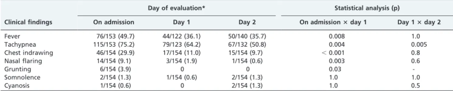
Table 1 shows an assessment of the frequency of overall clinical findings during the first 2 days of penicillin G treatment. Nobody presented seizure. Oxygen was pro-vided during the evolution to 16 (11.1%) patients of 144 admitted without initial oxygen supplement requirement. Penicillin G was substituted by other antibiotics in 28 (18.2%) patients, among whom the median (25th–75th percentile) duration of penicillin G administration was 3.5 (2-4) days (mean 4¡2). The subsequent antimicrobial agents were oxacillin plus ceftriaxone (n = 11), ceftriaxone (n = 11), erythromycin and oxacillin (n = 3 each). Patients treated with other antimicrobial agents stayed longer in the hospital than those treated solely with penicillin G (16¡6 vs. 8¡4 days, p,0.001, mean difference (95% confidence interval): 8 (6-10]). Oxygen supplement during evolution was more common among patients in whom substitution for penicillin G occurred (26.9% vs. 7.6%, p = 0.01).
12.7%, p,0.001) and nasal flaring (10.2% vs. 1.6%, p = 0.001). Differences were also found in the frequency of fever between the second and third day (26.8% vs. 13.7%, p = 0.001) and between the fourth and fifth day (17.8% vs. 10.3%, p = 0.006). Figures 2A and 2B present the daily evolution of clinical findings in the groups of patients without and with penicillin G substitution after 48 h of treatment. Antibiotic change was not associated with pleural effusion (25.0% vs. 15.8%, p = 0.2), severe (19.6% vs. 17.6%, p = 0.8) or very severe (15.8% vs. 18.5%, p = 1) CAP according to WHO, severe CAP according to BTS (18.7% vs. 17.0%, p = 0.8), severe malnutrition (16.7% vs. 18.2%, p = 1) or age (38¡29 vs. 30¡24 months, p = 0.2).
DISCUSSION
From the aforementioned data it can be seen that aqueous penicillin G successfully treated the great majority (82%) of
the studied hospitalized children aged $2 months with radiographically confirmed CAP. This result is in accor-dance with the expected therapeutic success rate (80%).15
addition to deterioration, no improvement after 48 h of therapy was used as the definition of clinical failure.18 By applying such a definition to the patients in this investiga-tion, the therapeutic failure rate of penicillin G would be much greater since on the fourth day of treatment over 40% presented tachypnea and around 20% presented fever (Figure 2A). Moreover, on the sixth day of treatment, over 30% still presented tachypnea (Figure 2A).
We found an association between the requirement for an oxygen supplement during treatment and penicillin G substitution. Therefore, the concept of deterioration was used by the pediatricians managing the studied patients. However, the concept of no improvement in the first 2 days of treatment was not used. By comparing the graphic evolution of children in whom penicillin G had been changed with the group receiving solely penicillin G
Table 1 -Assessment of the frequency of clinical findings during the first 2 days of penicillin G treatment among children hospitalized with community-acquired pneumonia.
Day of evaluation* Statistical analysis (p)
Clinical findings On admission Day 1 Day 2 On admission3day 1 Day 13day 2
Fever 76/153 (49.7) 44/122 (36.1) 50/140 (35.7) 0.008 1.0
Tachypnea 115/153 (75.2) 79/123 (64.2) 67/132 (50.8) 0.004 0.005
Chest indrawing 46/154 (29.9) 17/154 (11.0) 15/154 (9.7) ,0.001 0.8
Nasal flaring 14/154 (9.1) 3/154 (1.9) 1/154 (0.6) 0.003 0.6
Grunting 6/154 (3.9) 0 0 0.03
-Somnolence 2/154 (1.3) 1/154 (0.6) 2/154 (1.3) 1.0 1.0
Cyanosis 1/154 (0.6) 0 2/154 (1.3) 1.0 0.5
*Results are shown as n/N (%).
(Figures 2A and B), it can be seen that fever and tachypnea were steadily present during the studied period in the former group. Despite the statistical significance found for the decrease of tachypnea from the first to the second day of treatment in the former group, the increased frequency of tachypnea on the third day of treatment is in accordance with the observation of a marked absence of improvement among those patients.
The absence of an association between change of penicillin G and baseline aspects of the patient, such as age, severe or very severe CAP or severe malnutrition, was noteworthy. Severity has been identified as a risk factor for treatment failure among adults with CAP.19 Nonetheless, this was not found in this study. Moreover, pleural effusion was not associated with change of penicillin G. The main cause of bacterial CAP,S pneumoniae, has been identified as the most common bacterium among children with CAP and pleural effusion.20As a retrospective survey, this investiga-tion was influenced by the way in which the pediatrician handled the patients. Penicillin G has been widely used in the setting in which this survey was carried out because of the previous use of an investigational protocol on the association of pneumococcal resistance in vitro with peni-cillin G failurein vivo(the CARIBE Study).21Although the data for the latter protocol were collected between 1998 and 2000, data collection for our investigation included children hospitalized from 2002 to 2005. As the CARIBE Study concluded that there is no association between penicillin G therapeutic failure and pneumococcal minimal inhibitory concentration up to 4mg/mL,22 pediatricians are probably
confident about the success of penicillin G. By studying the pharmacokinetics of 200,000 IU/kg/day of penicillin G in children with CAP, serum penicillin G concentrations were .4 mg/mL for .40% of the interdose interval,23 which predicts therapeutic success in treating pulmonary infec-tions caused by pneumococcal strains with minimal inhibitory concentration up to 4mg/mL.24 In Brazil, up to
the present, the highest minimal inhibitory concentration described for pneumococcal strains is 4 mg/mL, which is
rare.25
Several methodological limitations should be recognized in this study: first, as data were collected retrospectively, there was no control on the measurement of the variables; second, as the patients were evaluated by different observers, standardization of the evaluations cannot be guaranteed; third, no attempt was made to determine the etiology and the causative agents of CAP were not known. Nevertheless, strict enrolment criteria were used, assuring that each included case had CAP defined by the ‘‘gold standard’’ parameter—chest x-ray diagnosis. Moreover, by taking into account that 26% of the cases presented pleural effusion and 98.4% of the pulmonary infiltrates were described as alveolar (95.1% as alveolar and 3.3% as interstitial–alveolar), the assumption was that the majority of the cases had a bacterial etiology.11As the patients were hospitalized in a teaching hospital where several research projects on CAP have been conducted during the past 13 years,26 it is highly probable that standardized measures were used. In daily practice, an antibiotic is chosen empirically to treat children with CAP and etiology is rarely established.17Therefore, the results presented herein may be generalized to similar situations.
In conclusion, penicillin G is highly effective in treating children hospitalized with CAP. Since patients’ disease does
not deteriorate during treatment, observation may continue for more than 48 h to document clinical improvement.
ACKNOWLEDGMENTS
The authors thank the medical chart unit of the Professor Hosannah de Oliveira Pediatric Center, Federal University of Bahia, Salvador, Brazil for its cooperation in reviewing the medical charts. Professor Nascimento-Carvalho is investigator of the Brazilian Council for Science and Technology Development (CNPq).
REFERENCES
1. Mulholland K. Childhood pneumonia mortality – a permanent global emergency. Lancet. 2007;370:285-9, doi: 10.1016/S0140-6736(07)61130-1. 2. Farha T, Thomson AH. The burden of pneumonia in children in the
developed world. Paediatr Respir Rev. 2005;6:76-82, doi: 10.1016/j.prrv. 2005.03.001.
3. Sazawal S, Black RE. Effect of pneumonia case management on mortality in neonates, infants, and preschool children: a meta-analysis of community-based trials. Lancet Infect Dis. 2003;3:547-56, doi: 10.1016/ S1473-3099(03)00737-0.
4. Dowell SF, Kupronis BA, Zell ER, Shay DK. Mortality from pneumonia in children in the United States, 1939 through 1996. N Engl J Med. 2000;342:1399-407, doi: 10.1056/NEJM200005113421904.
5. World Health Organization. ARI in children: case management in small hospitals in developing countries. A manual for doctors and other senior health workers. Programme for the Control of ARI. Geneva: WHO, 1990. 6. Addo-Yobo E, Chisaka N, Hassan M, Hibberd P, Lozano JM, Jeena P, et al. A randomized multicentre equivalency study of oral amoxicillin versus injectable penicillin in children aged 3 to 59 months with severe pneumonia. Lancet. 2004;364:1141-8, doi: 10.1016/S0140-6736(04)17100-6. 7. Hale KA, Isaacs D. Antibiotics in childhood pneumonia. Paediatr Respir
Rev. 2006;7:145-51, doi: 10.1016/j.prrv.2006.03.011.
8. Plouffe JF, Martin DR. Re-evaluation of the therapy of severe pneumonia caused by Streptococcus pneumoniae. Infect Dis Clin North Am. 2004;18:963-74, doi: 10.1016/j.idc.2004.07.010.
9. Jadavji T, Law B, Lebel MH, Kennedy WA, Gold R, Wang EE. A practical guide for the diagnosis and treatment of pediatric pneumonia. Can Med Assoc J. 1997;156(suppl):S703-11.
10. British Thoracic Society of Standards of Care Committee. BTS guideline for the management of community acquired pneumonia in children. Thorax. 2002;57:i1-24.
11. Cherian T, Mulholland EK, Carlin JB, Ostensen H, Amin R, de Campo M, et al. Standardized interpretation of paediatric chest radiographs for the diagnosis of pneumonia in epidemiological studies. Bull World Health Organ. 2005;83:353-9.
12. El-Radhi AS, Barry W. Thermometry in paediatric practice. Arch Dis Child. 2006;91:351-6, doi: 10.1136/adc.2005.088831.
13. World Health Organization. Integrated Management of Child-hood Illness chart booklet. (WC 503.2). Geneva: WHO, 2008. [WHO website]. Available at: http://whqlibdoc.who.int/publications/2008/ 9789241597289_eng.pdf (accessed 15 January 2009).
14. World Health Organization. Training Course on Child Growth Asses-sment. Geneva: WHO, 2008. [WHO website]. Available at: http:// whqlibdoc.who.int/publications/2008/9789241595070_A_eng.pdf (accessed 13 July 2009).
15. Ayieko P, English M. Case management of childdood pneumonia in developing countries. Pediatr Infect Dis J. 2007;26:432-40, doi: 10.1097/ 01.inf.0000260107.79355.7d.
16. Juve´n T, Mertsola J, Waris M, Leinonen M, Ruuskanen O. Clinical response to antibiotic therapy for community-acquired pneumonia. Eur J Pediatr. 2004;163:140-4, doi: 10.1007/s00431-003-1397-2.
17. Korppi M. Community-acquired pneumonia in children: issues in optimizing antibacterial treatment. Paediatr Drugs. 2003;5:821-32, doi: 10.2165/00148581-200305120-00005.
18. Straus WL, Qazi SA, Kundi Z, Nomani NK, Schwartz B. Antimicrobial resistance and clinical effectiveness of co-trimoxazole versus amoxicillin for pneumonia among children in Pakistan: randomized controlled trial. Pakistan Co-trimoxazole Study Group. Lancet. 1998;352:270-4. 19. Menendez R, Torres A. Treatment failure in community-acquired
pneumonia. Chest. 2007;132:1348-55, doi: 10.1378/chest.06-1995. 20. Nascimento-Carvalho CM, Oliveira JR, Cardoso MR, Arau´jo-Neto CA,
Barral A, Saukkoriipi A, et al. Pleural fluid and viral infections among children hospitalized with community-acquired pneumonia [Abstract A-181-0010-00431]. In: 6th World Congress of the World Society for Pediatric Infectious Diseases, Buenos Aires, Argentina, 18–22 November 2009. Buenos Aires: World Society for Pediatric Infectious Diseases; 2009.
and risk of treatment failure in pneumonia. Arch Dis Child. 2008;93:221-5, doi: 10.1136/adc.2006.111625.
22. Nascimento-Carvalho CM, Cardoso MR, Brandileone MC, Ferrero F, Camargos P, Berezin E, et al. Penicillin/ampicillin efficacy among children with severe pneumonia due to penicillin-resistant pneumococcus (MIC = 4mg/ml). J Med Microbiol. 2009;58:1390-2, doi: 10.1099/jmm.0.007765-0.
23. Giachetto G, Pirez MC, Nanni L, Martı´nez A, Montano A, Algorta G, et al. Ampicillin and penicillin concentration in serum and pleural fluid of hospitalized children with community-acquired pneumonia. Pediatr Infect Dis J. 2004;23:625-9, doi: 10.1097/01.inf.0000128783.11218.c9.
24. Nascimento-Carvalho CM, Ferrero F, Cardoso MR. New breakpoints to define resistance to penicillin among pneumococcal pneumonia strains [e-letter]. J Clin Invest. 2008;118:1291-300, doi: 10.1172/JCI33947. 25. Wolkers PC, Mantese OC, Paula A, Almeida VV, Aguiar PA, Alvares JR,
et al. New susceptibility breakpoints in antimicrobial resistance rates of invasive pneumococcal strains. J Pediatr (Rio J). 2009;85:421-5, doi: 10. 2223/JPED.1931.