Case Report
Relato de Caso
Retrocochlear impairments in systemic
sclerosis: a case report study
Alterações retrococleares na esclerose
sistêmica: relato de casos
Julia de Souza Pinto Valente1 Ana Paula Corona2
Keywords
Systemic Sclerosis Retrocochlear Disease Auditory Neuropathy Audiology BAEP
Descritores
Esclerose Sistêmica Doença Retrococlear Neuropatia Auditiva Audiologia PEATE
Correspondence address: Julia de Souza Pinto Valente Rua Pará, 466, ap. 302, Edf. Suzana, Pituba, Salvador (BA), Brazil, CEP: 41830-070.
E-mail: [email protected]
Received: December 09, 2016 Accepted: August 07, 2017
Study carried out at Instituto de Ciências da Saúde, Universidade Federal da Bahia – UFBA - Salvador (BA), Brazil.
1 Programa de Pós-graduação Processos Interativos dos Órgãos e Sistemas, Universidade Federal da Bahia –
UFBA - Salvador (BA), Brazil.
2 Departamento de Fonoaudiologia, Instituto de Ciências da Saúde, Universidade Federal da Bahia – UFBA -
Salvador (BA), Brazil.
Conlict of interests: nothing to declare. Financial support: nothing to declare.
ABSTRACT
Purpose: To report three cases of patients with Systemic Sclerosis (SSc) and retrocochlear impairments. Methods: This is a case report of three individuals with SSc and retrocochlear impairments assisted at a rheumatology outpatient clinic. All individuals underwent Brainstem Auditory Evoked Potential (BAEP) and, when necessary, audiometry. Results: All three individuals presented sensorineural hearing loss. Although no
retrocochlear impairment was identiied in the basic audiologic evaluation, the BAEP results were altered.
Conclusion: Retrocochlear impairments were present in the individuals under study, both in the absolute latencies and interpeak interval, thereby demanding the attention of rheumatologists and speech-language pathologists to such changes during the monitoring of SSc patients. The results also show a need for epidemiological studies on the theme.
RESUMO
Objetivo: Relatar três casos de pacientes portadores de Esclerose Sistêmica e que apresentaram alterações retrococleares. Método: Trata-se de um estudo de relato de casos de três indivíduos com esclerose sistêmica e alteração retrococlear, acompanhados em um serviço de reumatologia. Todos os pacientes realizaram o Potencial Evocado Auditivo de Tronco Encefálico e, quando necessário, nova audiometria. Resultado: Todos os indivíduos
apresentaram perda auditiva do tipo sensorioneural. Não foi identiicado na avaliação audiológica básica qualquer
INTRODUCTION
Systemic sclerosis (SSc) is a rare, multisystem disease
characterized by ibrosis in the organs, mainly on the skin, and
vasculopathy with Raynaud’s phenomenon. This disease of unknown etiology occurs in several ethnical groups worldwide, with estimated annual incidence of 19:1,000,000 inhabitants. There are no epidemiological data available on this disease in Brazil to date. It affects three times more women than men, and the mean age of disease onset is between 30 and 50 years(1-3).
SSc patients have been classiied into two categories
according to extent of skin involvement: limited cutaneous systemic sclerosis (lcSSc), which presents skin changes in the face and distal regions of the knees and elbows; diffuse cutaneous systemic sclerosis (dcSSc), which is characterized by
truncal and acral skin ibrosis in the torso and limbs, involving
the abdomen, thighs, face, and thorax(1-3).
SSc is an incurable autoimmune disease whose chronic symptoms become progressively worse. In addition to the limitations resulting from the disease, individuals with SSc
may present communication dificulties owing to the presence
of auditory impairments. Auditory complaints, such as tinnitus and hypoacusis, are frequent in patients with both types of SSc, and these symptoms may be associated with possible peripheral and retrocochlear auditory impairment(4-6).
Previous studies have reported the existence of inner ear involvement in SSc patients, probably as a result of vascular damage, considering that the cochlea is highly sensitive to these changes. Blood changes and hypoxia in the cochlea resulting from SSc culminate with death of the ciliated cells (cochlear sensory structure), which is clinically expressed as hypoacusis and tinnitus(3-8). This mechanism of hypoxia in
SSc is associated with activation of the endothelial cells by unknown factors, which promotes chronic endothelial injury
with platelet adhesion and activation of the ibrinolytic system,
thus generating increased vascular permeability and leukocyte
adhesion to blood vessel wall. This process generates ibrosis
and loss of elasticity, reducing the vascular lumen and causing progressive hypoxia and necrosis of the cochlear tissue(9).
Studies conducted to identify changes in the auditory system in SSc patients show that prevalence of auditory loss in these individuals varies considerably, but sensorineural hearing loss is the most frequently observed in these investigations. It is known that sensorineural changes can be cochlear and/or retrocochlear; however, only one case report study investigating the presence of retrocochlear impairment in SSc patients was
found in the speciic scientiic literature. Knowledge about the
possible retrocochlear impairments in this population may help speech-language therapists in the correct auditory rehabilitation of these patients, considering that each type of hearing impairment
requires a speciic auditory rehabilitation, thus providing adequate
treatment and, consequently, improvement in the quality of life of these individuals.
Under this perspective, this study aims to report three cases of patients with SSc and retrocochlear impairments.
METHODS
The cases herein reported refer to patients assisted at the rheumatology outpatient clinic of Ambulatório Magalhães Neto at the Universidade Federal da Bahia (UFBA). Study participants underwent audiological assessment through pure tone, speech and immittance audiometry and brainstem auditory evoked potential (BAEP) between September 2015 and July 2016.
This study was approved by the Research Ethics Committee of the Instituto de Ciências da Saúde under process no. 1.282.417. All participants signed an Informed Consent Form (ICF) prior to study commencement.
Initially, the patients were invited to participate in a broader research, entitled “Retrocochlear Changes in Systemic Sclerosis”, and those who agreed signed the ICF. Next, data of the basic audiological assessment were collected from those who presented valid audiometry in the medical record (performed less than 6 months before, with no new auditory complaints). After that, the selected individuals were instructed to attend the Speech-Language Pathology Teaching Assistance Center - CEDAF, according to individual schedule, for completion of anamnesis and examinations.
Individuals who presented valid basic audiological evaluation were submitted only to inspection of the auditory meatus and the BAEP assessment, whereas those whose medical records did not present audiometry conducted before 6 months or less in relation to the date of the BAEP underwent external inspection of the auditory meatus, pure tone threshold audiometry, speech audiometry, and the BAEP.
A MASBE Contronic manufactured device, properly calibrated in accordance with the ANSI S3.6-1996(10), ISO 8798(11),
ANSI S3.43-1992(12) norms, was used to conduct the BAEP.
The equipment was also biologically calibrated. To this end, the BAEP was conducted with 20 patients with normal
hearing (40 ears), 10 men and 10 women, both groups with ive individuals aged less than 40 years and ive individuals over
this age. In the calculation of normality patterns, a standard deviation of ±2.5 errors was considered for each variable in
order to obtain a 95% conidence interval(13).
The electrodes were placed according to the following pattern: the negative electrode at the vertex, the earth electrode on the side of the forehead, and the negative electrodes in the regions of the left and right mastoid muscles(9,13). Initial acoustic stimulus
intensity of 80 dB nNA was used, but it was increased in 10 dB nNA when needed, as to adapt to patients with hearing loss(9).
Regarding the other parameters of the examination, the
manufacturer’s suggestions were followed: rareied polarity;
acoustic stimulus generated through clicks; presentation rate
of 17.1 stimuli per sec; low-pass ilters of 5000 Hz and high-pass ilters of 1000 Hz, bilaterally.
Absolute latency values of waves were identiied by two evaluators. The values found from this identiication were recorded in a protocol and classiied as normal and altered
difference of the I-V interpeak interval, according to the biological calibration and audiometry of the patients(13-16). The
BAEP was considered altered when the values were greater than those obtained in the biological calibration, in relation to the analyzed patterns, on at least one side, or when they presented interaural intensity difference >0.3(13,15).
RESULTS
Case 1
This was a 78-year-old, female patient with a diagnosis of SSc for 12 years. She presented frequent, bilateral, high-pitched tonal tinnitus and mild, sporadic dizziness; both symptoms started six years after the onset of the disease.
In addition to SSc, the patient reported having hypertension
and gastroesophageal relux, and using medication to control them.
Pure tone audiometry showed sensorineural hearing loss as
of 3 and 4 kHz at the right and left ears, respectively. Speech
audiometry showed speech recognition threshold (SRT) compatible with the tritonal mean and normal speech recognition index (SRI) at both ears. Results of the tuning fork tests were also compatible with those of pure tone audiometry, with Weber indifferent and Rinne positive bilaterally.
Immittance audiometry showed type A tympanograms for both ears and presence of all contralateral stapedius muscle
acoustic relexes with differences within normality patterns. Acoustic relex threshold decay was negative at 500 and 1000 Hz
bilaterally.
The BAEP assessment showed normal absolute values of waves I, III, and V and normal I-III, III-V and I-V interpeak intervals for the right ear. As for the left ear, all these parameters were showed normal results, except for the absolute value of wave I, which presented a reception delay of 2.18 ms. Interaural attenuation values of wave V and I-V interpeak interval were adequate.
Case 2
This was a 62-year-old, male patient with a diagnosis of SSc
for seven years. He presented frequent, bilateral, high-pitched
tonal tinnitus and mild, sporadic vertigo. The otoneurological complaints started at 58 years of age, that is, three years after the onset of the disease.
In addition to SSc, the patient reported having hypertension
and gastroesophageal relux, and using medication to control them.
Pure tone audiometry showed sensorineural hearing loss as
of 6 and 8 kHz at the right and left ears, respectively. Speech
audiometry showed SRT compatible with the tritonal mean and normal SRI at both ears. Results of the tuning fork tests were also compatible with those of pure tone audiometry, with Weber indifferent and Rinne positive bilaterally.
Immittance audiometry showed type A tympanograms for both ears and presence of all contralateral stapedius muscle
acoustic relexes with differences within normality patterns.
Acoustic relex threshold decay was negative at 500 and 1000 Hz
bilaterally.
The BAEP assessment showed normal absolute values of waves I, III, and V and normal I-III, III-V and I-V interpeak intervals for the right ear. As for the left ear, all these parameters showed normal results, except for the absolute value of wave V and the III-V interpeak interval, which presented reception delays of 6.24 and 2.32 ms, respectively. Interaural attenuation values of wave V and I-V interpeak interval were adequate.
Case 3
This was a 51-year-old, female patient with a diagnosis of SSc for 11 years. She presented complaints about hearing loss in several situations of her daily life; bilateral, high-pitched tonal tinnitus; and mild, sporadic dizziness. The auditory complaints began one year after the medical diagnosis of SSc.
In addition to SSc, the patient reported having hypertension and using medication to control it. She also reported regular use
of medication to treat gastroesophageal relux and, sporadically,
of corticoids as a prophylactic treatment for SSc.
Pure tone audiometry showed unilateral sensorineural
hearing loss as of 3 kHz at the right ear. Speech audiometry
showed SRT compatible with the tritonal mean and normal SRI bilaterally. Results of the tuning fork tests were compatible with those of pure tone audiometry, with Weber indifferent and Rinne positive bilaterally.
A in the previous cases, immittance audiometry showed type A tympanograms for both ears and presence of all contralateral
stapedius muscle acoustic relexes with differences within normality patterns. Acoustic relex threshold decay was negative at 500 and 1000 Hz bilaterally.
The BAEP assessment showed normal absolute values of waves I, III, and V and normal I-III, III-V and I-V interpeak intervals with value of 2.27 ms for the right ear. As for the left ear, all these parameters presented normal results. Interaural attenuation values of wave V and I-V interpeak interval were adequate.
Because the patient presented unilateral hearing loss and
altered BAEP indings, she was referred to otorhinolaryngology
with the results of the examinations for investigation aiming to discard a diagnosis of tumor in the cranial nerve VII.
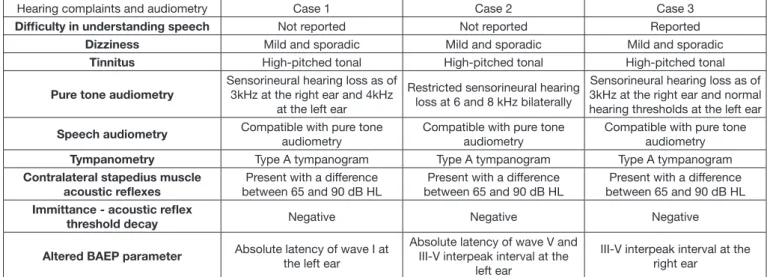
Chart 1 shows a summary of the indings of the audiological assessments conducted, where it is possible to observe that all study particpants presented sensorineural hearing loss. In the basic audiological evaluation, no results suggestive of retrocochlear
impairment were identiied, but the BAEP showed altered results.
Figures 1 and 2 depict the hearing thresholds with air
conduction obtained at frequencies ranging from 250 to 8000 Hz
for each Case on the right and left sides. When there was a need to measure the thresholds with bone conduction, the three Cases presented them coupled with air conduction.
Chart 1. Characterization of hearing complaints and audiometry of patients with altered BAEP results
Hearing complaints and audiometry Case 1 Case 2 Case 3
Difficulty in understanding speech Not reported Not reported Reported
Dizziness Mild and sporadic Mild and sporadic Mild and sporadic
Tinnitus High-pitched tonal High-pitched tonal High-pitched tonal
Pure tone audiometry
Sensorineural hearing loss as of 3kHz at the right ear and 4kHz
at the left ear
Restricted sensorineural hearing loss at 6 and 8 kHz bilaterally
Sensorineural hearing loss as of 3kHz at the right ear and normal hearing thresholds at the left ear
Speech audiometry Compatible with pure tone
audiometry
Compatible with pure tone audiometry
Compatible with pure tone audiometry
Tympanometry Type A tympanogram Type A tympanogram Type A tympanogram
Contralateral stapedius muscle acoustic reflexes
Present with a difference between 65 and 90 dB HL
Present with a difference between 65 and 90 dB HL
Present with a difference between 65 and 90 dB HL
Immittance - acoustic reflex
threshold decay Negative Negative Negative
Altered BAEP parameter Absolute latency of wave I at
the left ear
Absolute latency of wave V and III-V interpeak interval at the
left ear
III-V interpeak interval at the right ear
Caption: BAEP = Brainstem Auditory Evoked Potential
Figure 1. Right ear hearing thresholds with air conduction
Figure 2. Left ear hearing thresholds with air conduction Caption: BAEP = Brainstem Auditory Evoked Potential. *Altered results
Figure 3. BAEP parameters at the right ear
Caption: BAEP = Brainstem Auditory Evoked Potential. *Altered results
Figure 4. BAEP parameters at the left ear
DISCUSSION
Observation of the auditory complaints of the three patients investigated evidenced that dizziness and high-pitched tinnitus are present in all cases. The literature describes both symptoms as frequent in neuropathic patients and warning signs for possible retrocochlear impairments, considering that they are present in the main pathologies affecting the vestibulocochlear nerve(3,16-18).
Another point observed in the individuals analyzed is the fact that two of them are over 60 years old. There is still
controversy about the inluence of aging on the parameters of
the BAEP, and from what age it would occur(18,19). The studies
that consider the possibility of this inluence report that it occurs
mainly in the absolute latency of wave V(14,20) - a delay that was
occur in any absolute latency, with preservation of the interpeak intervals - characteristics also observed in one of the participants of this study(20,21). This increase in latency suggested by some
authors, either only in wave V or in all absolute latencies, would occur as a consequence of degeneration of the auditory pathway until the brainstem caused by the aging process. It is possible to observe delay in the synaptic transition, loss of neurons, change in neuronal membrane permeability, and loss of myelin sheath(14).
Another controversial point in the BAEP is the inluence of
gender in the parameters of this assessment. Some studies did
not ind signiicant differences between genders(16,18); however,
other authors have stated that the male gender presents higher latency of wave V and I-V interpeak interval compared with those of the female gender(21,22). In the present study, the patient
who presented impairment in the latency of wave V was male.
However, it is worth noting that biological calibration was
performed with half of the male patients, thus considering the possible differences between the genders.
In this scenario, it should be emphasized that it is not possible
to estimate the inluence of gender and age on the BAEP results,
not being possible to attribute the impairments found only to SSc. Nevertheless, it is important to stress that, in all of the cases, the auditory complaints started when the individuals had already been diagnosed with SSc.
Of the altered results found, two presented changes in absolute latency (waves I and V) and one in interpeak interval (III-V). Some authors suggest that interpeak intervals would be more
eficient in identifying retrocochlear pathologies(22). Changes in
absolute latency would be associated with impairments of the structures, with wave I associated with the distal portion of the auditory nerve in relation to the brainstem and wave V associated with the lateral lemniscus(23).
With respect to retrocochlear impairments in SSc, although there are no studies investigating the prevalence of this impairment using the BAEP, there is a case report study conducted in 2014(17)
which presents a patient with impairment in the cranial nerve VIII and emphasizes that the diagnosis was made through image examination. Most of the studies that addressed cranial nerve neuropathy described the trigeminal nerve as the most affected nerve. The pathophysiology of nerve injury is still not fully known, as opposed to peripheral injury, but the main explanation for trigeminal nerve injury is lack of nutrition due to poor blood supply as a consequence of vasculopathy(5,14). Therefore, it is
possible that the same pathophysiological process occurs with retrocochlear impairments in this population.
As previously mentioned, there are no studies on retrocochlear
impairments in SSc in the literature. However, studies conducted
with patients with systemic lupus erythematosus - another rheumatologic disease in which vasculopathy may occur, reported 6.7% of patients with altered BAEP results. Systemic lupus erythematosus presents secondary vasculopathy, less
important than that observed in SSc, but suficient to lead to retrocochlear impairments, suggesting that the signiicant
vascular impairment characteristic of SSc can lead to auditory neuropathy in patients(23,24).
CONCLUSION
The indings of the present study reveal occurrence of
retrocochlear impairments in the population investigated. In Brazil, there are no epidemiological data available on this disease to date, nor any studies assessing retrocochlear impairments. Therefore, epidemiological studies on this theme are needed
to ill this gap in knowledge.
Moreover, these results serve as a warning for rheumatologists and speech-language pathologists in their monitoring of SSc patients on the need to conducted Brainstem Auditory Evoked Potentials (BAEP) especially in individuals with hearing complaints, considering that retrocochlear impairments cause
signiicant impairment in communication and, for the most part,
present treatment different from that of other auditory changes.
ACKNOWLEDGEMENTS
The authors are grateful to the Speech-language Pathology Department of Universidade Federal da Bahia and the rheumatology outpatient clinic of Ambulatório Magalhães Neto of Universidade Federal da Bahia for all the support received during the research period, and to the study participants for their availability in this project.
REFERENCES
1. Samara AM. Esclerose sistêmica. Rev Bras Reumatol. 2004;44(1):9-10. http://dx.doi.org/10.1590/S0482-50042004000100001.
2. Amor-Dorado JC, Arias-Nuñez MC, Miranda-Filloy JA, Gonzalez-Juanatey C, Llorca J, Gonzalez-Gay MA. Audiovestibular manifestations in patients with limited systemic sclerosis and Centromere Protein-B (CENP-B) Antibodies. Medicine. 2008;87(3):131-41. PMid:18520322. http://dx.doi. org/10.1097/MD.0b013e318173aa56.
3. Allanore Y, Simms R, Distler O, Trojanowska M, Pope J, Denton CP, et al. Systemic sclerosis. Nat Rev Dis Primers. 2015;1:15002. PMid:27189141. http://dx.doi.org/10.1038/nrdp.2015.2.
4. Berrettini S, Ferri C, Pitaro N, Bruschini P, Latorraca A, Sellari-Franceschini S, et al. Audiovestibular involvement in systemic sclerosis. ORL J Otorhinolaryngol Relat Spec. 1994;56(4):195-8. PMid:8078672. http:// dx.doi.org/10.1159/000276655.
5. Deroee AF, Huang TC, Morita N, Hojjati M. Sudden hearing loss as the presenting symptom of systemic sclerosis. Otol Neurotol. 2009;30(3):277-9. PMid:19318884. http://dx.doi.org/10.1097/MAO.0b013e31819bda52.
6. Kastanioudakis I, Ziavra N, Politi E, Exarchakos G, Drosos A, Skevas A. Hearing loss in progressive systemic sclerosis patients: A comparative
study. Otolaryngol Head Neck Surg. 2001;124(5):522-5. PMid:11337656. http://dx.doi.org/10.1067/mhn.2001.115092.
7. Maciaszczyk K, Waszczykowska E, Pajor A, Bartkowiak-Dziankowska B, Durko T. Hearing organ disorders in patients with systemic sclerosis. Rheumatol Int. 2011;31(11):1423-8. http://dx.doi.org/10.1007/s00296-010-1503-5.
8. Monteiro T, Christmann R, Bonfá E, Bento R, Novalo-Goto E, Vasconcelos L. Hearing loss in diffuse cutaneous systemic scleroderma. Scand J Rheumatol. 2011;40(6):467-71. PMid:21916804. http://dx.doi.org/10.31 09/03009742.2011.588400.
9. Zimmermann A, Pizzichin MM. Atualização na etiopatogênese da esclerose sistêmica. Rev Bras Reumatol. 2013;53(6):516-24. PMid:24477730. http:// dx.doi.org/10.1016/j.rbr.2013.01.001.
11. ISO: International Organization for Standardization. ISO 8798:1987 - Acoustics Reference levels for narrow-band masking noise. Genebra: ISO; 1987. 12. ANSI: American, National Standards Institute. ANSI S3.43-1992: American
national standard: standard reference zero for the calibration of pure-tone bone-conduction audiometers. New York: ANSI; 1992.
13. Pedriali IVG, Kozlowski L. Infuência da intensidade e velocidade do clique no peate de ouvintes normais. Arq Int Otorrinolaringol. 2006;10(2):105-13. 14. Matas CG, Santos VAV Fa, Okada MMCP, Resque JR. Potenciais evocados auditivos em indivíduos acima de 50 anos de idade. Pró-Fono R Atual Cient. 2006;18(3):277-84. PMid:17180796. http://dx.doi.org/10.1590/ S0104-56872006000300007.
15. Soares IA, Menezes PL, Carnauba ATL, Pereira LD. Padronização do potencial evocado auditivo de tronco encefálico utilizando um novo equipamento. Pró-Fono R Atual Cient. 2010;4(22):421-6.
16. Iskandar SB, Loyd S, Roy TM. Cranial nerve VIII involvement in a patient with progressive systemic sclerosis. Tenn Med J Tenn Med Assoc. 2004;97(3):117-9. PMid:15054944.
17. Teasdall RD, Frayha RA, Shulman LE. Cranial nerve involvement in systemic sclerosis (scleroderma): a report of 10 cases. Medicine. 1980;59(2):149-59. PMid:6244477. http://dx.doi.org/10.1097/00005792-198003000-00006. 18. Assis CL, Souza FCR, Baraky LR, Azevedo Bernardi AP. Estudo da
audiometria de tronco encefálico em indivíduos de 20 a 30 anos com audição normal. Rev CEFAC. 2005;7(1):87-92.
19. Kaewsir SI, Waseenon W, Navacharoen N, Panyathong P, Phuackchantuc R. Correlation between age and gender, and parameters of auditory brainstem evoked response. Chiang Mai Med J. 2015;54(4):163-9.
20. Munhoz ASL, Silva MLG, Caovilla HH, Frazza MM, Ganança MG, Câmera JLS. Respostas auditivas de tronco encefálico. In Munhoz MSL, Caovilla
HH, Silva MLG, Ganança MM. Audiologia clínica. São Paulo: Atheneu;
2003. p. 191-220.
21. Esteves MCBN, Dell’Aringa AHB, Arruda GV, Dell’Aringa AR, Nardi JC. Brainstem evoked response audiometry in normal hearing subjects. Braz J Otorhinolaryngol Impresso. 2009;75(3):420-5. PMid:19649494. http:// dx.doi.org/10.1590/S1808-86942009000300018.
22. El Hassan S. Da influência do sexo, da intensidade do estímulo e do perímetro cefálico nas latências da audiometria de tronco encefálico [Internet] 1997 [citado em 2016 Ago 2]. Disponível em: http://repositorio.unifesp.br/ handle/11600/15252
23. Lima MAMT. Potencial evocado auditivo-eletrococleografia e audiometria de tronco encefálico. In: Frota S, organizador. Fundamentos em fonoaudiologia
audiologia. 2. ed. Rio de Janeiro: Guanabara Koogan; 2003. p. 157-72. 24. Klumb EM, Silva CAA, Lanna CCD, Sato EI, Borba EF, Brenol JCT, et al.
Consenso da sociedade brasileira de reumatologia para o diagnóstico, manejo e tratamento da nefrite lúpica. Rev Bras Reumatol. 2015;55(1):1-21. PMid:25595733. http://dx.doi.org/10.1016/j.rbr.2014.09.008.
Author contributions