Ricardo Machado Cruz1
Treatment of a Class III growing patient with mandibular
prognathism and severe anterior crossbite*
The treatment of growing patients with Class III skeletal pattern represents one of the greatest clinical chal-lenges for the orthodontist. Several treatment protocols have been proposed, almost all involving rapid maxil-lary expansion and maxilmaxil-lary protraction. However, there are cases where the maxilla is properly positioned in the anteroposterior direction and there is no transverse discrepancy, featuring only a mandibular prognathism. In such cases, when there is a set of favorable factors such as lack of laterognathism and lower mandibular plane angle, a viable option and which could prove quite interesting is the use of orthodontic chin cup during the night, aiming at trying to redirect the forward growth of the mandible. To have success, it is necessary that this proce-dure involves pubertal growth spurt and is extended to full skeletal maturation. This case was presented to the board of the Brazilian Board of Orthodontics and Dentofacial Orthopedics (BBO) as part of the requirements to become a BBO Diplomate.
Keywords: Class III. Chin cup. Anterior crossbite. Mandibular prognathism.
How to cite this article: Cruz RM. Treatment of a Class III growing patient with mandibular prognathism and severe anterior crossbite. Dental Press J Orthod. 2012 July-Aug;17(4):148-59.
Submitted: May 28, 2012 - Revised and accepted: July 4, 2012
» The author reports no commercial, proprietary or financial interest in the products or companies described in this article.
» Patients displayed in this article previously approved the use of their facial and in-traoral photographs.
Contact address: Ricardo Machado Cruz
Rua Shis, Q19/11 bloco L, sala 101 – Lago Sul, Brasília/DF – Brazil CEP: 71.625-210 – E-mail: [email protected] * Case report approved by the Brazilian Board of Orthodontics and Dentofacial
Orthopedics (BBO).
1 PhD in Animal Biology / Genetics, UNB. MSc in Orthodontics, UFRJ. Professor
of Orthodontics, UNIP, Brasília campus. President of the Brazilian Association of Orthodontics and Dentofacial Orthopedics (ABOR). President of the College of Graduates BBO. Diplomate Brazilian Board of Orthodontics and Dentofacial Orthopedics (BBO).
INTRODUCTION
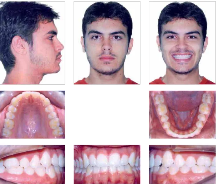
Caucasian male patient, 13 years old, in full pubertal growth spurt, with a chief complaint of anterior cross-bite and very unsatisfied with his smile appearance.
Ricardo Machado Cruz1
Tratamento de paciente Classe III em crescimento com
prognatismo mandibular e severa mordida cruzada anterior*
O tratamento de pacientes em crescimento com padrão esquelético de Classe III representa um dos maiores de-safios clínicos para o ortodontista. Vários protocolos de tratamento têm sido propostos, quase todos envolvendo expansão rápida da maxila e protração maxilar. Porém, existem casos onde a maxila está corretamente posicionada no sentido anteroposterior e não há discrepância transversa, caracterizando apenas um prognatismo mandibular. Nesses casos, quando há um conjunto de fatores favoráveis, tais como ausência de laterognatismo e menor ângulo do plano mandibular, uma opção viável e que pode se mostrar bastante interessante é o uso noturno da mentoneira, com o objetivo de tentar redirecionar o crescimento anterior da mandíbula. Para que haja sucesso, é necessário que esse procedimento envolva o surto de crescimento da puberdade e seja estendido até a completa maturação es-quelética. O presente caso clínico foi apresentado à diretoria do Board Brasileiro de Ortodontia e Ortopedia Facial (BBO), como parte dos requisitos para a obtenção do titulo de Diplomado pelo BBO.
Palavras-chave: Classe III. Mentoneira. Mordida cruzada anterior. Prognatismo mandibular.
Como citar este artigo: Cruz RM. Treatment of a Class III growing patient with mandibular prognathism and severe anterior crossbite. Dental Press J Orthod. 2012 July-Aug;17(4):148-59.
Enviado em: 28 de maio de 2012 - Revisado e aceito: 4 de julho de 2012
» O autor declara não ter interesses associativos, comerciais, de propriedade ou fi-nanceiros que representem conflito de interesse nos produtos e companhias descri-tos nesse artigo.
» O paciente que aparece no presente artigo autorizou previamente a publicação de suas fotografias faciais e intrabucais.
Endereço para correspondência: Ricardo Machado Cruz
Rua Shis, Q19/11 bloco L, sala 101 – Lago Sul, Brasília/DF – CEP: 71.625-210 E-mail: [email protected]
* Relato de caso clínico aprovado pelo Board Brasileiro de Ortodontia e Ortopedia Facial (BBO).
DIAGNOSIS
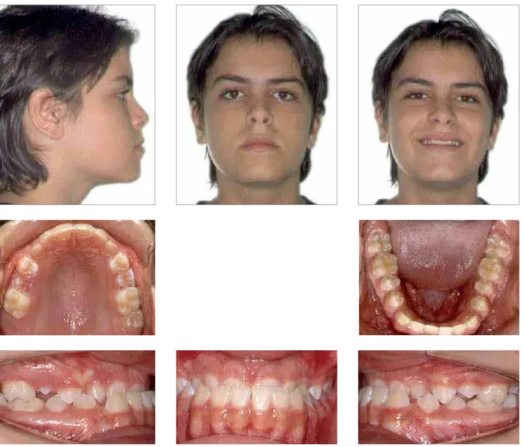
The patient had a slightly concave facial profile, with no obvious asymmetry, with a slight projection of the lower lip and unsatisfactory smile line, exposing mostly the lower anterior teeth and very little of the upper anterior teeth. He presented a good nasolabial angle, although slightly obtuse, and proper maxillary position in the anteroposterior direction (Fig 1).
In intraoral terms, he had a good dental and peri-odontal health, with the exception of the buccal sur-faces of the upper incisors, which had wear facets,
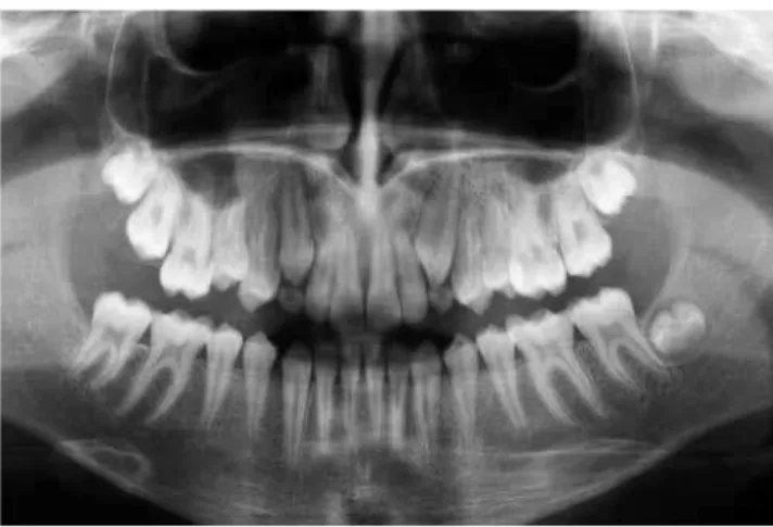
resulting from the dental crossbite. In addition, the maxillary central incisors were in over-eruption. He was in the late mixed dentition, with prolonged re-tention of deciduous canines and with permanent ca-nines successors still included. The second premolars and second molars were erupting. He also presented an Angle Class I malocclusion, with coincident den-tal midlines. He had a few diastemas in the lower arch and severe total crossbite in the anterior region (main complaint). All third molars were still included, but the lower right, absent by agenesis (Figs 1, 2, 3).
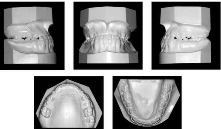
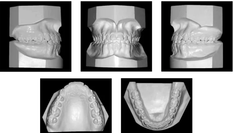
Figure 2 - Initial casts.
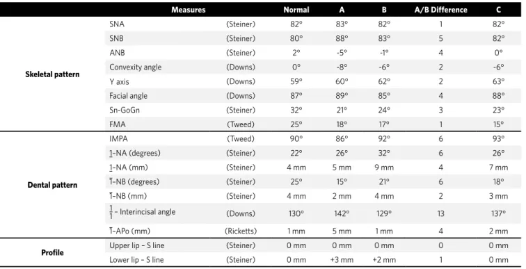
Cephalometrically, the patient had a low man-dibular plane angle and a tendency for predominant horizontal mandibular growth, in the counterclock-wise direction (SNGoGn = 21° and FMA= 18°). He was diagnosed with a Class III skeletal pattern, due to ex-cessive mandibular growth (ANB = -5°, SNA= 83° and SNB = 88°). The upper incisors were slightly buccal (1-NA = 26) and lower were retroclined (1-NB = 15°), seeking a natural dental compensation for the skeletal deficiency (Fig 4, Table 1).
TREATMENT OBJECTIVES
Considering the patient’s main complaint about the total anterior crossbite and the parent’s con-cern about the family history of mandibular prog-nathism, the main treatment goal was to try to re-direct mandibular growth, reducing, if possible, the SNB angle and at the same time, to maintain good maxillary anteroposterior position, because he had a satisfying nasolabial angle.
In the vertical and transverse aspects, the ob-jective was to maintain the dimensions and growth pattern. For this reason, rapid maxillary expan-sion and protraction using a face mask were not indicated. In dental terms, the basic objective was to perform treatment without extractions, since the patient had no arch perimeter deficiency, and there was concern in aligning, leveling and moni-toring the eruption of the teeth which had not erupted, especially the canines. In the incisor re-gion, the objective was to correct their compensa-tory inclinations in both arches, and to close the lower diastema by means of retraction of anterior teeth, correcting the anterior crossbite.
A B
Figure 3 -Initial panoramic and periapical radiographs.
Figure 4 - Initial lateral cephalometric radiograph and cephalometric tracing.
TREATMENT PLAN
A complete corrective Orthodontics a treatment with braces placed on both arches (metal brackets, slot 0.022 x 0.028-in, Straight-Wire system with Roth prescription) was proposed. In the lower arch, an-chorage was planned to allow diastema closure with retraction of the anterior teeth, without simultaneous mesial movement of the posterior teeth. The anchor-age proposal would use tip back bends on the poste-rior teeth associated, if necessary, with Class III elas-tics. The placement of braces would be made, initially, only in the lower arch, due to the impossibility to place orthodontic attachments on the buccal surfaces of upper anterior teeth. Leveling and aligning would be performed in the lower arch, starting from the 0.0175-in coaxial archwire, followed by 0.012-0.0175-in 0.014-0.0175-in and 0.016-in NiTi archwires and 0.016-in, 0.018-in and
Final space closure would be done with rectangular 0.019 x 0.025-in stainless steel archwires with T loops for incisor retraction. At this stage, anterior crossbite correction will be completed. The case would be fin-ished with 0.019 x 0.025-in stainless steel rectangu-lar archwires with individual bends and torques. If necessary, Class III intermaxillary elastics would be used. Throughout treatment, to control mandibu-lar growth, a chin cup with 500 g force was planned, only at night. As retention, in the upper arch, a wrap-around removable type plate would be used, and the lower intercanine fixed retainer, made with coaxial 0.0215-in wire. At the end, extraction of all three un-erupted third molars would be indicated. The patient would have to continue to use the chin cup at night until full skeletal maturation.
TREATMENT PROGRESS
Orthodontic bands were installed on the first four molars, with welded accessories (triple tubes in the upper teeth and double tubes in the lower teeth, all convertible). The chin cup was installed with 500 g of force for night use, and was custom made, after im-pression of the chin. Thanks to patient cooperation with the chin cup use, after aligning and leveling of the upper and lower arches, the bite was already edge to edge in the anterior region. Then, the retraction of the lower canines, and later, of the incisors was per-formed. During the entire space closure phase, pos-terior anchorage was performed by means of tip back bends on the lower molars and wire ligatures in tie-together posteriorly from the distal of the lower ca-nines. At the end of this phase, the patient had normal overjet and overbite. On completion, for the first time, intermaxillary Class III elastics were used in order to improve the intercuspidation, especially in the canine region. Appliance removal occurred as planned. It was recommended that in addition to the removable upper retention plate, chin cup was to be used every night to sleep, until the end of growth, which would be assessed at 18 years of age, by carpal radiography.
At the end of treatment, extraction of third molars was suggested and referral to an expert in Restorative Esthetic Dentistry, to improve the appearance of the buccal surfaces of maxillary incisors — worn after sev-eral years in crossbite — as well as for clinical evalua-tion of demineralizaevalua-tion white lesions.
RESULTS
The main treatment objectives were achieved, with correction of severe anterior crossbite and im-provement of the Class III skeletal pattern. The aes-thetic and functional results were quite satisfactory (Figs 5-8). The treatment time was of approximate-ly three years, and relativeapproximate-ly short for the complex-ity of the case. The decision of not performing rapid maxillary expansion, since there was no transverse deficiency on the perimeter, and maxillary protrac-tion, since it was well positioned in the anteroposte-rior direction, was correct. Cephalometric superim-positions (Fig 13) showed that there was a mandib-ular growth redirection, which was predominantly horizontal, to the vertical direction. Significant reduction of the SNB angle, possibly resulting from mandibular clockwise rotation, which increased the mandibular plane angle from 21° to 24°. These changes were the result of disciplined night use of the chin cup. As a final result the ANB angle went from -5° to -1°, a significant reduction (Tab 1).
The evaluation of periapical and panoramic final radiographs shows good root parallelism and discrete apical rounding of the upper incisors, consistent with the orthodontic movement (Fig 7).
Due to the family history of mandibular progna-thism, the periodic follow-ups were strictly observed in the phase of retention to assess the stability of the occlusion, the end of growth, the development and
eruption of third molars, and a possible relapse due to mandibular residual growth.
New follow up records, 26 months after the end of treatment, when the patient was eighteen years and two months old, was requested.
The occlusal situation remained stable and sat-isfactory, providing functional balance and excur-sive mandibular movements without interference,
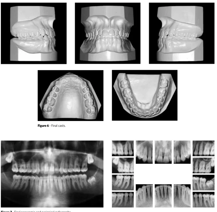
Figure 6 - Final casts.
Figure 7 -Final panoramic and periapical radiographs.
and good health of periodontal tissues (Figs 9, 10). Similarly, esthetic and facial smile remained appro-priate (Fig 9). The comparison of cephalometric mea-surements (Tab 1) showed the improvement of some parameters to values closer to normality, as the SNB angle, which decreased 1°, generating an ANB with 0°; the inclination of upper incisors, which decreased 6° and returned to its value from the beginning of treat-ment without compromising the final occlusion; the inclination of the lower incisors, which reduced 3°,
also approaching the initial value, and the facial pro-file, where there was greater reduction in the projec-tion of the lower lip (Figs 12, 13 and Tab 1).
The removable upper retention was used day and night for two years and intercanine fixed retainer re-mained, with suggestion of indefinite use.
A B
Figure 8 - Final lateral cephalometric radiograph and final cephalometric tracing.
Figure 10 - Two years and two months post-treatment casts.
A
A
B
B
Figure 12 - Two years and two months post-treatment lateral cephalometric radiograph and cepha-lometric tracing.
Figure 13 - Total (A) and partial (B) superimpositions of initial cephalometric tracing (black), final (red) and two years and two months post-treatment (green).
This disciplined use of the chin cup, even after the end of active orthodontic treatment was important to con-trol the residual growth of the mandible. Despite the orientation, the patient did not carry out the proce-dures for esthetic wear on the buccal surfaces of upper anterior teeth or removal of third molars (Figs 9 to 11).
Despite the family history of mandibular prog-nathism, it is believed that the case is stable.
However, the patient remains under control, with periodic annual reassessments.
FINAL CONSIDERATIONS
growth and morphology of the mandible itself.1
Stud-ies have shown that the force of the chin cup can change the shape of the jaw, slow condylar growth, improve the position of the chin in the anteroposte-rior direction and inhibit some maxillary vertical de-velopment.1,5,6,8,9,11 For this to occur, a minimum of two
years of therapy with night use is necessary, because the condyles should be at rest when the compressive force is applied.5,6 The daytime use could lead to a risk
of articular disk displacement.5,6.11 It is recommended
that previous records of temporomandibular disor-ders are performed and that, at the appearance of any sign of joint dysfunction, forces and amount of hours are decreased or even suspend chin cup use.9
Results of previous studies have shown that the use of chin cups applying a force of 500 to 600 g,11,12 hours,
on average, can lead to significant changes in man-dibular growth patterns during puberty. However, the force duration can vary from 6 to 15 hours/day, and it would not interfere much in the amount of growth that would be inhibited or redirected. The different responses found are mainly due to individual differ-ences to the effects of orthopedic applied forces.6,11
However, the changes achieved in the jaw will not be maintained if the use of the chin cup is inter-rupted before skeletal maturity.5 Condylar growth
is really slowed during the first two years of therapy, but once the force is removed, there is still growth potential in the condylar cartilage, there seems to be an acceleration in condylar growth and a recov-ery in growth to the initial level. The degree of re-covery depends on how much change has already been achieved in the chin cup therapy and how much growth potential is still left.1,8,9,11
There is no consensus in the literature regard-ing the best time to start orthodontic/orthopedic treatment of Class III subjects, but all agree that this
Initial Final Post-treatment
Upper intercanine distance 33.0 36.4 36.4
Lower intercanine distance 29.6 28.0 33.0
Upper intermolar distance 55.2 54.6 54.0
Lower intermolar distance 47.8 46.5 47.0
Table 2 -Intercanine and intermolar distances (mm).
Table 1 -Summary of cephalometric measurements.
Measures Normal A B A/B Difference C
Skeletal pattern
SNA (Steiner) 82° 83° 82° 1 82°
SNB (Steiner) 80° 88° 83° 5 82°
ANB (Steiner) 2° -5° -1° 4 0°
Convexity angle (Downs) 0° -8° -6° 2 -6°
Y axis (Downs) 59° 60° 62° 2 63°
Facial angle (Downs) 87° 89° 85° 4 88°
Sn-GoGn (Steiner) 32° 21° 24° 3 23°
FMA (Tweed) 25° 18° 17° 1 15°
Dental pattern
IMPA (Tweed) 90° 86° 92° 6 93°
1–NA (degrees) (Steiner) 22° 26° 32° 6 26°
1–NA (mm) (Steiner) 4 mm 5 mm 9 mm 4 7 mm
1–NB (degrees) (Steiner) 25° 15° 21° 6 18°
1–NB (mm) (Steiner) 4 mm 2 mm 4 mm 2 3 mm
1
1 – Interincisal angle (Downs) 130° 142° 129° 13 137°
1–APo (mm) (Ricketts) 1 mm 5 mm 1 mm 4 2 mm
Profile Upper lip – S line (Steiner) 0 mm 0 mm 0 mm 0 0 mm
1. Abu Alhaija ES, Richardson A. Long-term effect of the chincap on hard and soft tissues. Eur J Orthod. 1999 Jun;21(3):291-8.
2. Baccetti T, Franchi L, McNamara JA. Cephalometric variables predicting the long-term successor failure of combined rapid maxillary expansion and facial mask therapy. Am J Orthod Dentofacial Orthop. 2004;126:16-22.
3. Franchi L, Baccetti T, McNamara JA. Postpubertal assessment of treatment timing for maxillary expansion and protraction therapy followed by fixed appliances. Am J Orthod Dentofacial Orthop. 2004;126:555-68.
4. Malgorzata K, Baccetti T. Duration of the pubertal peak in skeletal Class I and Class III subjects. Angle Orthod. 2010 Jan:80(1):54-57.
5. Mitani H. Early application of chincap therapy to skeletal Class III malocclusion. Am J Orthod Dentofacial Orthop. 2002;121(6):584-5.
6. Mitani H, Fukazawa H. Effects of chincap force on the timing and amount of mandibular growth associated with anterior reversed occlusion (Class III malocclusion) during puberty. Am J Orthod Dentofacial Orthop. 1986 Dec;90(6):454-63.
REFERENCES
7. Mitani H, Sato K, Sugawara J. Growth of mandibular prognathism after pubertal growth peak. Am J Orthod Dentofacial Orthop. 1993 Oct;104(4):330-6. 8. Sugawara J, Asano T, Endo N, Mitani H. Long-term effects of chincap therapy on
skeletal profile in mandibular prognathism. Am J Orthod Dentofacial Orthop. 1990; 98(2):127-33.
9. Sugawara J, Mitani H. Facial growth of skeletal Class III malocclusion and the effects, limitations and long-term dentofacial adaptations to chincap therapy. Semin Orthod. 1997;3(4):244-54. Review.
10. Uçüncü N, Uçem TT, Yüksel S.A comparison of chincap and maxillary protraction appliances in the treatment of skeletal Class III malocclusions. Eur J Orthod. 2000;22(1):43-51.
11. Uner O, Yüksel S, Uçüncü N. Long-term evaluation after chincap treatment. Eur J Orthod. 1995;17(2):135-41.
should involve pubertal growth spurt.3 There seems
to be no difference between the final skeletal profiles of individuals who started treatment early, around 7 years old, and those who left to start treatment later in the early pubertal growth spurt.3,8 When planning
such treatment, the clinician must take into account the likely duration of the growth spurt, to establish the time of use of orthopedic mechanics. A recent study showed that, in Class III, the duration of the growth peak is on average longer than five months in the Class I. This difference is statistically signifi-cant and, according to the authors, it could explain the greater increase in mandibular length in Class III individuals.4 In any event, the recommendation is
that the use of chin cup should not be less than 12 months.11 However, after the end of the growth spurt,
there seems to be no difference between the growth pattern of a patient with mandibular prognathism and a normal individual.7