Luana Paz Sampaio*, Dirceu Barnabé Raveli**, Ary dos Santos-Pinto**, Denise Rocha Goes Landázuri***, Savana de Alencar Maia****
How to cite this article: Sampaio LP, Raveli DB, Santos-Pinto A, Landázuri DRG, Maia SA. Inluence of the banded Herbst appliance on dental changes in mixed dentition. Dental Press J Orthod. 2012 Jan-Feb;17(1):44.e1-10.
» The author reports no commercial, proprietary, or inancial interest in the products or companies described in this article.
Influence of the banded Herbst appliance on
dental changes in mixed dentition
Objective: This prospective clinical study was conducted with the purpose of evaluating the influence of the banded Herbst appliance on dental changes during the early treatment of Class II malocclusion. Method: The sample consisted of 15 prepubertal subjects (12 boys and 3 girls, initial age: 9 years and 6 months) who were treated with the Herbst appliance. Treatment effects were compared with those of a Class II Division 1 group of 15 subjects (8 boys and 7 girls, mean initial age 9 years and 1 month), not treated orthodontically. Sta-tistical analysis was performed using Student t-test with 5% significance level. Results: The results showed that treatment with the banded Herbst appliance in the mixed dentition stage tended to upright maxillary incisors (mean: 4.14°). The maxillary molars were distalized and intruded significantly (mean 2.65 mm and 1.24 mm, respectively), the lower incisors slightly protruded anteriorly (mean 1.64 mm) and the molars showed no significant changes in the horizontal and vertical directions. Furthermore, significant improvements were noted in over-bite (1.26 mm), overjet (4.8 mm) and molar relationship (12.08 mm). Conclusions: Changes in the upper dental arch were found to be greater than changes in the lower arch. Further-more, mandibular anchorage loss was reduced due to the anchorage system used in the study.
Abstract
Keywords: Herbst appliance. Anchorage system. Mixed dentition. Orthodontics. Cephalometry.
* PhD in Orthodontics, Araraquara Dental School (FOAr-UNESP). Assistant professor, specialization course in Orthodontics, GESTOS/FA-MOSP, Araraquara.
** Full professor, FOAr-UNESP. Associate professor in Orthodontics, FOAr-UNESP. *** MSc and PhD student in Orthodontics, FOAr-UNESP.
IntROduCtIOn
Class II malocclusion is a maxillomandibu-lar discrepancy that not only affects a signifi-cant percentage of the population26 but is also
considered one of the most frequent problems in orthodontic practice as it triggers a wide range of aesthetic and functional problems. During correction of this malocclusion, a va-riety of alterations may occur in the antero-posterior relationship, including distalization of maxillary teeth, mesialization of mandibu-lar teeth, growth and/or orthopedic changes in the apical bases. Within this context, the Herbst appliance, developed by Emil Herbst in 1905,18 has been used in an attempt to change
the amount and direction of growth of the maxilla and mandible.
Having as its main goal to stimulate mandib-ular growth and correct Class II malocclusion, it is reasonable to assume that tooth movement during treatment with the Herbst appliance is not desirable. However, it is difficult to avoid anchorage loss in both upper and lower teeth,15
which therefore renders anchorage with the Herbst appliance a daunting task.
Thus, throughout all these years the Herbst appliance has prompted researchers to develop different types of anchorage that are comfort-able for the patient while reducing side effects to a great extent. Fixed anchorage, as proposed by Pancherz,18 has been significantly modified
in an attempt to enhance treatment effective-ness. For example, bands have been replaced by steel crowns on the anchorage teeth.11 The
use of metal15 or acrylic6 splints has also been
suggested in order to enhance anchorage and reduce breakage in regions where attachments are welded to bands. Mayes12 recommended
that anchorage be achieved with the aid of a cantilever if treatment is performed early, prior to the eruption of first premolars. Therefore, this prospective clinical study was conducted in order to evaluate the influence of the banded
Herbst appliance on dental changes during the early treatment of Class II malocclusion.
MAteRIAl And MethOds sample characterization
The treatment group comprised 15 Cauca-sian children (12 boys and 3 girls), aged 8-10 years (mean initial age of 9.4±0.64 years, mean final age of 10.1± 0.64 years). Subjects were selected according to the following criteria:
» Class II, Division 1 facial pattern associ-ated with mandibular retrusion.
» Class II, Division 1 dental relationship. » Central and lateral permanent maxillary and mandibular incisors either erupted or erupting.
» Mixed dentition.
» Absence of severe crowding in the lower arch and absence of transversal issues.
To determine Class II facial pattern and Class II dental relationship a clinical analysis of the face and occlusion was performed. Thus, a cer-tain amount of subjectivity was present, as no measurable data obtained from lateral cephalo-grams were used. Facial analysis disclosed some features that helped to determine the Class II facial pattern, such as: Morphological evalua-tion of the nasolabial angle (straight, acute and obtuse) and length of the chin-neck line. Thus, individuals who had a convex profile, a straight or slightly acute angle, and a short chin-neck line, were classified as Class II facial pattern. Dental Class II, Division 1 relationship was determined by the sagittal position of the first permanent molars and deciduous or permanent canines, and overjet. Subjects with molar Class II equal to or greater than half a cusp, and overjet equal to or greater than 4 mm, were included in the sample.
Facial morphological pattern was determined by Jarabak’s Facial Index. In this study, 60% of the subjects had mesofacial pattern, 33.33%, brachyfacial and 6.66%, dolichofacial pattern.
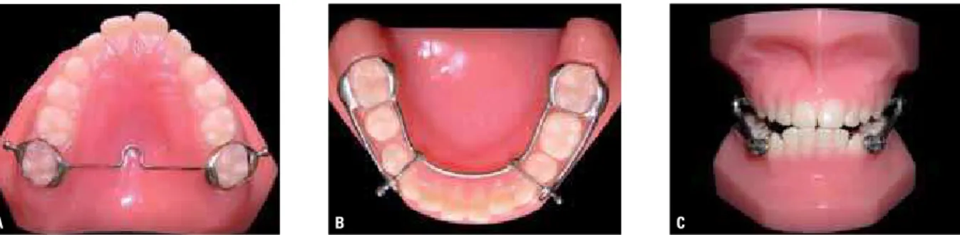
A B C banded and bound together by a transpalatal
arch welded to the bands and 2 mm away from the palate6 (Fig 1A). A modified Nance lingual
arch made with 1.2 mm steel wire welded to the bands of the first permanent molars was used for anchorage. A cantilever extending to the region of primary or permanent canines was welded to the buccal side of mandibular first molar bands. The cantilever was attached to the lingual arch in the region of primary canines and first molars or permanent canines and first premolars, using 0.9 mm wire in or-der to avoid occlusal interference22 (Fig 1B).
The anterior protrusion of the mandible with the Herbst appliance was performed as recom-mended by Pancherz16, i.e., single mandibular
advancement to an extent that an incisor end-to-end bite was achieved (Fig 1C). The appli-ance was used for a period of 7 months.
The control group was selected among the patient records of the Burlington Growth Cen-tre, located at the University of Toronto, and comprised 15 children (7 girls and 8 boys). The criteria for selection of the control group were: » Class II, Division 1 facial pattern associ-ated with mandibular retrusion.
» Class II, Division 1 dental relationship. » Erupted maxillary and mandibular perma-nent central incisors.
» No prior orthodontic treatment.
The mean initial age in the control group was
9 years and 1 month (SD=0.09) and mean final age was 10 years (SD=0.05). Regarding facial morphology in the control group, 73% of the subjects had mesofacial pattern, 20 % brachyfa-cial and 7% dolichofabrachyfa-cial patterns.
Skeletal age in both groups was assessed by means of lateral cephalometric radiogra-phy, using cervical vertebrae indicators of skel-etal maturity. Bone age was determined by the same operator and in the form of a blind study (without identifying the patient being as-sessed), which reduces the subjectivity effect in the evaluation. The subjects of this study were in maturation stages 1 and 2, i.e., before the period of pubertal growth spurt, according to Baccetti et al2 and O’Reilly and Yanniello.14
Methods
For each subject in the experimental group two lateral cephalograms were obtained at maximum intercuspation. They were named T1 (Beginning of treatment) and T2 (Seven months after treat-ment). Radiographs were obtained using an X-ray machine (model MR05 Rotograph Plus) set at 85 kVp and 10 mA and exposure time of 0.5 seconds. For the control group two lateral cephalo-grams were obtained at maximum intercuspation. They were named T1, at 9 years of age, and T2 at age 10. The radiographs were taken in a Keleket device set to 120 kVp, 25 mA and exposure time of 0.3 seconds.
Go
Me 7
8
9 10
13 11
12 4
3 2
5 1
6
C
A B
Although these radiographs were obtained by different X-ray machines, image magnifica-tion was not corrected. Image magnificamagnifica-tion, i.e., the percentage of magnification of the ex-perimental sample, was 10%, equivalent to an increase of 0.1000 cm (1.000 mm), according to Sakima.23 In the control group, a 9.84%
magni-fication was reported, according to the records provided by the Burlington Growth Centre, as reported by Popovich and Thompson.21 Since
the percentage difference in magnification be-tween the samples was 0.16%, it would not af-fect the comparison of variables obtained by ra-diographs taken with different X-ray machines. This difference in magnification corresponds to a difference of 0.0016 cm (0.016 mm) be-tween the radiographs. All radiographs were traced manually by one operator and then the cephalometric points (landmarks) were entered into a Numonics AccuGrid tablet and evalu-ated through computer software Dentofacial Planner Plus 1.2 to obtain the cephalometric measurements (Fig 2 and Table 1).
statistical planning
To assess the potential occurrence of errors in measurements attributable to the observer or the measurement process all tracings were once again digitized and measured by the same operator with an interval of 2 weeks between the first and second evaluation. Intraclass cor-relation coefficient (ICC) was used to evaluate method error (reproducibility).
To compare the changes that occurred in the cephalometric measures, with and without treatment, it was necessary to eliminate the ef-fect of the difference in time between measure-ments performed in the experimental and con-trol groups. To this end, changes in measures were annualized.
To evaluate the data the following statistical tests were conducted:
a) Student t-test for equality of means of two independent populations: To examine the hypothesis that the mean of each cephalomet-ric measure is equal to the experimental group at baseline (Table 2).
FIGURE 2 - Dental cephalometric measures: A) 1= IIs-PNS, 2= IIs-PP, 3= IIs.PP, 4= IIi-Pog, 5= IIi-MP, 6= IIi.MP; B) 7= CMs-PNS, 8= CMs-PP, 9= CMi-Pog, 10= CMi-MP; C) 11= overbite, 12= overjet, 13= molar relationship.
PL. OCLUSAL
PNS PNS
Go
Me
ANS ANS
TABLE 1 - Linear and angular cephalometric measures.
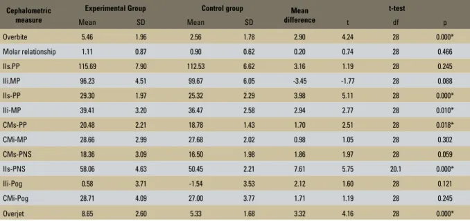
TABLE 2 - Mean and standard deviation of cephalometric measurements in experimental and control groups and mean differences between distances, before treatment, and Student t-test for differences in cephalometric measures.
* Statistically significant at 5% significance level.
Source: SPSS software package (Statistical Package for the Social Sciences, for Windows, version 10.0, SPSS Inc., Chicago, IL, USA).
Cephalometric measure Deinition
1) IIs-PNS Linear distance from points IIs and PNS reflected perpendicularly in the occlusal plane. Represents the anteroposterior relationship between incisors and PNS
2) IIs-PP Linear distance from the edge of the maxillary incisor perpendicularly to the palatal plane. Represents the vertical position of the incisors relative to PP
3) IIs.PP Angle formed by the long axis of the maxillary incisors relative to the palatal plane. Represents incisor inclination with PP
4) IIi-Pog Linear distance from points IIi and Pog reflected perpendicularly in the occlusal plane. Represents the anteroposterior relationship between incisor and pogonion
5) IIi-MP Linear distance from the edge of the mandibular incisor perpendicularly to the mandibular plane. Represents the vertical position of the incisor relative to MP
6) IIi.MP Angle formed by the long axis of the mandibular incisor and the mandibular plane. Represents incisor inclination relative to the mandibular plane
7) CMs-PNS Linear distance from points CMs and PNS reflected perpendicularly in the occlusal plane. Represents the anteroposterior relationship between first molar and PNS
8) CMs-PP Linear distance from the tip of the mesiobuccal cusp of the first permanent maxillary molar perpendicularly to the palatal plane. Represents the vertical position of the first molar relative to PP
9) CMi-Pog Linear distance from points CMi and Pog reflected perpendicularly in the occlusal plane. Represents the anteroposterior relationship between molar and pogonion
10) CMi-MP Linear distance from the tip of the mesiobuccal cusp of the first permanent mandibular molar perpendicularly to the mandibular plane. Represents the vertical position of first molar relative to MP
11) Overbite Linear measurement which represents the vertical relationship between the edges of the maxillary and mandibular central incisors relative to the occlusal plane
12) Overjet Linear measurement which represents the horizontal relationship between the edges of the maxillary and mandibular central incisors relative to the occlusal plane
13) Molar relationship Linear distance from points CMs and CMi reflected perpendicularly in the occlusal plane. Represents the horizontal relationship between maxillary and mandibular molars
Cephalometric measure
Experimental Group Control group Mean
difference
t-test
Mean SD Mean SD t df p
Overbite 5.46 1.96 2.56 1.78 2.90 4.24 28 0.000*
Molar relationship 1.11 0.87 0.90 0.62 0.20 0.74 28 0.466
IIs.PP 115.69 7.90 112.53 6.62 3.16 1.19 28 0.245
IIi.MP 96.23 4.51 99.67 6.05 -3.45 -1.77 28 0.088
IIs-PP 29.30 1.97 25.32 2.29 3.98 5.11 28 0.000*
IIi-MP 39.41 3.20 36.47 2.58 2.94 2.77 28 0.010*
CMs-PP 20.48 2.21 18.78 1.43 1.70 2.51 28 0.018*
CMi-MP 28.66 2.99 27.68 2.02 0.98 1.05 28 0.302
CMs-PNS 18.36 3.09 16.50 1.98 1.86 1.97 28 0.059
IIs-PNS 58.06 4.63 50.45 2.21 7.61 5.75 20.1 0.000*
IIi-Pog 0.58 3.71 -1.54 3.53 2.12 1.60 28 0.121
CMi-Pog 28.71 4.09 27.00 3.77 1.71 1.19 28 0.245
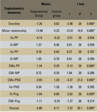
b) Student t-test for equality of means of two populations with independent samples: To examine the hypothesis that the observed changes in a given cephalometric measure be-tween times 1 and 2 are identical, on average, in the control and experimental groups (Table 3).
Results
The results indicated that the measurement process was highly accurate since the expected ICC value was at least 0.983 and, for most variables, the ICC was above 0.99. Given the high degree of agreement between the two measurements, it was decided that the mean obtained in both measure-ments would be used for each variable at each time. Assessment of equivalence between the experimental and control groups regarding the variables of interest at baseline (Table 2) showed that the experimental group had great-er ovgreat-erjet and ovgreat-erbite.
According to Table 3, it can be seen that in 6 (overbite, molar relationship, PP, CMs-PNS, IIi-Pog, overjet) of the 13 variables stud-ied, the mean changes that occurred in the treated population is statistically different from the mean changes which occurred in the un-treated population.
dIsCussIOn
Orthopedic appliances are meant to provide maximum orthopedic effect, so it is reasonable to assume that tooth movement during treatment is not desirable. However, no matter how good the performance of the appliance, it is difficult to avoid anchorage loss in both maxillary and man-dibular teeth.15 This limitation in the skeletal
ef-fect of orthopedic devices is due in part to what is referred to as “distance” anchorage, i.e., includ-ing both the upper and lower dental arches.25
Therefore, when the telescoping system of the Herbst appliance is fitted, it produces a force in the superior and posterior direction in maxillary teeth, and a force in the anterior and inferior di-rection in mandibular teeth.8
Therefore, when analyzing the changes oc-curred in maxillary incisors, it was found that while in the control group variable IIs-PNS in-creased significantly (1.28 mm/year) in the ex-perimental group the change was not significant in the same direction (0.34 mm), although much smaller than in the untreated group. This find-ing suggests that since in the untreated group the upper incisors tended to follow maxillary growth, the experimental group tended to show inhibited anterior drift of these teeth. By observ-ing variable IIs.PP, it was found that in the ex-perimental group it showed a significant –4.14°/ year decrease, whereas in the control group it showed no significant alteration. This finding confirms that in this study maxillary incisors were uprighted during treatment. This outcome stood out due to the fact that the maxillary in-cisors were not incorporated in the anchorage
TABLE 3 - Means and results of Student t-tests of equality of means in the experimental group and control group, for each of the vari-ables under study.
(*) Statistically significant at 5% significance level.
Source: SPSS software package (Statistical Package for the Social Sci-ences, for Windows, version 10.0, SPSS Inc., Chicago, IL, USA).
Cephalometric measures
Means t-test
Experimental Group
Control
group t gl p
Overbite -1.26 0.62 -2.98 28 0.006*
Molar relationship -12.08 0.23 -13.41 14.8 0.000*
IIs.PP -4.14 -0.32 -2.01 28 0.054
IIi.MP 1.37 0.46 0.61 28 0.550
IIs-PP 0.76 0.64 0.27 28 0.787
IIi-MP 1.05 0.70 0.91 28 0.369
CMs-PP -1.24 0.29 -5.12 28 0.000*
CMi-MP 0.72 0.29 1.04 28 0.306
CMs-PNS -2.65 1.20 -4.37 21.0 0.000*
IIs-PNS 0.34 1.28 -1.30 28 0.205
IIi-Pog -1.64 0.60 -2.82 28 0.009*
CMi-Pog -1.11 -0.24 -1.27 28 0.214
system. It is believed that this uprighting was caused by vertical mandibular postural change toward the anterior region, thereby yielding im-proved lip competence. Rego22 found in their
study that maxillary incisors were uprighted by about 6º in patients subjected to early treat-ment with the Herbst appliance. Several stud-ies1,3,7,16,20,29,30 have reported a similar movement
in maxillary incisors. However, other authors ob-served no changes in the position of these teeth during treatment with the Herbst appliance.17,27
As regards the vertical behavior of maxillary incisors, it was observed that while in the control group variable IIs-PP increased significantly (0.64 mm/year), in the experimental group it experi-enced no significant change during the treatment period. Lai and McNamara9 also observed no
change in upper incisors in the vertical plane in subjects treated with the Herbst appliance. Like-wise, Valant and Sinclair27 found that maxillary
incisors did not change position during treatment. Regarding changes in maxillary molars, it was observed that in the experimental group there was a significant change in the sagittal position of these teeth when variable CMs-PNS was mea-sured (-2.65 mm/year). This alteration in the treated group occurred in opposite direction of the untreated group (1.2 mm/year). This rever-sal in the direction of the change showed that in the experimental group maxillary molars were distalized during treatment with the Herbst ap-pliance while in the control group these teeth followed maxillary growth, thus worsening the Class II. This finding was also reported by Franchi et al,5 who found an upper molar distalization
of -1.71 mm during treatment with the Herbst appliance. Konik et al7 also noted a distalization
of –2.6 mm in patients undergoing early treat-ment with the Herbst appliance, and Pancherz16
found a –2.8 mm distalization of maxillary mo-lars during treatment with the Herbst appliance. In most studies, upper molar distalization con-tributes significantly to the correction of molar
relationship. There is, however, substantial varia-tion in the amount of distalizavaria-tion between stud-ies, ranging from 1.8 mm5 to 2.8 mm.16 When
using the Herbst appliance with bands, distal-ization is responsible for 25% to 40% of molar correction.7,20 On the other hand, when using
the Herbst appliance combined with an acrylic splint, distalization is responsible for 20% to 25% of molar correction.5,9,30
Regarding the behavior of upper molars in the vertical plane, a significant decrease (–1.24 mm/year) was observed in the CMs-PP measure, while the control group showed that a non-significant change took place in this measure, but in the opposite direction. It is therefore reasonable to conclude that the Herbst appliance acted by restricting molar eruption. Almeida et al1 also found that the
Herbst appliance produced greater inhibition of upper molar eruption (–0.7 mm) in subjects undergoing treatment with mixed dentition. Likewise, Pancherz17 found that molar
erup-tion was inhibited by –1.0 mm during treat-ment, and Flores-Mir et al4 identified a change
of –0.9 mm in the vertical plane of maxillary molars during treatment with the Herbst ap-pliance. These effects, which were observed in molars in this study, may be explained by the fact that the telescopic mechanism of the Herbst appliance, once installed, produces a force in the posterior and superior direction in the upper dental arch, thus simulating the ef-fect of a high-pull heagear.5,13,17,30
group and the control group (0.60 mm/year), showing that the lower incisors moved ante-riorly relative to the pogonion. Anterior man-dibular incisor inclination occurred as a result of anchorage loss due to the anterior force ex-erted by the telescoping system in mandibular teeth.18,20 These data are consistent with other
studies.1,4,7,9,13,16,19,20,22,24,29,30 In these studies, the
degree of inclination of mandibular incisors was very high and extremely variable (ranging from 2.0° to 8.4°). In this study, we found a smaller amount of flaring, an outcome similar to that recorded by Valant, Sinclair27 and Croft et al.3
This slight flaring of lower incisors is probably due to the mandibular anchorage structure that was used. This mandibular structure consisted of a lingual arch placed 3 mm away from the anterior mandibular teeth, i.e., there was no direct contact between the anchorage system and these teeth, thereby reducing the force pro-duced by the telescoping system in this region.
As regards the vertical behavior of mandibu-lar incisors, it was observed that variable Iii-MP showed a significant change both in the experi-mental group (1.05 mm/year) and in the control group (0.70 mm/year). However, a comparison between the experimental and control groups showed that there was no statistically significant change between them. According to this finding, it was noted that the Herbst appliance does not influence the process of mandibular incisor erup-tion. Rego22 concluded that mandibular incisors
did not suffer any change in the vertical direction after 12 months of treatment with the Herbst appliance. Similarly, McNamara et al13 and
Pan-cherz16,17 found no vertical changes in these teeth.
In assessing the molar, a significant change was identified in variable CMi-Pog (–1.11 mm/year), indicating that the molar was mesialized dur-ing treatment. Compardur-ing this outcome with that of the control group, a change in the same direction was identified, although more intense in the treated group. However, no statistically
significant difference was found between the groups. Konik et al7 found that the
mandibu-lar momandibu-lar was mesialized by 1.3 mm in patients subjected to an early treatment. Franchi et al5
also found a 1.44 mm movement in the anterior direction in the lower molar, while Pancherz and Hägg19 found a change of 1.5 mm in these
same teeth in patients treated with the Herbst appliance. Mesialization of mandibular molars also contributes to the correction of Class II molar relationship, ranging from 0.8 mm5 to
2.2 mm.30 In studies that use the banded Herbst
appliance, mesialization of mandibular molars is usually smaller than maxillary molar distaliza-tion, and contributes about 20 to 30% to the correction of malocclusion as a whole. In stud-ies using the Herbst appliance with an acrylic splint these changes are similar and contribute 25% to correcting molar relationship.5,9,30
In analyzing measure CMi-MP, it was found that there was no significant vertical change in the mandibular molar in both groups. This find-ing is consistent with the findfind-ings of Flores-Mir et al,4 Valant and Sinclair.27 However, the results
of this study disagreed with those of Almeida et al;1 Lai, McNamara9; Pancherz17 and Rego,22
who found significant molar eruption during treatment with the Herbst appliance.
In assessing the effects caused by the use of the Herbst appliance on the horizontal relationship of incisors, it was observed that while the control group tended to increase this variable, but not significantly, in the experimental group there was a statistically significant decrease of –4.8 mm/ year in this measure. Pancherz and Hansen20
–1.26 mm was observed in this measure. This re-sult is in agreement with Croft et al,3 who found
a decrease of –2.0 mm in the treated group. Pan-cherz17 also reported a –2.5 mm decrease in
over-bite during treatment with the Herbst appliance. An analysis of molar relationship showed that there was a statistically significant change for this measure, equivalent to –12.08 mm/year. By comparing these changes with those observed in the control group, which had been in the op-posite direction to this group (0.23 mm/year), it can be argued that the Herbst appliance caused a positive change in the correction of molar re-lationship in the present study.
According to the results, it was noted that dental changes did occur, and therefore to avoid such changes, it is recommended that more teeth be incorporated in the anchorage system. However, no anchorage system exists
today which is capable of preventing dental changes. Weschler and Pancherz28 evaluated the
effects of three different anchorage systems and concluded that anchorage loss was inevitable, regardless of the type of anchorage used. They further argued that anchorage loss in Class II treatment with the Herbst appliance always oc-curs, and orthodontists should therefore learn to cope with this reality.
COnClusIOns
Contact address
Luana Paz Sampaio Av. Portugal, 887, Centro
Zip code: 14.801-075 – Araraquara/SP, Brazil E-mail: [email protected]
1. Almeida MR, Henriques JF, Almeida RR, Weber U, McNamara JA Jr. Short-term treatment effects produced by the Herbst appliance in the mixed dentition. Angle Orthod. 2005;75(4):540-7.
2. Baccetti T, Franchi L, McNamara JA. The cervical vertebral maturation (CVM) method for the assessment of optimal treatment timing in dentofacial orthopedics. Semin Orthod. 2005;11(3):119-29.
3. Croft RS, Buschang PH, English JD, Meyer R. A cephalometric and tomographic evaluation of Herbst treatment in the mixed dentition. Am J Orthod Dentofacial Orthop. 1999;116(4):435-43.
4. Flores-Mir C, Ayeh A, Goswani A, Charkhandeh S. Skeletal and dental changes in Class II division 1 malocclusions treated with splint-type Herbst appliance. A systematic review. Angle Orthod. 2007;77(2):376-81.
5. Franchi L, Baccetti T, McNamara JA Jr. Treatment and post treatment effects of acrylic splint Herbst appliance therapy. Am J Orthod Dentofacial Orthop. 1999 Apr;115(4):429-38. 6. Howe RP. The bonded Herbst appliance. J Clin Orthod.
1982;16(10):663-67.
7. Konik M, Pancherz H, Hansen K. The mechanics of Class II corrections in late Herbst treatment. Am J Orthod Dentofacial Orthop. 1997;112(1):87-91.
8. Lai M. Molar distalization with the Herbst appliance. Semin Orthod. 2000;6(2):119-28.
9. Lai M, McNamara JA Jr. A evaluation of two-phase treatment with the Herbst appliance and pre-adjusted Edgewise therapy. Semin Orthod. 1998;4(1):46-58.
10. Lamparski DG. Skeletal age assessment utilizing cervical vertebrae. Am J Orthod. 1975;67(4):458-9.
11. Langford NM Jr. Updating fabrication of the Herbst appliance. J Clin Orthod. 1982;16(3):173-4.
12. Mayes JH. Improving appliance eficiency with the cantilever Herbst. A new answer to old problems. Clin Impressions. 1994;3(2):2-5,17-9.
13. McNamara JA Jr, Howe RP, Dischinger TG. A comparison of the Herbst and Fränkel appliances in the treatment of Class II malocclusion. Am J Orthod Dentofacial Orthop. 1990;98(2):134-44.
14. O’Reilly MT, Yanniello GJ. Mandibular growth changes and maturation of cervical vertebrae — A longitudinal cephalometric study. Angle Orthod. 1988;58(2):179-84. 15. Pancherz H. History, background, and development of the
Herbst appliance. Semin Orthod. 2003;9(1):3-11.
16. Pancherz H. The mechanism of Class II correction in Herbst appliance treatment. Am J Orthod. 1982;82(2):104-13. RefeRenCes
17. Pancherz H. The Herbst appliance — Its biologic effects and clinical use. Am J Orthod. 1985;87(1):1-20.
18. Pancherz H. Treatment of Class II malocclusions by jumping the bite with the Herbst appliance. Am J Orthod. 1979;76(4):423-42.
19. Pancherz H, Hägg U. Dentofacial orthopedics in relation to somatic maturation. An analysis of 70 consecutive cases treated with the Herbst appliance. Am J Orthod. 1985;88(4):273-87.
20. Pancherz H, Hansen K. Occlusal changes during and after Herbst treatment: a cephalometric investigation. Eur J Orthod. 1986;8(4):215-28.
21. Popovich F, Thompson GW. Craniofacial templates for orthodontic case analysis. In: Clark JW. Clinical dentistry. Philadelphia: Harper & Row; 1983. p. 1-24.
22. Rego MVNN. Estudo cefalométrico das alterações esqueléticas, dentárias e tegumentares induzidas pelo aparelho Herbst no tratamento da má oclusão Classe II, 1ª divisão de Angle. [dissertação]. Porto Alegre (RS): Pontifícia Universidade Católica; 2003.
23. Sakima PRT. Efeitos dos erros de projeção sobre as grandezas cefalométricas das análises de Steiner e McNamara. [dissertação]. Araraquara (SP): Universidade Estadual de São Paulo; 2001.
24. Schiavoni R, Grenga V, Macri V. Treatment of Class II high angle malocclusions with the Herbst appliance. A cephalometric investigation. Am J Orthod Dentofacial Orthop. 1992;102(5):393-409.
25. Silva Filho OG. Aparelho de Herbst - variação para uso na dentadura mista. Rev Dental Press Ortod Ortop Facial. 2000; 5(5):58-67.
26. Silva Filho OG, Freitas SF, Cavassan AO. Prevalência de oclusão normal e má oclusão na dentadura mista em escolares da cidade de Bauru (São Paulo). Rev Assoc Paul Cir Dent. 1989; 43(6):287-90.
27. Valant JR, Sinclair P. Treatment effects of the Herbst appliance. Am J Orthod Dentofacial Orthop, 1989; 95(2):138-47.
28. Weschler D, Pancherz H. Eficiency of three mandibular anchorage forms in Herbst treatment: a cephalometric investigation. Angle Orthod. 2005;75(1):23-7. 29. Wieslander L. Intensive treatment of severe Class II
malocclusion with a headgear-Herbst appliance in the early mixed dentition. Am J Orthod. 1984;86(1):1-13.
30. Windmiller EC. The acrylic-splint Herbst appliance: a cephalometric evaluation. Am J Orthod Dentofacial Orthop. 1993;104(1):73-84.