An interview with
How to cite this article: Rino Neto J. Interview. Dental Press J Orthod. 2013 Jan-Feb;18(1):8-29. Submitted: December 13, 2012 - Revised and accepted: December 19, 2012
» Patients displayed in this article previously approved the use of their facial and intraoral photographs.
José Rino Neto
It is an honor for me to present this interview with Professor José Rino Neto. Famous professor of the University of São Paulo (USP), José Rino Neto has built his academic career founded on solid basis at the University of São Paulo (USP), attending there from dental school (FOB-USP) until his complete training as a professor (MSc, PhD and Full Professor of FOUSP). For this reason, he is a great messenger of the works of memorable professors, such as Sebastião Interlandi and Julio Wilson Vigorito. Besides being a legitimate representative of orthodontic quality in Brazil, our interviewee has particular academic and personal characteristics worth mentioning. His career path has enabled him to create a summarized coherent understanding of “Dentofacial Morphology”, which lead him to be the clinic coordinator of the Orthognathic Surgery graduate program at USP. We are convinced that orthodontics has gone forward under the critical eyes of this young and wise educator, who ponders over the probabilities of sci-ence, in order to achieve permanent success and demystify innovations. The intimate coexistence with his mentors has allowed him to become a skillful professor, who knows how to be strict, yet humble, kind and a true perfection-ist in science and in the clinic, enabling him to build noble relationships with his students. Professor Rino kindly granted us with the following interview, where he expresses his relections about orthodontics as one of the most signiicant professors in Brazil and based on his own clinic and scientiic experience.
José Valladares Neto
• Graduated by the School of Dentistry of Bauru, São Paulo University
(FOB-USP), Brazil.
• MSc and PhD in Orthodontics, School of Dentistry, University of São
Paulo(FOUSP), São Paulo, Brazil.
• Full Professor in Orthodontics, FOUSP.
• Associate Professor of the Department of Orthodontics, University of São
The Department of Orthodontics of the Univer-sity of São Paulo (FOUSP) has recently published an improved version of the USP Cephalometric Analysis. What are the main improvements of
the USP-2 Analysis? (José Valladares Neto)
The USP cephalometric analysis was published in 1968 by Professor Interlandi and comprises the set of cephalometric landmarks originated from the analysis of Downs, Tweed, Steiner, Bjork, Holdaway, Inter-landi and Vigorito. According to the author, the main objective of this cephalometric analysis was to assist the orthodontist in diagnosis and treatment planning. The establishment of the USP analysis was an un-questionable milestone in the history of Orthodontics in Brazil, and nowadays it is a key diagnostic tool in orthodontic decisions.
The department of Orthodontics of the Universi-ty of São Paulo understands that there has been a new diagnostic methodology being shaped in the last few years and the USP-2 analysis was developed to better correlate to this new orthodontic scenario.
The USP-2 cephalometric analysis is divided in three categories (soft tissue, skeletal and dental struc-tures) to simplify the readability and interpretation of the cephalometric measurements. The new cephalo-metric landmarks are originally from the Ricketts, Jarabak, Jacobson and Epker and Fish Analysis and its main improvements are in the vertical and sagittal soft tissue evaluation, inner relationship of the cranial base anatomic structures and vertical dental relation-ship assessment (Fig 1).
In surgical cases, what are the indications for occlusal plane change? What reference do you use to determine the inclination of the occlusal
plane? (Adilson Ramos)
The changes in inclination of the occlusal plane have been performed routinely as part of the orthodon-tic-surgical treatment. The decision to change the angle of the occlusal plane cannot be arbitrary and should be made according to esthetic and functional goals.
Increased inclination of the occlusal plane is indi-cated in Class I cases with dentofacial deformities such as reduced occlusal and mandibular plane; in short fac-es with Class II, division 2 malocclusion, deep overbite and pronounced chin; and occasionally in the Class III
dentofacial deformities. The procedure reduces chin Figure 1 - USP-2 standard cephalometric analysis.
SKELETAL EVALUATION Cranial base
S-N 75 mm 3
Ba-N.FH 27° 3
Facial type and Growth pattern
Facial axis 90° 3
FNP 90° 3
FMA 26° 4
Lower facial height 47° 4
Mandibular arch 26° 4
N.S.Ar 123° 5
S.Ar.Go 143° 6
S-N : Go-Me 1:1
--S-Ar : Ar-Go 3:4
--Ar.Go.N 52–55%
--N.Go.Me 70–75%
--S-Go/N-GMe 60–64%
--Maxillomandibular relationship
SNA 82° 2
SNB 80° 2
ANB 2° 2
A-N.Perp 1 mm
--P-N.Perp -4 / +2 mm
--Co-A -
--Co-Gn -
--ENA-Me -
--A point convexity 2.0 mm 2
Wits (AO–BO) 0 mm
---1.0 mm
--N-ENA : ENA-Me 1:1
--N.CF.A 53° 3
DENTAL RELATIONSHIPS Sagittal
1.1 131° 6
1.NA 22° 2
1-NA 4 mm
--1.NB 25° 2
1-NB 4 mm
--IMPA 90° 5
1.PP 110°
--Vertical
1-Stms 1–3 mm
--Overbite 3.0 mm 2
1-Occlusal plane 1.25 mm 2
Clinical norm Clinical deviation SOFT TISSUE EVALUATION
vertical
G-Sn : Sn-Me 1:1
--Sn-Stms : Stmi-Me 1:2
--Stms-Stmi 0–3 mm
--Sn-Stms 19–22 mm
--1-Stms 1–3 mm
--horizontal
Sn.Perp–Prn 16–20 mm
--Sn.Perp–Ul 2–4 mm
--Sn.Perp–Ll 0–2 mm
--Sn.Perp–Pg’ 0 / -4 mm
--Sn-Gn’ : C-Gn’ 1:0.8
--Nasolabial angle 90–110°
--projection and posterior face height, promotes the ad-vancement of paranasal structures, as well as upright-ing upper incisors and larupright-ing the lower incisors.
The reduction of the occlusal plane inclination, i.e., the counterclockwise rotation of the occlusal plane, can be done in two ways. The first consists of the autorotation of the mandible after the superi-or repositioning of the maxilla that is not surgically manipulated. The autorotation of the mandible is in-dicated to individuals that have increased lower face height, excessive exposure of the upper front teeth in rest or smiling, increased interlabial distance in-creased and retrusive chin. The counterclockwise ro-tation of the maxillomandibular complex should be planned for cases with high occlusal and mandibular planes, anterior excess height and/or posterior defi-cient height of the maxilla, increased anterior height of the mandible and/or reduced posterior height of the mandible, mandible with deficient chins and in severe cases of obstructive sleep apnea.
As for determinant references for the planning of the occlusal plane inclination change, the assessment of the craniocervical muscle anatomy and physiolo-gy, the respiratory function and the esthetic goals for the case should be mentioned. I believe the maxillary occlusal plane positioning is an important step in the planning of occlusal plane manipulation, since this position determines the incisor display in rest posi-tion, as well as the gum and incisor display in smile position. Another relevant factor is the chin, since the occlusal plane rotation can increase or reduce its prominence according to the treatment objectives and initial profile convexity.
Regarding the diagnosis and planning of den-tofacial deformities, do you prefer to use Frankfort Horizontal Plane (Larry Wolford) or Soft Tissue Cephalometric Analysis (William
Ar-nett)? (Roberto Macoto)
The use of a particular diagnosis and planning method is determined by the orthodontist’s prefer-ence. The greatness of Dr. Wolford’s surgical cases results is unquestionable; his cases are planned based on skeletal and dental cephalometric landmarks, which use the Frankfort Horizontal Plane as the main reference. I have been using a different approach to diagnose and plan Orthognathic Surgery cases since
1998, this approach enhances the importance of face evaluation and cephalometric analysis of the soft tis-sues, which is assessed when the patient’s head is in Natural Position, the facial muscles are relaxed and bite is preferably in centric relation. Therefore, my preference for diagnosing and planning surgical cases is the Soft Tissue Cephalometric Analysis from Ar -nett. I believe it better correlates facial esthetics and orthodontic and surgical decisions. Moreover, it is based on the Natural Head position, which I consider as the ideal position for facial assessment and deter-mination of cephalometric dentofacial relationships. Furthermore, the evaluation of soft tissues is outlined by extra cranial vertical and horizontal lines, which are ideal references for evaluation of patients with dentofacial deformities, since those patients often have discrepant relationships of internal parts of the cranial base, which may affect the interpretation of cephalometric measurements.
Virtual planning of Orthodontic and Orthog-nathic surgery cases has become a reality in nowadays practice. What is your opinion on
this technology? (Adilson Ramos)
The virtual planning is already a reality for the Orthognathic Surgery patients because it provides the benefits generated by the technology in favor of better treatment outcomes. Considering that dentofacial de-formities compromise the relationship of skeletal and dental structures, and soft tissue in the three planes of space, nothing is more appropriate than the use of a technology that can diagnose and plan these cases in three dimensions. The virtual method of orthodontic and surgical treatment planning enables high results accuracy, as well as reliably reproducing the virtual plane at the operating room.
This includes the construction of 3D virtual models generated from cone beam computed to-mography, from which it is possible to observe an-atomic structures of interest, a dynamic cephalo-metric diagnosis and planning, surgical movements simulation, construction of surgical guides, size and position of plates and screws for the rigid internal fixation and allows the access and navigation in the database during the surgery.
structures important in defining the treatment goals, osteotomy design and accurate positioning of oste-otomized bone segments; furthermore it enables the surgeon to estimate the difficulty level of the surgery before the surgical procedure.
CONE BEAM COMPUTED TOMOGRAPHY
Some scientific publications have questioned the routine use of CBCT scans in orthodontics due to its high ionizing radiation doses, but we know that CBCT scans provide important information when compared conventional or-thodontic records. What is your opinion on this matter? Are the risks worth the benefits?
(João Batista de Paiva)
The use of CBCT imaging is already established in orthodontics and its use has been widely accept-ed. Even though there is not a guideline from ortho-dontics associative entities in respect to the use of the 3D images generated from the ionizing radiation, it is important to consider some aspects that may allow us to use those examinations in a rational and objec-tive way, and take advantage of its high detail capacity that cannot be found in conventional x-rays.
The request of CBCT scans should be done con-sidering the potential risk of radiation, especially in children and adolescents, and should be based on pa-tient history, clinical examination and the presence of a speciic clinical condition that ensures its beneit to the diagnosis and or treatment plan of the patient. At this time, it is important to consider the speciic image capture protocol design; which includes Voxel size, ield of view size, type of equipment used, and
oth-ers — noting that all these items inluence the image quality and radiation dosage. Actions must be taken to minimize radiation dosage to exposed patients fol-lowing the ALARA principle, “as low as reasonably achievable”. Special care must be devoted to children due to their active growth and development, peri-od which they have a great cell growth, and greater radio-sensitivity than adults. The main indications of CBCT imaging in orthodontics are the diagnosis and treatment planning of the dentofacial deformities, such as morphological evaluation of the airway, TMJ, cortical bone thickness and orthodontic-surgical vir-tual planning. Furthermore, it is indicated when the patient has supernumerary teeth; internal and external root resorption; impacted teeth, close relationship be-tween molars and the alveolar nerve, TMJ pathologies and size abnormalities; such as abnormal shape and position of skeletal structures, as condylar hyperpla-sia, hypoplasia or aplasia; degenerative conditions, as rheumatoid arthritis; adaptive abnormal conditions, as condyle reabsorption and remodeling; trauma, as con-dyle fracture; in addition to allowing the precise loca-tion of skeletal anchorage devices (Fig 2).
However, the request for precise examinations such as CBCT images are useless if the professional is not able to interpret it. From a legal standpoint, it is important that these images are read and ana-lyzed by a radiologist so that the responsibilities of incidental findings, which are common, are not the orthodontist’s responsibility. To finalize, I believe the use of 3D images generated from the CBCT scans are worth the risks when used with responsibility and knowledge by the orthodontist.
Figure 2 - Indications for the use of CBCT: Evaluation of alveolar and cortical bone thickness (A); TMJ evaluation (B); Airway evaluation (C).
Considering your research and clinical experi-ence with cone beam computed tomography, what changes can we expect in the airways after procedures such as rapid maxillary expansion, surgical maxillary advancement (Pattern III), surgical mandibular advancement (Pattern II), maxillomandibular advancement (Pattern I) or another that you may find relevant to the pa-tients with obstructive problems?
(Adilson Ramos)
In the last two decades, numerous scientific pub-lications have reported changes in airway dimen-sions after orthognathic surgery and its relation to the treatment of obstructive sleep apnea (OSA). Re -cent advances in airway observation based on imag-es generated from cone beam computed tomography (CBCT) have helped us in understanding normal and abnormal airways and their changes in response to surgery. The anatomic knowledge of the airway and its characteristics based on preoperative studies allow us to define a precise surgical treatment plan focused on the restriction areas.
After surgically assisted rapid maxillary expansions (SARME), it is possible to observe in the 3D images, a swelling on the lower portion of the nasal cavity, es-pecially where the nasal valve is located, which is the region of greatest constriction. This swelling happens due to the opening of V-shaped sutures, which can be observed in the coronal and axial planes. In the coronal plane, this opening becomes smaller towards the cranium; in the axial plane, the larger opening is anteriorly and progressively decreases posteriorly. These changes produce minimal, yet positive effects on the respiratory parameters during sleep.
The literature does not recognize specific chang-es in the airway space size due to surgical maxillary advancement only. In most studies, the maxillary ad-vancement was performed in association with man-dibular setback for the correction of Class III dento-facial deformity, which is considered the factor that decreases the risk of occurrence of OSA after surgery. An increase in volume of the nasopharynx and oro -pharynx area can be observed as well, however the long-term benefits is still questionable due to the possibility of adaptive changes in the soft palate mor-phology after the advancement of the maxilla to keep the oropharyngeal closure.
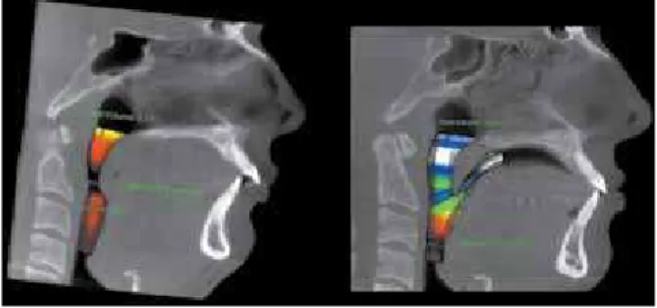
Patients who have mandibular advancement sur-gery show an increase in the airway volume space in general, especially in the retroglossal portion of the oropharynx, which extends from the soft palate to the epiglottis. This alteration happens due to the advancement of all the structures that are connect-ed somehow to the mandible (like the hyoid bone, the suprahyoid musculature, the genioglossus and the tendons), which promotes increased muscle tension and generates greater tissue collapse resistance in this region during sleep (Fig 3).
The maxillomandibular advancement is consid-ered the method of choice in orthognathic surgery for the treatment of OSA. The justification for this procedure is based on the fact that the advancement of the maxilla and the repositioning of the mandi-ble enamandi-bles anterior positioning of the suprahyoid and velopharyngeal muscles, which leads to an anterior movement of the soft palate, tongue and tissues of the anterior wall of the pharynx, resulting in an increase in the size of the nasopharynx, oropharynx and hypo-pharynx. This significant increase of the anteroposte-rior and lateral dimensions of the palatine and lingual regions, observed in the CBCT image, associated with increased tension of the walls of the orophar-ynx, is responsible for the improvement reported by patients as indexes assessed in polysomnography and subjective symptoms postoperatively.
ORTHOGNATHIC SURGERY
The standard orthodontic treatment prior to orthognathic surgery addresses mostly dental corrections. However, the anticipated bene-fit orthodontic-surgical treatment procedures have been recently introduced to orthodontics
and it proposes orthognathic surgery before orthodontic treatment. Are there any particular dentofacial deformities to which this technique should be recommended?
(Gilberto Vilanova Queiroz)
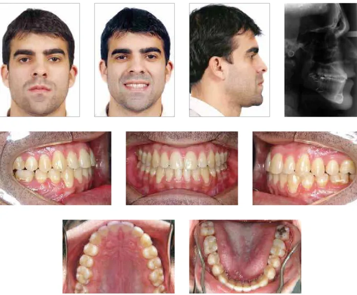
Considering the conventional and anticipated benefit technique for the treatment of dentofacial de-formities, I believe there are specific circumstances in which one prevails in relation to the other. However, regardless of the approach to be taken, it is essential to achieve balanced facial esthetics, chewing efficien-cy, longevity of dental and periodontal tissues, cor-rection stability, patient satisfaction, and acceptable respiratory and sleep function. The conventional sur-gical-orthodontic treatment is indicated for correc-tion of all types of dentofacial deformities; therefore its mechanics is theoretically simpler and within the knowledge of most well prepared professionals. An -other advantage is that this method provides greater result prediction, since after the aggravation of the patient’s deformity through presurgical orthodon-tics, the planning of the skeletal and integumentary modifications of the face becomes less complex to be executed (Figs 4 A, B, C). The disadvantage of this therapeutic approach is the possible temporary wors-ening of facial esthetics prior to surgery.
The anticipated benefit technique is indicated when there is an immediate need for surgery because of functional problems, especially those related to severe obstructive sleep apnea and to treat esthetic problems at the beginning of treatment. In my opin-ion, the most adequate cases for this procedure are those with well-aligned teeth or light crowding with flat or average Curve of Spee, or well positioned in-cisors with slight or no transverse discrepancies. The cases where this technique is not indicated are those with severe crowding, asymmetries, differences in the occlusal plane, decreased lower face height or even Class II patients with deep overbite and deep Curve of Spee. As advantages of the anticipated benefit, I mention the treatment duration reduction due to the accelerated dental movement phenomenon. On the other hand, I understand there are many disadvan-tages in the anticipated benefit method, such as less stability of occlusion immediately after surgery, less predictable results and the need to be performed by highly experienced and trained orthodontists.
When correction of sagittal skeletal discrep-ancies is performed by orthognathic surgery, what criteria do you use to define the amount of overjet prior to surgery?
(Gilberto Vilanova Queiroz)
The answer to this question is complex and awakens some questions relevant to the topic, such as: What is the impact of this treatment on the pa-tient’s face? Is there a need for tooth extractions for decompensation of the buccolingual inclinations of upper and lower incisors? How much orthodontic presurgical preparation will be required? How much movement of the osteotomized bone segments is re-quired for the correction of the skeletal and soft tis-sue components of the face?
These responses begin to be answered during the interview with the patient, but for this data to be rel-evant and helpful in the decision-making process, it is important to have specific forms to identify how the deformity impacts the patient’s quality of life. These forms allow the patient to self assess his dentofacial esthetics and the correlation between social issues and the deformity. The use of psychometric instruments has as main objective to evaluate quantitatively their self-image and their perception of the deformity in a more elaborate way.
The patient facial esthetics planning is an extreme complex process and its decisions require the ortho-dontist and oral surgeon to fully comprehend facial harmony and it’s ethnic characteristics, as well its relationship with functional aspects of craniofacial components. Therefore, this process requires prop-er records, such as facial and intraoral clinical exam, 2D and 3D images. The face clinical exam allows the orthodontist and oral surgeon to recognize which structures are balanced and which are out of balance, enabling them to determine if the maxilla, the man-dible or both caused the deformity.
The intraoral exam is not only important to assess the dental positioning status, but also to evaluate the pa-tient’s periodontal status, integrity and structure, since these tissues will have to support the planned dental de-compensation caused by orthodontic treatment.
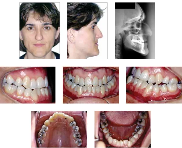
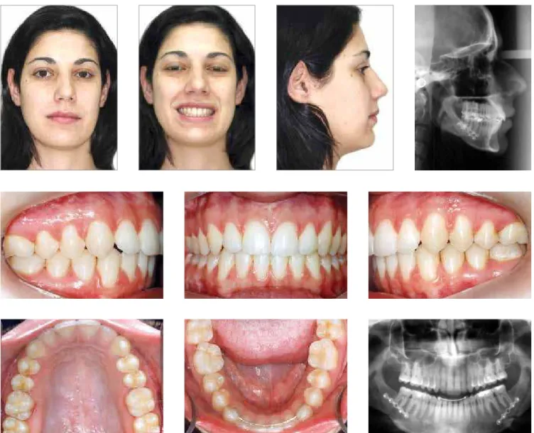
Figure 4B -Combined orthodontic and surgical treatment: Presurgical photographs.
Figure 4A - Combined orthodontic and surgical treatment: Initial photographs.
position of the upper and lower incisor. This is done through CBCT images observation, speciically by sag-ittal tomographic cuts on the upper and lower incisor area, which enables the orthodontist to evaluate the al-veolar bone limits and to understand possible
Figure 4C - Combined orthodontic and surgical treatment: Final photographs.
In short, the amount of presurgical overjet will be determined by considering important factors, such as: 1) the chief complaint of the patient and his den-tofacial esthetic concerns, 2) middle and lower face problem severity and its correction options, 3) teeth movement limitations; 4) need for extractions to eliminate preexisting dental compensations, and 5) pathology involving the TMJ and the airways.
In patients with transverse relationship prob-lems and considering the current therapeutic possibilities to treat it, which criteria do you use to choose between: A) Two stage surgical treatment comprising initial Surgically Assist-ed Rapid Maxillary Expansion and final
Sagit-tal and Vertical Orthognathic Surgery; B) One stage surgical treatment comprising Surgically Assisted Rapid Maxillary Expansion only; and C) Orthodontic dentoalveolar archwire
expan-sion? (Gilberto Vilanova Queiroz)
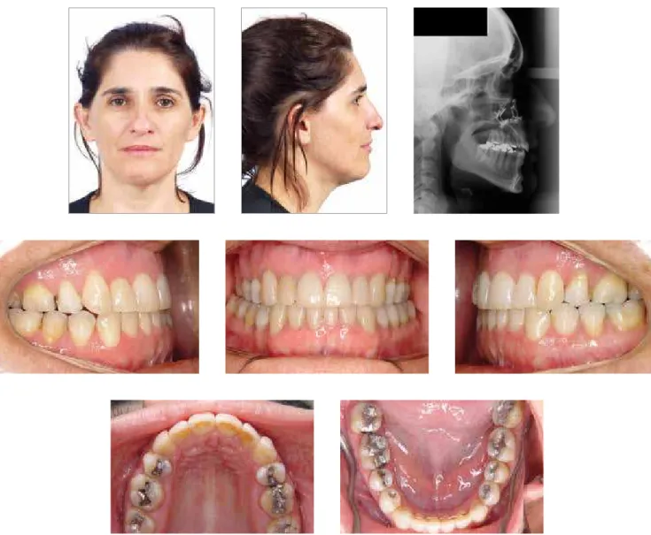
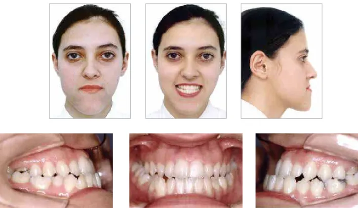
Figure 5A -Combined orthodontic and surgical treatment with minimal facial esthetic changes: Initial photographs.
Figure 5B -Combined orthodontic and surgical treatment with minimal facial esthetic changes: Photographs of the presurgical orthodontic preparation.
The two stage surgical treatment, when the SARME and the orthognathic surgery are performed in two separate steps, is indicated for treatment of maxillary narrowing since it allows greater expansion (Fig 6A, B, C), this procedure also shows greater long term stabil-ity. It is important to emphasize that with the advent
of bone supported expanders, some limitations of con-ventional palate expansion, which required SARME, were overcome, such as patients missing multiple teeth and poor periodontal status.
Figure 5C -Combined orthodontic and surgical treatment with minimal facial esthetic changes: Final photographs.
undergo a single surgical procedure. In such cases, the Le Fort I maxillary osteotomy in two or three pieces is indicated to promote lateral repositioning of palate, which allows the full maxillary narrowing correction in only one surgical procedure. This pro-cedure, however, demands greater experience from the surgeon and proper presurgical orthodontic cedures, such as intentional root divergence to pro-vide access for the osteotomy.
The main advantage of this procedure is that it is done in one single step, where the transverse, sagittal and vertical deformities are corrected simultaneous-ly. However, despite being an excellent problem solv-ing technique, it is only indicated for patients who need mild maxillary expansion, the indication for this
procedure is limited to cases where mild maxillary expansion is needed, due to the minimal elasticity of the palatal mucosa. Furthermore, this procedure presents higher morbidity, increased risk for compli-cations and greater relapse potential.
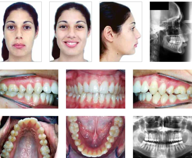
Figure 5D -Combined orthodontic and surgical treatment with signiicant facial esthetic changes: Initial photographs.
In your opinion, what is the role of the ortho-dontist and the oral surgeon in regards to the diagnosis and treatment of obstructive sleep
apnea? (João Batista de Paiva)
Orthodontists and oral surgeons have an import-ant role in both the diagnosis and treatment of ob-structive sleep apnea (OSA). Their contribution to therapeutic conduct of OSA begins in the clinical examination of the patients, more specifically in the achievement of the complete medical and sleep histo-ry, especially in adult patients. In this stage,
subjec-tive questionnaires can be used, such as the Epworth Sleep Scale, which provides some information on the sleep and waking of the patient. It is essential to also ask questions to the patient’s spouse before the com-pletion of the initial exam. The next step is collecting records including 3D images, which allows craniofa-cial complex analysis in the three planes of space, as well as the nasopharynx, oropharynx and hipophar-ynx volume and area estimation (Fig 7).
images and may require more specific tests, such as nasofibrolaryngoscopy and polysomnography, to make a diagnostic conclusion. It’s hard to ascertain the best treatment option for this condition, since there are many different severity levels of OSA and the treatment options are also different for each of these levels. In cases of mild to moderate apnea an intraoral device for mandibular advancement during sleep may be indicated, which is a responsibility of the orthodontist and/or oral surgeon. In cases of se-vere apnea, orthognathic surgery can be indicated, and in those cases both the orthodontist and the oral surgeon are protagonists in the treatment of OSA.
How much should the orthodontist interfere and participate on the surgical planning?
(Roberto Macoto)
The treatment planning of orthodontic-surgical cases requires a collaborative knowledge from all the specialties involved. I believe orthodontists and oral sur-geons should work together, so that the details pertinent to each specialty can be applied to beneit the patient
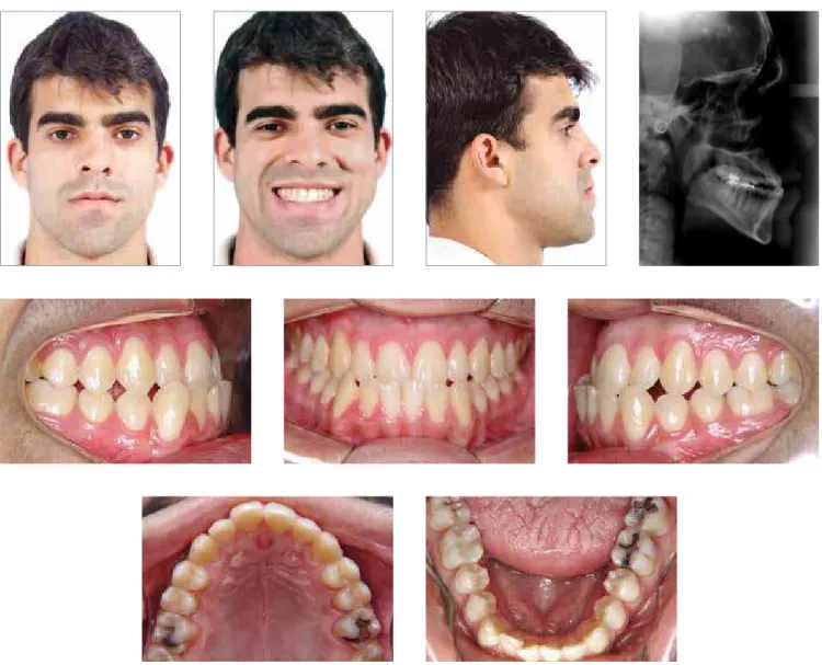
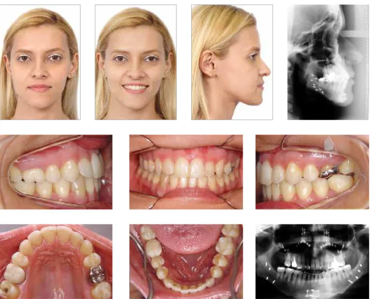
Figure 6A -Combined orthodontic and surgical treatment of Class II dentofacial deformity by two separate surgical procedures: Initial photographs.
Figure 6C -Treatment of Class II dentofacial deformity by two separate surgical procedures: Final photographs after mandibular advancement.
The orthodontist’s ability to deal with these and other questions, as well as solving dental and skeletal problems, earns the oral surgeon’s respect and trust, facilitating their interaction.
Based on this principle, the orthodontist must par-ticipate positively in the treatment planning of ortho-dontic-surgical cases. For that reason, the orthodon-tist must fully understand and consider the patient’s main concerns, facial harmony and esthetics, ortho-dontic possibilities, as well as the soft tissues changes caused by orthodontic and surgical procedures. The risks and benefits of the chosen surgical technique to obtain determined results should be also discussed.
TMJ
There is great controversy regarding the surgi-cal management of temporomandibular joint disorders in patients with painful symptoms. From an orthodontic point of view, when do you believe TMD surgical interventions are in-dicated? Are you favorable to the use of other methods, such as neuromuscular deprogram-ming plates, arthrocentesis, etc?
(Roberto Macoto)
As an orthodontist who’s been involved in multi -disciplinary approach for TMD treatment, my actions and recommendations are to always perform accurate differential diagnosis, to identify the causes of the TMD and to treat them as conservatively as possible. In my private practice, I have been using Michigan plates since the early 90s, either on its own or in com-bination with drug therapy, when acute pain is pres-ent. I also recommend physiotherapy, acupuncture, cognitive behavioral psychology, and other recovery and muscle relaxation methods. Other procedures, such as occlusal adjustment, orthodontic treatment, occlusal rehabilitation or even orthognathic surgery may be indicated for TMD treatment.
I have observed a great deal of controversy in the literature regarding the therapeutic approach to temporomandibular joint disorders (TMD) in symptomatic patients, especially regarding issues related to whether therapeutic or surgical treatment should be performed. This controversy has gener-ated mistaken conducts by many professionals, thus negative results can come from a bad indication of such determined surgical procedure. However, we
also can also find well-defined TMD clinical and surgical treatment options in the literature.
There are several well-defined clinical situations in which the surgical TMD treatment is unquestionably indicated. These are the cases of ankylosis, fractures, hemifacial microssomia, total idiopathic condylar re-sorption (due to arthritis or autoimmune disorders), traumatic condylar destruction, malignant and be-nign tumors and condylar hyperplasia (Figs 8A-E). The purpose of this treatment method is to modify the internal anatomic structure of the TMJ, such as in cases of recurrent mandibular dislodgement and surgical disk recapturing (which is the reattachment of articular displaced discs). The greatest risk of the surgical assessment is in its irreversible character.
Arthrocentesis is a less invasive low morbidity pro -cedure indicated in cases of acute and chronic pain with decreased joint movement range, which provides good results — especially when used as auxiliary ther-apy in cases of orthognathic surgery with mandibular alterations. This procedure is indicated to reduce the negative pressures that interfere in the mobility of the articulation disk, as well as eliminates chemical medi-ators that cause pain and inlammation. Most discus-sions in relation to the indications of this isolated pro-cedure reside in the duration of its beneits.
A very important issue to be considered is related to TMD patients with chronic pain, for those sufering from central nervous system sensitization, the contin-ued stimulation of aferent pathways, associated with changes in descending inhibitory pain system. In these cases, even ater surgery, the pain or even the physio-logical deiciency to inhibit it, may remain or worsen. Therefore, surgical treatment for patients with chronic pain may not be the best treatment option.
In my opinion, there are insufficient longitudinal studies and randomized clinical trials to prove the ef-fectiveness of each surgical method and thus elimi-nate controversies.
SELF-LIGATING BRACKETS
In your opinion, what has a larger impact on self-ligating systems: the self-ligating brackets or the super elastic archwires? What is your ex-perience in orthodontic treatment of the defor-mities using self-ligating brackets?
Figure 8A -Combined orthodontic and surgical treatment of a short face patient associated with con-dylectomy due to active condylar hyperplasia: Initial photographs.
In a self-ligating system, what really makes the difference is exactly the combination of the self-li-gating brackets and the super elastic memory arch-wires. Nowadays, it is the knowledge of the or-thodontic community that there is no scientific evidence to prove the clinical superiority of self-li-gating when compared to conventional brackets. A real benefit of the use of self-ligating systems is the shorter chair time during patient care and less lower incisor inclination during teeth leveling. However, many in vitro studies have demonstrated the signifi-cant friction and binding reduction between self-li-gating brackets when compared to conventional bracket systems, which are ligated either by stainless steel or elastic ties.
Figure 8C -Combined orthodontic and surgical treatment of short face patient associated with condylectomy due to active condylar hyperplasia: Surgery – condylectomy and disk ixation.
Figure 8E -Combined orthodontic and surgical treatment of short face patient associated with condylectomy, due to active condylar hyperplasia: 5 years post-treatment.
Despite having similar biological orthodontic force response, in spite of the bracket system, there is an important aspect to be considered is that the combination of self-ligating brackets and superelas-tic archwires, due to it’s low friction character, gen-erates lower magnitude forces on the teeth for a lon-ger period of time, which allows an ideal biological response, control of the desired orthodontic and less reported pain by the patients, particularly at early stages of treatment (Fig 9).
Approximately 3 years ago, I started using self-li -gating brackets and superelastic archwires in
Figure 9 -Self-ligating bracket systems/super elastic memory archwires: A) 0,016-in Power Cable archwire, B) 0,016-in Heat Activated NiTi archwire;
C) 0,016 x 0,022-in Heat Activated NiTi archwire; D) 0,019 x 0,025-in Heat Activated NiTi archwire.
A
C
B
D EDUCATION
How do you see the training of radiographic cephalometry in the current clinical practices? (Neto José Valladares)
Since 1991, when I joined the discipline of ortho-dontics at FOUSP, I was assigned to teach under-graduate and under-graduate classes on radiographic cepha-lometry, so it’s been accurately 22 years dedicated to the study and teaching of radiographic cephalometry.
Over the years, many concepts have changed in this field, especially the paradigm shift regarding the importance of soft tissues in the diagnosis and treat-ment planning. Unquestionably, this new concept has changed significantly the methods for the diagnosis and treatment planning of cases. Another important evolution over the years was the use of computer
soft-ware to perform computerized cephalometric trac-ings and to predict surgical and orthodontic results.
Considering the facts and the current stage of radio-graphic cephalometry, I understand this still constitutes one of the pillars for the understanding of orthodontics as a whole. Therefore, from an academic standpoint, even aware of the limitations of the method, I believe in the importance of teaching radiographic cephalometry, as regard to the knowledge of the anatomic relationship of skull and the face, of the main cephalometric analysis,
and mostly in the dynamic application of these concepts in addition to information obtained from other diag-nostic elements. My opinion is supported by the Ortho-dontic Faculty at FOUSP, so we have published a new cephalometric analysis called USP-2 Standard Analysis, which is already being used by our orthodontic resi-dents and will soon be available for examination and use by the whole orthodontic community.
The orthodontic training is still deicient regard-ing the diagnosis and treatment plannregard-ing of den-tofacial deformities. How is this subject taught in the program in which you participate and what’s your suggestion to improve this training?
(José Valladares Neto)
I fully agree with your statement about the lack of proper training of Brazilian orthodontists regarding the diagnosis and treatment planning of dentofacial de-formities, although this educational deiciency is also present in other countries. On the other hand, there is a clear trend to improve this situation as the most inluent orthodontic professors in Brazil have certainly ofered the students proper knowledge to diagnose and treat patients with dentofacial deformities.
Orthognathic surgery is a ield of orthodontics that fascinates me and attracts considerable interest on my part, both in private practice and in academics. In 1995, I had the opportunity and the incentive to create and be responsible for the treatment of patients with dentofacial deformities who needed orthognathic surgery, at the Dental School of the University of São Paulo. Ater 18 years, the presurgical orthodontic preparation clinic at FOUSP is a reality and is in full speed, open on a weekly
basis and it has a modern infrastructure and is operat-ed with the help of orthodontic residents, students of Masters Program in Orthodontics and uses all the sup-port from administration oicials and lab technicians. We have an institutional ailiation with the University Hospital (HU-USP), which enables us to refer surgery cases to competent oral surgeons, who are assisted by the residents of the HU-USP and by our students.
Adilson Luis Ramos
» MSc in Orthodontics, University of São Paulo, Bauru School of Dentistry, Bauru, Brazil (FOB-USP). » PhD in Orthodontics, FOAR – UNESP, Brazil. » Associate Professor, Department of Orthodontics,
Maringá State University (UEM).
» Former Editor in Chief of the Dental Press Journal of Orthodontics (2003 – 2006).
» Diplomate of the Brazilian Board of Orthodontics and Dentofacial Orthopedics.
Gilberto Vilanova Queiroz
» Graduated from the University of São Paulo, Bauru School of Dentistry, Bauru, Brazil.
» Specialist in Orthodontics at PROFIS, Bauru, Brazil. » MSc and PhD in Orthodontics, University of São Paulo
(FOUSP), São Paulo, Brazil.
» Coordinator of the Specialization Course in Orthodontics, ABENO-SP.
João Batista de Paiva
» Graduated from the University of São Paulo, Ribeirão Preto School of Dentistry, Ribeirão Preto, Brazil. » MSc and PhD in Orthodontics, University of São
Paulo(FOUSP), São Paulo, Brazil.
» Professor of the Department of Orthodontics, University of São Paulo (FOUSP), São Paulo Brazil.
José Valladares Neto
» Specialist in Orthodontics and Facial Orthopedic of the Craniofacial Anomalies Rehabilitation Hospital, University of São Paulo, Bauru, Brazil.
» MSc in Morphology, ICB-UFG, Goiânia, Brazil. » PhD Student in Orthodontics, University of São Paulo,
Bauru School of Dentistry, Bauru, Brazil.
» Associate Professor of the Department of Orthodontics, Federal University of Goiás, Goiania, Brazil.
» Diplomate of the Brazilian Board of Orthodontics and Dentofacial Orthopedics.
Roberto Macoto
» Graduated from the University of São Paulo, Bauru School of Dentistry, Bauru, Brazil.
» Specialist in Orthodontics at PROFIS, Bauru, Brazil. » MSc and PhD in Oral Surgery, UNESP, Araçatuba, Brazil. » Head Surgeon, Craniofacial Anomalies Rehabilitation