Instituto de Farmacologia e Neurociências, Faculdade de Medicina, e Unidade de Neurociências, Instituto de Medicina Molecular
Modulation of glutamate AMPA receptors by adenosine,
in physiological and hypoxic/ischemic conditions
Raquel Alice da Silva Baptista Dias
Doutoramento em Ciências Biomédicas Especialidade em Neurociências
Instituto de Farmacologia e Neurociências, Faculdade de Medicina, e Unidade de Neurociências, Instituto de Medicina Molecular
Modulation of glutamate AMPA receptors by adenosine, in
physiological and hypoxic/ischemic conditions
Raquel Alice da Silva Baptista Dias
Tese Orientada pela Professora Doutora Ana Maria Sebastião
Doutoramento em Ciências Biomédicas Especialidade em Neurociências
Todas as afirmações efectuadas no presente documento são da exclusiva responsabilidade do seu autor, não cabendo qualquer responsabilidade à Faculdade de Medicina pelos conteúdos nele apresentados.
A impressão desta dissertação foi aprovada pelo
Concelho Científico da Faculdade de Medicina de Lisboa
em reunião de 19 de Abril de 2011.
The experimental work contained in this thesis was performed at the Institute of Pharmacology and Neuroscience, Faculty of Medicine and Unit of Neurosciences, Institute of Molecular Medicine, under the supervision of Professor Ana Maria Ferreira de Sousa Sebastião.
O trabalho experimental constante da presente tese foi realizado no Instituto de Farmacologia e Neurociências, Faculdade de Medicina e Unidade de Neurociências, Instituto de Medicina Molecular, sob orientação da Professora Doutora Ana Maria Ferreira de Sousa Sebastião.
Publications
The scientific content described in the present thesis has been the subject of two original articles, listed below. Regarding the second manuscript, only the experiments performed by the author were included in the corresponding results chapter, although reference to complementary results is made in their discussion.
• Dias RB, Ribeiro JA, Sebastião AM. Enhancement of AMPA currents and GluR1 membrane expression through PKA-coupled adenosine A2A receptors. Hippocampus, 2010, epub
ahead of print, PMID: 21080412
• Moidunny S, Dias RB, Wesseling E, Sekino Y, Boddeke HW, Sebastião AM, Biber K. Interleukin-6-type cytokines in neuroprotection and neuromodulation: oncostatin M, but not leukemia inhibitory factor, requires neuronal adenosine A1 receptor function. Journal of Neurochemistry, 2010, 114:1667-77
1 Introduction ... 1
1.1 AMPA receptors... 4
1.1.1 Alternative splicing of AMPA receptor subunits ... 6
1.1.2 RNA Editing of AMPA Receptor subunits ... 8
1.1.3 AMPA receptor subunit phosphorylation... 11
1.1.4 AMPA receptor trafficking ... 14
1.1.5 Synaptic Plasticity ... 21
1.1.5.1 AMPA receptors in Synaptic Plasticity: NMDA receptor-dependent LTP in the CA1 area ... 25
1.2 Excitotoxicity: AMPA receptors in ischemia ... 30
1.3 Neuromodulation by adenosine ... 38
1.3.1 Adenosine receptors ... 39
1.3.2 Regulation of extracellular adenosine levels ... 43
2 Aim... 50
3 Techniques... 51
3.1 Patch-clamp Recordings... 51
3.1.1 Applications and technical pitfalls of the patch-clamp technique .... 56
3.2 Acute brain slice preparations ... 66
3.2.1 The hippocampal slice model... 67
3.3 Western Blot analysis ... 73
3.3.2 Protein Transfer to a membrane (blot) ... 78
3.3.3 Protein Detection... 80
3.4 Protein Biotinylation ... 81
4 Materials and Methods ... 86
4.1 Tissue Preparation ... 86
4.2 Patch-clamp recordings ... 87
4.2.1 AMPA-evoked postsynaptic currents... 88
4.2.2 mEPSC Recordings ... 88
4.2.3 EPSC recordings and LTP... 89
4.2.4 Hypoxia induction ... 90
4.2.5 Oxygen/glucose deprivation... 90
4.3 Protein biotinylation ... 91
4.4 Immunoblot analysis ... 92
4.5 Drugs ... 93
4.6 Preparation of recombinant cytokine samples ... 93
4.7 Statistical Analysis ... 94
5 Results... 95
5.1 Activation of A2A Adenosine Receptors Facilitates AMPA receptor-mediated responses in CA1 Pyramidal Neurons with consequences for synaptic plasticity ... 95
5.2 A2A Adenosine receptor activation Modulates Ischemia-induced Plasticity in the CA1 area ... 133
5.3 Crosstalk between immunoregulatory cytokines of the Interleukin-6 family and neuronal adenosine A1 receptor function: implications for synaptic transmission regulation and neuroprotection from excitotoxic damage ... 150
8 Acknowledgements ... 171 9 References ... 175
Figure index
Figure 1.1. Steps in the process of chemical synaptic transmission………...5
Figure 1.1.1. Structure and composition of AMPA receptors………...7
Figure 1.1.1.1. Localization of protein binding and phosphorylation sites in the C terminal of AMPA receptor subunits………...9
Figure 1.1.2.1. Model accounting for the differential dependence of calcium permeability and inward rectification on GluR2 abundance...………11
Figure 1.1.4.1. Schematic representation of the role played by different interacting proteins upon AMPAR trafficking………...……..16
Figure 1.1.4.2. Constitutive and regulated trafficking of AMPARs at synapses……….…….18
Figure 1.1.4.3. A Model of Basal AMPA Receptor Trafficking……….20
Figure 1.1.4.4. Two-step model for synaptic delivery of AMPARs during LTP……….…….21
Figure 1.1.5.1.1. NMDA and AMPA receptors regulate synapse formation, growth and stabilization……….……..28
Figure 1.1.5.1.2. Changes in the subunit composition of AMPARs during LTP in CA1 pyramidal neurons………..…….………29
Figure 1.2.1. Excitotoxic cell death……….33
Figure 1.3.1.1. Adenosine receptors can couple to several G proteins………43
Figure 1.3.1.2. Distribution of high affinity adenosine receptors (A1, A2A and
ectonucleotidases………...………..45
Figure 1.3.2.2. Receptor complexes activated by different members of the IL-6 cytokine family……….48
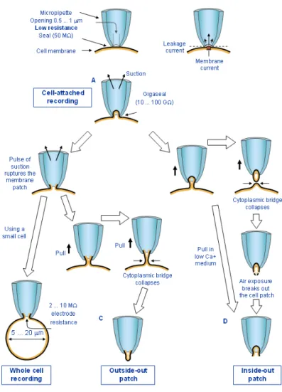
Figure 3.1.1. Patch-clamp recordings can be performed under four different configurations………...…………..……….56
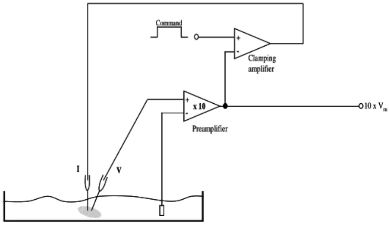
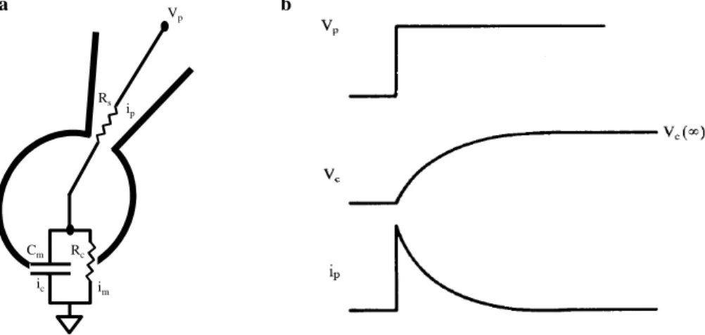
Figure 3.1.2. Two-electrode voltage-clamp schematic circuit………...…….59
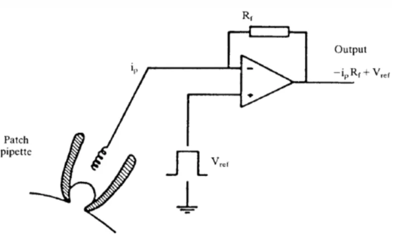
Figure 3.1.3. Schematic diagram of the headstage current/voltage amplifier……….……..60
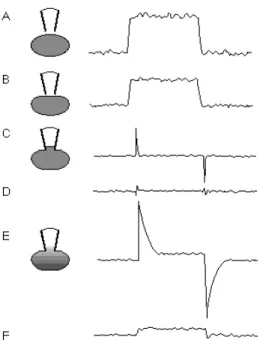
Figure 3.1.4. Test pulses produce different current responses as one proceeds through the establishment of a whole-cell voltage clamp recording………...62
Figure 3.1.5. Equivalent circuit of whole-cell recording………...64
Figure 3.2.1.1. The great limbic lobe of Broca………69
Figure 3.2.1.2. Diagram of a transverse hippocampal slice……….72
Figure 3.3.1. Effect of the anionic detergent SDS on proteins………....77
Figure 3.3.1.1. Discontinuous SDS-PAGE electrophoresis procedure……...78
Figure 3.3.2.1. Protein Blotting procedure…………...………...80
Figure 3.4.1. Reaction of Sulfo-NHS-LC-biotin with a primary amine……..84
Figure 5.1.1. Patch-clamp recordings of AMPAR-mediated currents……...99
Figure 5.1.2. Activation of A2A adenosine receptors potentiates the amplitude of AMPAR-mediated currents recorded from CA1 pyramidal cells…...…..102
Figure 5.1.3. A2A receptor- induced potentiation of AMPA currents does not
depend on NMDA or GABAA receptor activation, nor does it depend on synaptic activity………..…...105
Figure 5.1.4. Activation of A1 adenosine receptors inhibits the amplitude of
AMPA-evoked postsynaptic currents in CA1 pyramidal cells………..107
Figure 5.1.5. Activation of A2A adenosine receptors does not affect the
amplitude of AMPA-evoked postsynaptic currents recorded from stratum radiatum or stratum oriens interneurons………109
Figure 5.1.6. Activation of A2A receptors increases the amplitude, but not the
frequency, of spontaneous miniature excitatory postsynaptic currents (mEPSCs)……….….112
Figure 5.1.7. Facilitation of AMPA-evoked currents by A2A receptor
activation is dependent on postsynaptic PKA, but not PKC, activity……...115
Figure 5.1.8. AMPA-evoked currents are potentiated by superfusion of an adenylate cyclase activator………...……….117
Figure 5.1.9. Activation of A2A receptors enhances membrane expression of
phospho Ser845 GluR1….……….………119
Figure 5.1.10. Facilitation of AMPA-evoked currents by A2A receptor
activation is not dependent on postsynaptic protein synthesis…….……….121
Figure 5.1.11. Activation of A2A receptors facilitates afferent-evoked EPSCs
and Long Term Potentiation (LTP)……….…………..…124
Figure 5.1.12 Endogenous modulation of LTP expression by A2A receptor
activation………...126
Figure 5.2.1. Transient in vitro ischemia causes a significant increase in afferent-evoked Excitatory Postsynaptic Current (EPSC) amplitude……....138
component……….139
Figure 5.2.3. Intracellular spermine prevents ischemia-induced increase in afferent evoked EPSCs………..………141
Figure 5.2.4. A2A receptor blockade prevents ischemia-induced increase in
afferent evoked EPSCs……….………….142
Figure 5.2.5. Prevention of ischemia-induced facilitation of EPSC amplitude by A2A receptor blockade is preserved in the presence of internal spermine..
………...143
Figure 5.3.1 OSM potentiates inhibition of synaptic transmission caused by A1 receptor activation………156
Figure 5.3.2. LIF does not alter inhibition of synaptic transmission caused by A1 receptor activation………157
Figure 5.3.3. Oncostatin M potentiates hypoxia-induced inhibition of synaptic transmission………..………...….159
Figure 5.3.4. Hypoxia-induced inhibition of synaptic transmission is dependent on adenosine A1 receptor activation…………..………...………160
List of abbreviations
ABP: AMPA receptor binding protein AC: adenylate cyclase
aCSF: artificial cerebrospinal fluid ADAC: adenosine amine congener
AIDA: 1-Amino-2,3-dihydro-1H-indene-1,5-dicarboxylic acid AMPA: α-amino-3-hydroxy-5-methyl-4-isoxazolepropionic acid
AMPAR: AMPA receptor
AMP: adenosine 5´-monophosphate ANOVA: analysis of variance AP: alkaline phosphatase ATP: adenosine 5´-triphosphate
BDNF: brain-derived neurotrophic factor Bicc: bicuculline
BSA: bovine serum albumin CA: cornu ammonis
CaMKII: calcium/calmodulin-dependent protein kinase cAMP: 3´, 5´ -cyclic AMP; adenosine 3´, 5´ -cyclophosphate CGRP: calcitonin gene-related peptide
CGS 21680: 2-[4-(2-p-carboxyethyl)phenylamino]-50-N-ethylcarboxamidoadenosine CNQX: 6-cyano-7-nitroquinoxaline-2,3-dione
CNTF: ciliary neurotrophic factor CPA: N6-cyclopentyladenosine
CREB: cAMP response element-binding protein CT-1: cardiotrophin 1
DIC: differential infra-red interference contrast DL-APV: DL-2-amino-5-phosphonovaleric acid DMSO: dimethylsulfoxide
DNA: deoxyribonucleic acid
DPCPX: 8-cyclopentyl-1,3-dipropylxanthine DTT: dithiothreitol
EAAT: excitatory amino acid transporter Ecto-5′-NT: Ecto-5′-nucleotidase EDTA: ethylenediaminetetraacetic acid EGTA: ethylene glycol tetraacetic acid
E-NPP: ectonucleotide pyrophosphatase/phosphodiesterase ENT: equilibrative nucleoside transporter
E-NTPDase: ectonucleoside triphosphate diphosphohydrolase EPSC: excitatory postsynaptic current
GABA: γ-aminobutyric acid
GAT: GABA transporter
GF109203X: bisindolylmaleimide I
GIRK: G-protein dependent inwardly rectifying K+ channel
GRIP: glutamate receptor interacting protein GTP: guanosine 5'-triphosphate
H-89: N-[2-((p-bromocinnamyl)amino)ethyl]5-isoquinolinesulfonamide IL-6: interleukin-6
HEPES: N-2-hydroxyethylpiperazine-N'-2-ethanesulfonic acid HRP: horseradish peroxidase
JAK-STAT: Janus-activated kinase–signal transducer and activator of transcription Kd: equilibrium dissociation constant
L-AP4: 2-amino-4-phosphonobutyrate LIF: Leukemia inhibitory factor LIFr: LIF receptor
LTD: long term depression LTP: long term potentiation
LY 354740: (1S,2S,5R,6S)-2-aminobicyclo[3.1.0]hexane-2,6-dicarboxylic acid MAPK: mitogen-activated protein kinase
MCA: middle cerebral artery
mEPSC: miniature excitatory postsynaptic current
NBQX: 1,2,3,4-tetrahydro-6-nitro-2,3-dioxo-benzo[f]quinoxaline-7-sulfonamide NHS: n-hydroxysuccinimide
NKCC: sodium/potassium/chloride co-transporters NMDA: N-methyl-D-aspartate
NMDAR: NMDA receptor NO: nitric oxide
NSF: N-ethylmaleimide-sensitive fusion protein OSM: oncostatin M
OSMr: OSM receptor
PACAP: pituitary adenylate cyclase-activating polypeptide PBS: phosphate-buffered saline
PICK1: protein interacting with C kinase-1 PKA: protein kinase A
PKC: protein kinase C PPF: paired-pulse facilitation PSC: postsynaptic current PSD-95: post-synaptic density 95 PTP: post-tetanic potentiation PVDF: polyvinylidene fluoride
RIPA: radio-immunoprecipitation assay RNA: ribonucleic acid
(RS)-APICA: (RS)-1-amino-5-phosphonoindan-1-carboxylic acid RT-PCR: reverse transcription-polymerase chain reaction SEM: standard error of the mean
SCH 58261:
7-(2-phenylethyl)-5-amino-2-(2-furyl)-pyrazolo-[4,3-e]-1,2,4-triazolo[1,5-c]pyrimidine
SDS: sodium dodecyl sulfate
SDS-PAGE: SDS polyacrylamide gel electrophoresis Ser: Serine
SNAP: soluble NSF attachment protein
TARP: transmembrane AMPA receptor regulatory protein TEA: tetraethylammonium
TEMED: 1,2-bis(dimethylamino)ethane TBS: tris-buffered saline
Tris: tris-hydroxymethyl-aminomethane Trk: tropomyosin-related kinase TTX: tetrodotoxin
Most of the fast excitatory transmission in the brain is conveyed by ionotropic glutamate α -amino-3-hydroxy-5-methyl-4-isoxazolepropionic acid (AMPA) receptors, formed by tetrameric assemblies of different subunit (GluR1-GluR4) composition. Modulation of AMPA receptors enables profound changes in synaptic efficiency, underlying the maturation of neuronal networks throughout development and plasticity, but also glutamate-mediated excitotoxicity. Accurate tuning of AMPA function can be attained by subunit phosphorylation, affecting channel properties and receptor trafficking rates. Accordingly, activation of noradrenergic and dopaminergic metabotropic receptors positively modulates AMPA receptor function through increased PKA activity and GluR1 phosphorylation, an effect restricted to brain areas targeted by pathways relying on these neurotransmitters. In contrast, adenosine is ubiquitously present throughout the nervous system, being released by glia and neurons or derived from the extracellular catabolism of adenine nucleotides. The present work thus aimed at evaluating the modulation of postsynaptic AMPA receptors by high-affinity, G-coupled A1 and A2A adenosine
receptors and its implications for long term potentiation (LTP), widely perceived as the cellular correlate for memory formation (chapter 5.1). The involvement of A2A (chapter 5.2) and A1 (chapter 5.3)
receptor-mediated tuning of glutamatergic transmission was further addressed in excitotoxicity conditions. Exogenous activation of A2A receptors by
2-[4-(2-p-carboxyethyl)phenylamino]-50-N-ethylcarboxamidoadenosine (CGS21680) was found to significantly facilitate AMPA-evoked currents in CA1 pyramidal neurons, by a postsynaptic PKA-dependent mechanism leading to increased GluR1 membrane expression. The
functional impact of this modulation was evidenced by LTP facilitation at the CA3-CA1 synapse, following brief CGS21680 application. Moreover, endogenous A2A receptor activation was required for
ischemia-induced facilitation of glutamatergic transmission, revealing a conserved regulatory mechanism between both forms of plasticity, which may be of interest for functional recovery (through circuit rewiring) from stroke. Additionally, results suggests that some (OSM), but not all (LIF) immunoregulatory cytokines of the IL-6 family can exert neuroprotection from hypoxia through upregulation of A1
receptors, to tone down synaptic transmission and consequently, energy expenditure.
Os receptores ionotrópicos para o glutamato do tipo AMPA são responsáveis por mediar grande parte da transmissão excitatória rápida que tem lugar no sistema nervoso central. Estes receptores são constituídos por tetrâmeros, contendo diferentes combinações de subunidades (GluR1-GluR4) e a sua regulação é responsável por consideráveis alterações na eficiência da transmissão sináptica, inerentes à maturação de redes neuronais ao longo do desenvolvimento bem como a fenómenos de plasticidade. Contudo, os receptores AMPA são também moléculas-chave em situações de excitotoxicidade mediada por glutamato. Tanto a alteração da composição do tetrâmero, como variações no estado de fosforilação das suas subunidades, constituem mecanismos que permitem uma regulação eficaz da função destes receptores. De facto, há evidência de que à activação de receptores metabotrópicos para a dopamina ou noradrenalina se associa um aumento de função AMPA, através de um aumento na actividade da PKA e consequente exarcebamento do estado de fosforilação de subunidades GluR1. No entanto, este tipo de regulação da função AMPA encontra-se necessariamente restrito às áreas cerebrais que recebem inervação dopaminérgica ou noradrenérgica. O mesmo não acontece com a modulação pela adenosina, cuja presença é ubíqua no sistema nervoso, uma vez que pode ser libertada por células da glia e neurónios, através de locus pré-, pós- e não- sinápticos. Além disso, o catabolismo de nucleótidos de adenina, co-libertados com diferentes neurotransmissores, representa uma fonte de adenosina extracelular adicional. O trabalho experimental de que trata a presente tese teve assim como objectivo investigar uma possível modulação da componente AMPA pós-sináptica, através da activação dos receptores
de alta afinidade para a adenosina, do tipo A1 e A2A. Foram ainda
abordadas as implicações deste mecanismo de regulação para o estabelecimento de potenciação a longo prazo (LTP) da eficiência sináptica, a qual é encarada como o substrato celular para a formação de novas memórias (capítulo 5.1). A ocorrência de uma possível modulação da transmissão glutamatérgica pelos receptores A2A
(capítulo 5.2) e A1 (capítulo 5.3) em situações de excitotoxicidade foi
estudada através da aplicação de modelos de isquémia e hipóxia. A grande maioria do trabalho experimental foi realizado em células piramidais da área CA1 do hipocampo, que representa uma população neuronal particularmente propensa a expressar alterações de eficiência sináptica de acordo com os níveis de actividade, mas que é também especialmente susceptível a morte por excitotoxicidade, após isquémia.
As observações mais relevantes descritas na presente tese dizem respeito à facilitação significativa de correntes evocadas por ejecção de AMPA (correntes AMPA), observada após activação dos receptores A2A com um agonista selectivo (CGS 21680). Esta facilitação teve
expressão por meio de um mecanismo dependente de PKA, mas não de síntese proteica, tendo sido reproduzida pela perfusão de um activador da adenilato ciclase (forskolina). Além disso, as acções do agonista A2A
não foram afectadas aquando do bloqueio simultâneo dos receptores GABAA, NMDA e canais de sódio dependentes da voltagem, pelo que
nenhum dos últimos parece desempenhar um papel na mesma. É ainda possível concluir que o efeito do agonista A2A terá um locus de
expressão pós-sináptico, na medida em que se manteve, mesmo em condições de bloqueio do disparo de potenciais de acção. Efectivamente, observou-se que a perfusão de CGS 21680 conduziu a
espontâneas, o que sugere um acréscimo na capacidade de resposta (pós-sináptica) ao glutamato libertado. É sabido que a fosforilação pela PKA de subunidades GluR1 aumenta a probabilidade de abertura de receptores AMPA, além do que leva a um aumento do seu tráfego (em receptores que contenham subunidades GluR1) para porções extra-sinápticas da membrana. Desta forma, quando se avaliou a expressão destas subunidades ao nível da membrana, verificou-se que tanto a expressão total, como a de subunidades fosforiladas se encontrava aumentada após tratamento com o agonista A2A. A relevância deste
acréscimo na expressão membranar de receptores AMPA traduziu-se numa facilitação da LTP entre neurónios das zonas CA3 e CA1, após um curto tratamento com CGS 21680. Além disso, a modulação endógena da componente AMPA pós-sináptica revelou-se necessária à expressão de LTP, já que a última se encontrou francamente diminuída em situações de bloqueio dos receptores A2A. Face aos resultados
obtidos na primeira parte deste trabalho, é pois possível inferir que, em condições de aumento súbito da adenosina extracelular proporcionadas por um exarcebamento da actividade neuronal, a activação de receptores A2A, facilita a fosforilação pela PKA de subunidades GluR1
– aumentando a disponibilidade de receptores nas reservas membranares a partir das quais estes são recrutados para a sinapse, permitindo reforçar a transmissão sináptica.
A exposição a períodos transitórios de hipóxia ou isquémia é suficiente para induzir um aumento mantido na eficiência sináptica que partilha muitos dos mecanismos inerentes à expressão de LTP, incluindo um ganho de função pela componente AMPA pós-sináptica.
Com o objectivo de investigar a contribuição por uma possível modulação pelos receptores A2A da transmissão glutamatérgica para
esta forma de plasticidade, compararam-se os efeitos de breves insultos isquémicos, na presença e na ausência (controlo) de um antagonista selectivo dos receptores A2A da adenosina (SCH 58261). A sujeição ao
episódio isquémico transitório começou por anular a transmissão sináptica, a qual recuperou após reoxigenação, progressivamente, até valores acima dos valores iniciais de referência. Estas observações validam portanto o recurso a este tipo de insulto isquémico transitório para o estudo de formas de plasticidade induzidas por isquémia. Para além disso, observou-se que esta facilitação da transmissão glutamatérgica era completamente perdida, aquando da inclusão de espermina na solução intracelular (com acesso ao compartimento citosólico), o que sugere que requererá a activação de receptores AMPA permeáveis ao cálcio (bloqueados por espermina intracelular). Estes resultados revelam assim mais um mecanismo comum à chamada “LTP induzida por isquémia” e à sua correspondente fisiológica (LTP), a qual depende também da inserção sináptica, transitória, de receptores AMPA permeáveis ao cálcio. Quando o insulto isquémico foi realizado na presença de um antagonista A2A, tão pouco se observou uma
facilitação da transmissão glutamatérgica após isquémia. Estes dados são consistentes com um envolvimento endógeno dos receptores A2A
nesta forma de plasticidade, à semelhança da contribuição que têm para a expressão de LTP. A recuperação da transmissão sináptica em condições de bloqueio dos receptores A2A manteve-se inalterada na
presença de espermina intracelular, sugerindo o recrutamento de um mecanismo comum, em ambos os casos (bloqueio dos receptores AMPA permeáveis ao cálcio e bloqueio dos receptores A2A). Para uma
plasticidade, será necessária uma avaliação do intervalo de tempo pelo qual os receptores AMPA permeáveis ao cálcio permanecem na sinapse, bem como testar directamente a hipótese de que os receptores A2A modulem a inserção de receptores AMPA permeáveis ao cálcio.
Ainda assim, esta segunda parte do presente trabalho tem o mérito de revelar um (novo) mecanismo comum a duas formas de plasticidade consideradas completamente distintas até recentemente, colocando em evidência um envolvimento relevante dos receptores A2A nas alterações
da eficiência sináptica desencadeadas por isquémia.
É desde há muito reconhecido o potencial neuroprotector que a activação de receptores A1 da adenosina oferece contra insultos de
hipóxia e/ou isquémia, ainda que a aplicação terapêutica de agonistas A1 se encontre muito dificultada pela ocorrência de graves efeitos
secundários. É plausível que a administração de moléculas capazes de modular a função destes receptores possa oferecer uma alternativa viável ao uso de agonistas A1. De facto, esta aplicação foi recentemente
sugerida para a interleucina 6 (IL-6), capaz de proteger neurónios de dano por excitotoxicidade, através de um aumento nos níveis de receptores A1 e um consequente aumento da sua sensibilidade ao tónus
adenosinérgico. Com o objectivo de avaliar se esta acção seria extensível a outros membros da mesma família de interleucinas, analisaram-se os efeitos do tratamento com oncostatina (OSM) e factor inibidor de leucemia (LIF) sobre a modulação da transmissão glutamatérgica dependente de receptores A1. Após tratamento com
OSM, mas não com LIF, observou-se uma potenciação da inbição das respostas sinápticas glutamatérgicas induzida por perfusão de um
agonista selectivo de receptores A1 (CPA), o que é consistente com um
ganho de função destes receptores. A relevância desta interacção para uma situação de modulação da transmissão sináptica aquando de um insulto excitotóxico, reflectiu-se numa maior inibição da transmissão glutamatérgica, dependente da activação de receptores A1, após
sujeição a um episódio hipóxico transitório. Estes dados funcionais corroboram os resultados de colaboradores, que mostram que apenas o tratamento com OSM consegue aumentar os níveis de proteína e mRNA correspondentes ao receptor A1. No seu conjunto, os resultados
sugerem que o acréscimo de função dos receptores A1 da adenosina é
necessário à neuroprotecção exercida pelas citocinas IL-6 e OSM, mas não para a protecção mediada por LIF. No entanto, a pleiotropia de efeitos conhecida para esta família de interleucinas, bem como o tipo de protocolo usado para testar o seu envolvimento na neuroprotecção contra hipóxia (pré-tratamento), tornam prematura qualquer sugestão do seu uso como potenciais agentes terapêuticos.
Do presente trabalho emerge desta forma a noção da modulação de receptores A1 como mecanismo redundante capaz de conferir
propriedades de neuroprotecção a citocinas imunorreguladoras. Por outro lado e através da activação de receptors A2A, com consequente
ganho de função AMPA, este trabalho mostrou como a adenosina é capaz de modular a plasticidade sináptica, o que se poderá revelar de particular interesse no estabelecimento de novos contactos sinápticos que ocorre em resposta a insultos isquémicos.
1
Introduction
In the beginning of the 20th century, it was commonly accepted by the scientific community that synaptic communication relied upon bio-electricity. One of the first clues that this might not be the case emerged from Ramón y Cajal´s cutting edge discoveries, revealing distinguishable gaps between communicating neurons, which questioned the possibility of electrical transmission through them. In 1921, the seminal work by Otto Loewi with ex-vivo vagus nerve preparations came to confirm the notion that neurons could indeed communicate by releasing unknown chemical substances (Loewi, 1921). Loewi dissected two beating hearts out of frogs, maintained them in saline solution and severed the autonomous innervation to one of them. By electrical stimulation of the vagus, the first heart was made to beat slower and when the liquid bathing it was applied to the second heart, a comparable decrease in heart rate was observed. This experiment elegantly showed that there had to be some soluble chemical, released upon vagus nerve stimulation, which was controlling the heart rate. Loewi´s Vagusstoff was later found to be acetylcholine, which was thus the first identified neurotransmitter. As for glutamate, its role as the principal excitatory neurotransmitter in the mammalian brain would slowly become established over the course of more than twenty years, perhaps because it seemed unlikely that this non-essential, highly abundant amino-acid, central to energy metabolism, could in addition function as a tightly regulated neurotransmitter. In 1954, Hayashi observed that glutamate injection into the brain or carotid arteries produced convulsions, leading him to
hypothesize it might also work as a transmitter molecule (Hayashi, 1954). On the following years, Watkins and his co-workers further explored the actions of glutamate and related amino-acids upon populations of neurons (Curtis and Watkins, 1959; Curtis et al., 1960; Curtis and Watkins, 1961), which prompted them to propose the existence of different types of glutamate receptors, according to ligand selectivity. Interestingly, the realization that glutamate and other excitatory amino-acids exerted their excitatory actions via multiple receptors preceded their establishment as synaptic neurotransmitter receptors. Indeed, glutamate receptors were only definitely shown to be synaptic receptors in the late 1970´s, by the sensitivity of certain excitatory pathways in the spinal cord to specific NMDA receptor antagonists (Biscoe et al., 1977). By the early 1980´s, glutamate was commonly accepted to exert postsynaptic actions upon three families of ionotropic receptors which were named after their preferred agonists:
N-methyl-D-aspartate (NMDA), α -amino-3-hydroxy-5-methyl-4-isoxazolepropionic acid (AMPA) and kainate receptors. All of these constitute intrinsic cation permeable channels, albeit with variable permeabilities to sodium and calcium ions, depending on the type and subunit composition of the receptor (reviewed in Meldrum, 2000). Metabotropic, G-protein coupled, glutamate receptors (mGluRs) were not described until 1987, having been cloned in the early 1990´s (Schoepp and Conn, 1993) (Table 1).
Table 1 – Glutamate Receptor Classification, Subunit Composition and Pharmacology CPPG Glutamate L-AP4 mGluR4 mGluR6-8 Metabotropic group III (RS)-APICA Glutamate LY 354740 mGluR2,3 Metabotropic group II AIDA Fenobam Glutamate L-quisqualate mGluR1 mGluR5 Metabotropic group I CNQX Topiramate Glutamate Kainate Domoate GluK1-5 GluR5-7 KA1,2 Kainate DL-APV Glutamate NMDA GluN1 GluN2A-D GluN3A/B NR1 NR2A-D NR3A,B NMDA CNQX NBQX Glutamate AMPA Kainate L-quisqualate GluA1-4 GluR1-4 AMPA Antagonists Agonists Subunit Nomenclature (see Collingridge et al., 2009) Subunit
Nomenclature (e.g., Watkins and Jane, 2006) Receptor family CPPG Glutamate L-AP4 mGluR4 mGluR6-8 Metabotropic group III (RS)-APICA Glutamate LY 354740 mGluR2,3 Metabotropic group II AIDA Fenobam Glutamate L-quisqualate mGluR1 mGluR5 Metabotropic group I CNQX Topiramate Glutamate Kainate Domoate GluK1-5 GluR5-7 KA1,2 Kainate DL-APV Glutamate NMDA GluN1 GluN2A-D GluN3A/B NR1 NR2A-D NR3A,B NMDA CNQX NBQX Glutamate AMPA Kainate L-quisqualate GluA1-4 GluR1-4 AMPA Antagonists Agonists Subunit Nomenclature (see Collingridge et al., 2009) Subunit
Nomenclature (e.g., Watkins and Jane, 2006) Receptor family Io n o tr o p ic r ec e p to rs M et a b o tr o p ic r e ce p to r s
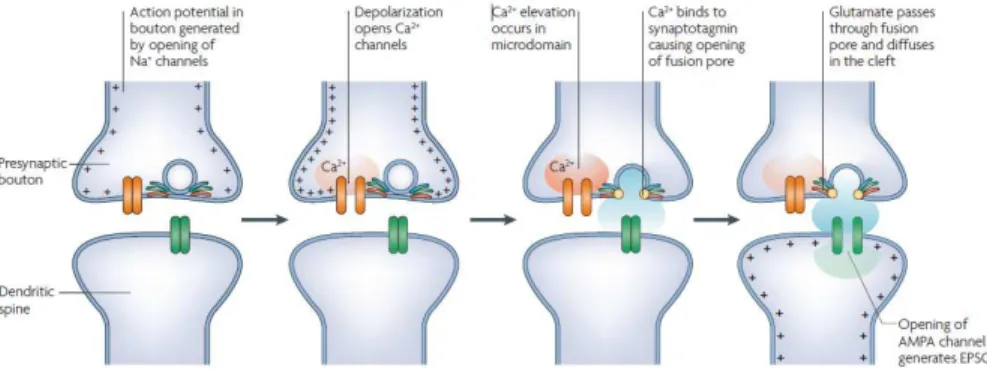
Despite this wide variability of glutamate receptors, most of the fast excitatory transmission that takes place in the mammalian central nervous system is conveyed by AMPA receptors (Figure 1.1). Therefore, the presence and function of these receptors at synapses must be carefully regulated in order to ensure not only correct neuronal communication, as also the reinforcement of synaptic efficacy that is thought to underlie the formation of new memories (Bliss and Collingridge, 1993; Martin et al., 2000).
Figure 1.1.Steps in the process of chemical synaptic transmission. These steps occur in both
vertebrates and invertebrates, during chemical neurotransmission at the neuromuscular junction as well as in central synapses. The example given applies to fast synaptic glutamatergic transmission, but also to that mediated by other neurotransmitters and corresponding postsynaptic ionotropic receptors. Adapted from Lisman et al., 2007.
1.1 AMPA receptors
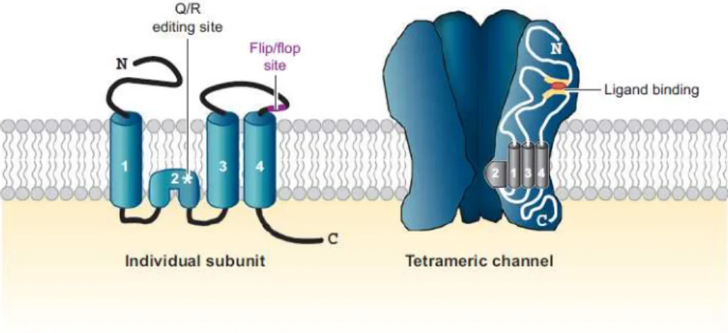
AMPA receptors are ionotropic, ligand-gated receptors which undergo conformational changes upon glutamate binding, rendering them transiently permeable to cation influx and causing a net depolarization of the postsynaptic membrane that is responsible for propagating information at excitatory synapses throughout the brain. They are tetramers composed of four subunits (GluR1-GluR4), which combine in different stoichiometries to form ion channels with distinct functional properties (Hollmann and Heinemann, 1994) (see Figure 1.1.1). Accordingly, expression of receptor subunits is developmentally regulated and is brain region specific (reviewed in Shepherd and Huganir, 2007). Furthermore, receptor subunit composition determines not only channel properties (single-channel conductance, desensitization kinetics), but also stoichiometric association with transmembrane AMPA receptor regulatory proteins (TARPs), which regulate receptor maturation and trafficking (Gereau and Swanson,
2008). Aside from that introduced from different subunit combinations, AMPA receptor diversity is also generated within each subunit, by variations arising from alternative splicing, RNA editing and post-transcriptional modifications, such as phosphorylation and/or palmitoylation. Each AMPA receptor subunit comprises four hydrophobic domains, three of which traverse the membrane (TM1, TM3 and TM4), while one (M2) forms a re-entrant loop that faces the cytoplasm and contributes to the assembly of the cation permeable channel pore (reviewed by Santos et al., 2009) (Figure 1.1). Reflecting their most likely evolutionary origin, the structure of AMPA receptors is in accordance with the fusion of three gene segments that were once individual bacterial proteins. As such, the amino terminal is homologous to the bacterial leucine-isoleucine-valine–binding protein while the ligand-binding domain resembles a bacterial lysine-arginine-orthinine binding protein (Gereau and Swanson, 2008). In turn, the two segments (S1 and S2) of the ligand-binding domain are interrupted by the ion channel pore, whose structure is similar to that of bacterial K+ channels. Both the extracellular and transmembrane regions share a high degree of homology between different AMPAR subunits (Gereau and Swanson, 2008). The same is not true for their carboxyl terminal (C terminal), which is the most variable region among different subunits and the site of subunit-governed protein interactions. Within the C terminal, different phosphorylation sites can also be found, which has implications for receptor trafficking and synaptic delivery (Esteban, 2003).
Figure 1.2. Structure and composition of AMPA receptors. Structure of AMPAR subunits
and the tetrameric channel they form. Individual subunits are composed of four transmembrane domains, and the channel consists of four subunits, which are usually two dimers. In the endoplasmic reticulum, two subunits combine to form an initial dimer, which requires interactions between amino terminals; this is followed by a second dimerization process relying upon association of membrane-spanning and extracellular loop regions (e.g., Zipp, 2007). Adapted from Shepherd and Huganir, 2007.
1.1.1 Alternative splicing of AMPA receptor subunits
Different post-transcriptional modifications can amplify the functional diversity of AMPA receptors, by generating multiple subunit isoforms. For instance, the mRNA for each AMPA receptor subunit can be alternatively spliced to produce either a flip or a flop isoform, a process which introduces variation within the extracellular portion of the protein, close to the final transmembrane domain (TM4). Although flip and flop subunit forms only differ in a few aminoacid residues, their incorporation affects receptor desensitization, as well as export from the endoplasmic reticulum (reviewed by Santos et al., 2009) and possibly even receptor formation and stoichiometry (Brorson et al., 2004). Accordingly, the expression of flip and forms varies throughout development and across different neuronal types. The flop versions, which predominate in the adult brain, generally desensitize much more
rapidly than the flip forms in response to glutamate (Sommer et al., 1990). In addition, alternative splicing affects the pharmacologic properties of the AMPA receptor channel, conditioning its differential sensitivity to allosteric modulators (Monyer et al., 1991).
The intracellular, C terminal domains of GluR2 and GluR4 subunits may also undergo alternative splicing (Gallo et al., 1997). As a consequence, GluR2 and GluR4 can be expressed as both short- and long- tailed proteins. As such, GluR1, the predominant splice form of GluR4 and an alternative splice form of GluR2 (GluR2L) have longer cytoplasmic tails than GluR3, the predominant splice form of GluR2 and the alternative splice form of GluR4 (GluR4c) (Santos et al., 2009) (see Figure 1.1.1.1). Importantly, receptors which comprise only short-tailed subunits (e.g., GluR2/GluR3) are subject to constitutive trafficking to and from the synaptic membrane, unlike what occurs with those containing long-tailed subunits (e.g., GluR1/GluR2), whose synaptic targeting occurs in an activity-dependent way (Esteban et al., 2003). Finally, alternative splicing of the C terminal regulates AMPA receptor function by conditioning binding to specific interacting proteins, as well as regulation by protein phosphorylation (Song and Huganir, 2002).
Figure 1.1.1.1. Localization of protein binding and phosphorylation sites in the C terminal of AMPA receptor subunits. In addition to the predominanf subunit forms expressed,
alternatively spliced forms of GluR2 (Glu2L) and GluR4 (GluR4c) are also considered. Protein binding sites are indicated by boxes, adjacent to proteins with which they have been described to interact. Phosphorylation sites are underlined and evidenced by a larger font size. Adapted from Santos et al., 2009.
1.1.2 RNA Editing of AMPA Receptor subunits
Additional AMPA receptor diversity is generated by RNA editing, a process involving enzymatic deamination of ribonucleotides (adenosine residues) in pre-spliced mRNA encoding glutamate receptor subunits (Bass, 2002). This mechanism brings about the replacement of a gene-encoded aminoacid by a different, non-coded one, thus allowing the assembly of receptors with novel physiological properties. In AMPA receptors, RNA editing mostly concerns the GluR2 subunit, where it mediates the conversion of a glutamine (CAG) to an arginine (CGG) codon in the ion channel pore region. As a result, edited GluR2 subunits possess an arginine (R) in the M2 membrane spanning
segment at position 586, whereas GluR1, GluR3 and GluR4 subunits have a glutamine (Q) in the homologous position (Hammond, 2008). This process affects retention of GluR2 subunits at the endoplasmic reticulum and also the properties of the AMPA receptors they come to integrate. In fact, Q/R editing constitutes a major quality control checkpoint, detaining GluR2 subunits in the endoplasmic reticulum and diminishing the formation of GluR2 homomeric tetramers (see Santos et al., 2009). RNA editing is also a widespread mechanism for regulating both the calcium permeability and channel rectification properties of the ion channel. AMPA receptors containing Q/R edited GluR2 subunits display low single channel conductance and little permeability to calcium ions, dictated by the size and positive charge of the substitute arginine residue (Burnashev et al., 1992; Jonas and Burnashev, 1995). Most GluR2 subunits in the adult brain exist in their edited form and evidence suggests that deficient RNA editing of this subunit correlates with neuronal death in amyotrophic lateral sclerosis (Kwak and Kawahara, 2005), as well as with dysfunctional AMPA function after ischemia (Peng et al., 2006). In contrast, the remaining subunits mostly exist in their non-edited forms (Burnashev et al., 1992), which, when assembled into GluR2-free homomers or heteromers, display permeability to calcium and inward-rectifying properties due to the glutamine present in the pore-forming region. Calcium permeability is thus governed by the relative expression of GluR2 subunits, and the (GluR1+GluR3)/GluR2 ratio has been used as a predictor of the assembly of calcium-permeable AMPA receptors (see Pelligrini-Gianpetro et al., 1997). Calcium permeable, GluR2-lacking AMPA receptors are also susceptible to blockade by endogenous, intracellular polyamines, which mediate channel blockade in a voltage-dependent
manner (Donevan and Rogawski., 1995; Washburn et al., 1997) (see Figure 1.1.2.1). In agreement with its elemental contribution for overall AMPA receptor function, the relative abundance of GluR2 at synaptic AMPARs has been shown to increase with development, in cultured CA1 neurons (Pickard et al., 2000). Indeed, in the adult hippocampus, the AMPA receptor pool is mostly composed of GluR2/GluR3 and GluR1/GluR2 complexes (Wenthold et al., 1996), causing approximately linear current-voltage relationships, with a mean reversal potential of 0 mV (Jonas and Sakmann, 1992).
Figure 1.1.2.1. Model accounting for the differential dependence of calcium permeability and inward rectification on GluR2 abundance. The essence of the model is that the ring of
carbonyl oxygens in GluR2-lacking AMPA receptors (A) contributes to or forms a binding site for permeating divalent cations. Internal polyamines also interact with this ring of polar residues. Incorporation of a single positively charged arginine into the Q/R site (B) disrupts the ring of carbonyl groups, which is postulated to eliminate the divalent ion binding site. This arginine also neutralizes a negative charge at the internal channel mouth by formation of a salt bridge between its guanidinium group and the carboxyl group of the aspartate, which reduces the number of anionic binding sites for internal polyamines and thus decreases their affinity for the channel. As more arginines are incorporated into the Q/R site (C), the number of negative charges at the channel mouth decreases, and hence polyamine affinity for the internal blocking site, is progressively reduced. Adapted from Washburn et al., 1997.
1.1.3 AMPA receptor subunit phosphorylation
After synthesis, AMPAR subunits are subject to post-translational modifications such as glycosylation, phosphorylation and palmitoylation. Each AMPA receptor subunit can be N-glycosylated at 4-6 different sites of its extracellular domain, a modification which protects from proteolytic clivage but that is not required for ligand recognition and does not seem to significantly affect receptor assembly or trafficking (Jiang, 2006). Although it has been proposed that this sort of protein modification may be involved in receptor maturation and transport, its functional repercussions are still unclear. AMPA receptor subunits can also undergo palmitoylation of cysteine residues in their C terminal and M2 domain, with consequences for postsynaptic receptor trafficking and membrane expression (see Santos et al., 2009). Phosphorylation of AMPA receptor subunit residues can occur under resting conditions or as a reponse to changes in synaptic activity. With the exception of GluR3, all AMPAR subunits have been reported to undergo phosphorylation on several amino acid residues of their C terminal, by a variety of kinases (Jiang et al., 2006). For instance, phosphorylation of GluR2 subunits at Serine 880 by PKC has been implicated in AMPA receptor internalization during NMDA-independent Long Term Depression (LTD) of synaptic transmission in the cerebellum (Chung et al., 2003). Interestingly, phosphorylation of Tyrosine 876 (Tyr 876) by Src family tyrosine kinases can also facilitate rapid internalization of GluR2-containing membrane AMPA receptors (Hayashi and Huganir, 2004). In both cases, subunit phosphorylation modulates the receptor´s ability to bind regulatory proteins that assist in its trafficking to and from the membrane. The
function of a third potential PKC phosphorylation site (Ser 863) identified on the C-terminal of the GluR2 subunit (Hirai et al., 2000) remains unknown. GluR4 subunits comprise two phosphorylation sites in their intracellular C terminals. One of them, Serine 842, can be phosphorylated in vitro by PKA, PKC and CaMKII (Carvalho et al., 1999) and its phosphorylation by PKA has been shown to be both required and sufficient for synaptic delivery of homomeric receptors (Esteban et al., 2003). Phosphorylation of Ser 842 by PKC can also increase calcium influx through activated AMPA receptors, in cultured retinal neurons (Carvalho et al., 1998). In addition, threonine 830 represents a potential phosphorylation site for PKC (Carvalho et al., 1999). As for GluR1 subunit phosphorylation, it has been the subject of intense research, prompted by the pivotal role that GluR1-containing receptors have been shown to play in hippocampal plasticity (Zamanillo et al., 1999; Shi et al., 1999). As a result of such research, four phosphorylation sites have been described to occur in the C-terminal of GluR1 subunits. These are serine 831 (Ser 831), which can be phosphorylated by both PKC (Roche et al., 1996) and CaMKII (Mammen et al., 1997); serine 845 (Ser 845), a PKA phosphorylation site (Roche et al., 1996); serine 818 (Ser 818, Boehm et al., 2006) and threonine 840 (Thr 840, Munton et al., 2007; Lee et al., 2007), which are two additional PKC phosphorylation sites that were recently discovered. Phosphorylation of Ser 845 by PKA has been shown to facilitate receptor peak open probability (Banke et al., 2000) and lead to long term potentiation (LTP) of naïve synapses (Lee et al., 2000), while its dephosphorylation associates with decreased synaptic strength underlying LTD (Ehlers, 2000). Phosphorylation of Ser 831 by CamKII enhances single channel conductance (Derkach et al., 1999) and
intervenes in the recruitment of silent synapses, as well as in synaptic transmission facilitation during LTP (reviewed by Palmer et al., 2005). Of note is the fact that although both Serine 831 and Serine 845 are required for AMPAR delivery into synapses, transgenic mice with mutations in both phosphorylation sites still display reduced LTP (Lee et al., 2003a). Recently, a third regulatory phosphorylation site has been implicated in AMPA receptor trafficking underlying LTP. Phosphorylation of Serine 818 (by PKC) was shown to increase after LTP and pharmacological induction of its phosphorylation could enable synaptic incorporation of GluR1-containing AMPA receptors, possibly through modified interaction with candidate proteins capable of modulating receptor trafficking (Boehm et al., 2006). Although it is still not clear how do the above identified phosphorylation events on GluR1 subunits interact to produce synaptic potentiation, it is possible that a conjunctive phosphorylation by Ser 845, Ser 831 and Ser 818 may be required (Boehm et al., 2006). Finally, LTP induction has no effect on GluR1 phosphorylation at Threonine 840 (Delgado et al., 2007). In turn, dephosphorylation of this site, secondary to NMDA receptor activation and protein phosphatase 1/2A activity, has been shown to correlate with changes in synaptic strength during hippocampal LTD (Delgado et al., 2007). As these examples illustrate, different phosphorylation sites are associated with distinct alterations in AMPA receptor properties and/or trafficking pattern. However, in what concerns the role played by phosphorylation sites in the C terminal of GluR1 subunits, all of them have been shown to affect some form of synaptic plasticity (Lee et al., 2000; Esteban, 2003; Boehm et al., 2006; Delgado et al., 2007).
1.1.4 AMPA receptor trafficking
Most AMPARs are thought to be synthesized in the neuronal cell body, far away from the dendritic spines whose receptor population they ultimately integrate. Before reaching this synaptic target, they must thus accomplish a series of trafficking steps, which include regulated exit from the endoplasmic reticulum and active transport through the microtubular cytoskeleton. All of these processes involve intricate networks of protein-protein interactions, between subunit domains and different sets of anchoring and regulatory proteins. The impact that interaction with these regulatory proteins had for AMPA receptor trafficking became clear with the study of the stargazer mouse, a spontaneous mutant that displays head tossing and ataxic gate and absence epilepsy (Ziff, 2007). In these animals, lack of functional stargazin compromises surface delivery of functional AMPA receptors in cerebellar granule cells, a phenotype that could be rescued by expressing the wild-type protein. Interestingly, when using a version of stargazing that lacked a binding site for post-synaptic density 95 (PSD-95), only AMPA receptor delivery to non-synaptic sites was recovered, demonstrating that stargazin´s ability to restore receptor delivery to synapses requires a secondary interaction with this scaffolding protein (Chen et al., 2000). Besides stargazin (or γ−2), seven other related proteins (γ−1, γ−3 to 8) constitute the transmembrane AMPA receptor regulatory protein (TARP) family (see Ziff, 2007). TARPs belong to the group of interacting proteins that lack a PDZ (post-synaptic density 95-discs large-zona occludens 1) domain, as well as NSF (N-ethylmaleimide-sensitive fusion protein) or the 4.1 protein. The second major group of proteins that govern AMPA receptor surface expression
is constituted by proteins with PDZ domains. PDZ domains are one of the best characterized protein interaction modules, responsible for mediating protein interactions by recognizing and binding peptide epitopes within their interacting partners. Widely expressed, PDZ domains were first recognized as sequence repeats in the primary structures of the PSD-95, disk-large and zona occludens-1 proteins and were named accordingly (Kurakin et al., 2007). Each PDZ doman binds only one ligand and binding selectivity is thought to result from variations in the size and geometry of the peptide-binding region (Palmer et al., 2005). Well known examples of PDZ-domain containing proteins are GRIP/ABP (glutamate receptor interacting protein/AMPA receptor binding protein) and PICK1 (protein interacting with C kinase-1). Frequently, PDZ-domain containing proteins function as scaffolds at the post-synaptic density, where they mediate organization and maintenance of large macromolecular complexes (see Figure 1.1.4.1).
Figure 1.1.4.1. Schematic representation of the role played by different interacting proteins upon AMPAR trafficking. Pathway (a) represents GluR1 trafficking and pathway
stargazin (1). GluR1-containing AMPARs are inserted at extrasynaptic sites through an activity-dependent mechanism (3a). Stargazin binds PSD95, which localizes the complex at the synapse where the receptor can bind other scaffolding proteins such as 4.1N (4a). Stargazin can then be released. GluR2-containing AMPAR are inserted at synaptic sites through a constitutive mechanism (3b). PICK1 facilitates the transport of the receptor possibly both to and from the plasma membrane but is removed from the AMPAR by the NSF/SNAP (SNAP, soluble NSF attachment protein) when the receptor reaches the plasma membrane. NSF/SNAP/PICK1 form a transient complex with GluR2-containing AMPAR (not shown on this schematic). AMPA receptors are stabilized at the membrane by scaffolding proteins. Phosphorylation by PKC prevents interaction between ABP/GRIP and GluR2 (4b) and favors the PICK1/GluR2 interaction (5b). AMPA receptors are then internalized following a coupling with PICK1, and AMPAR/PICK1 complex are destabilized by NSF/SNAP when the receptor reaches its destination (6b). GluR2-containing AMPARs can then interact with an intracellular pool of ABP/GRIP, possibly in the endosome (7b), and get recycled to the membrane through interaction with PICK1 (8b) or targeted to lysosomes (9b) and degraded (10b). Adapted from Kittler and Moss, 2006.
As illustrated in Figure 1.1.4.1, local receptor insertion and removal from synapses is a dynamic process. In fact, AMPA receptors are continuously being delivered and removed in and out of synapses in response to neuronal activity, adjusting synaptic strength during brain development and experience-dependent plasticity (reviewed by Esteban, 2003). Interestingly, receptor cycling dynamics between intracellular receptor stores and the cell surface is largely dependent upon receptor subunit composition. In fact, while GluR2–GluR3 oligomers are continuously being shipped to synapses in a manner independent of neuronal activity (Shi et al., 2001), synaptic delivery of GluR1–GluR2 (Esteban et al., 2003) and GluR4-containing receptors (Zhu et al., 2000) requires increased synaptic activity and consequent NMDA receptor activation. The first trafficking pathway thus allows the number of AMPARs at synapses to be preserved despite protein turnover (constitutive pathway), whereas the second (regulated pathway) enables activity-dependent regulated synaptic delivery of
GluR1–GluR2 receptors to synapses, during plasticity (Malinow et al., 2000) (Figure 1.1.4.2).
Figure 1.1.4.2. Constitutive and regulated trafficking of AMPARs at synapses. Left.
AMPAR oligomers containing GluR1 or GluR4 subunits are added into synapses in an activity-dependent manner (regulated delivery) during long-term potentiation (LTP). GluR2–GluR3 oligomers are continuously cycling (constitutive pathway) in and out of synapses. Right. The activity-dependent (regulated) removal of AMPARs from synapses (LTD) is likely to affect all receptor populations. Adapted from Esteban, 2003.
There is also a marked discrepancy between the trafficking profile of extrasynaptic and synaptic receptors. Indeed, if extrasynaptic receptors constitute a highly mobile population, synaptic AMPA receptors behave as a rather immobile pool under basal conditions. This notion can be derived from elegant experiments using focal glutamate uncaging and irreversible AMPAR blockade, coupled to patch-clamp recordings, which have shown that rapid delivery of AMPARs from internal stores is restricted to nonsynaptic sites (Adesnik and Nicoll, 2005). In these experiments, the authors used a photoreactive AMPA receptor antagonist which, when irradiated with UV light, promptly and irreversibly blocks surface receptors. Therefore, the unsilencing of AMPA receptor-mediated responses can only occur when these are
replaced by spare AMPA receptors, present at intracellular stores. Hence, by using patch-clamp recordings to follow the ‘recovery’ of AMPA receptor-mediated currents immediately after a global or focal photoinactivation of surface receptors, a direct and quantitative measurement of the exocytosis and lateral diffusion of native AMPA receptors could be attained. When applying this method to evaluate delivery of internal AMPA receptors to synapses, recovery of sucrose-evoked synaptic AMPA currents was found not to occur until several hours after the insult. Furthermore, this timecourse was independent of neuronal activity, since it was unchanged in the presence of tetrodotoxin. Interestingly, when measuring the recovery of AMPA-mediated currents from extrasynaptic membrane patches (pulled from the soma), a ~ 40% recovery of the initial current value was observed within 30 min. Subsequenty, it was shown that recovery of extrasynaptic currents could also occur by lateral diffusion, in the second timescale, as was observed when blocking receptors from only a small patch of membrane and measuring recovery of responses to focal glutamate release in whole-cell configuration (previously shown to impair receptor delivery from internal pools). Comparably fast lateral diffusion has also been shown by others to occur in dendrites (Tardin et al., 2003). Overall, the work of Adesnik and Nicoll (2005) helps devise a model in which diffusion along the surface is a primary route for targeting AMPA receptors to the synapse, with which receptors from internal stores cycle on a much slower timescale, possibly because of the closely packed structure of the postsynaptic density (see Figure 1.1.4.3). This trafficking pattern has most obvious implications for LTP, where regulated delivery of receptors is crucial for synaptic reinforcement (Triller and Choquet, 2005).
Figure 1.1.4.3. A Model of Basal AMPA Receptor Trafficking. A large intracellular
pool of AMPA receptors exchanges rapidly (1) with extrasynaptic somatic AMPA receptors, and these newly inserted AMPA receptors then travel laterally (2) out to dendrites to reside stably at synapses. The lateral diffusion of perisynaptic receptors into the synapse may be regulated by accessory synaptic proteins (3). The exchange of intracellular receptors with synaptic receptors is slow (4). Adapted from Adesnik and Nicoll, 2005.
In CA1 pyramidal neurons, the extrasynaptic pool of AMPARs is almost exclusively composed of GluR1-containing receptors and knock-out animals for this subunit display significant plasticity impairments (Andràsfalvy et al., 2003). One may hypothesize that any mechanism that brings about increased delivery of AMPARs to the extrasynaptic contingent can potentially enhance the ability of that synapse to be reinforced, by increasing the number of receptors available for synaptic tagging. Accordingly, D1 dopamine receptor
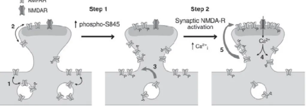
activation has been shown to significantly promote LTP in cultured hippocampal neurons, by increasing the size of the GluR1 extrasynaptic pool in a PKA-dependent way (Gao et al., 2006). Likewise, exposure to noradrenaline and emotional stress can drive GluR1 phosphorylation at Ser 845 and Ser 831, leading to synaptic delivery of GluR1-containing AMPA receptors, thus lowering the threshold for LTP (Hu et al., 2007). PKA phosphorylation of GluR1 subunits has been shown to cause AMPA receptor externalization from internal stores to membrane extrasynaptic pools (Oh et al., 2006). In fact, a two-step model for
delivery of GluR1-containing AMPARs to synapses during activity-dependent LTP proposes that phosphorylation of the Ser 845 residue by PKA can traffic AMPARs to extrasynaptic sites, making them available for subsequent delivery to synapses during activity-dependent plasticity, by lateral diffusion (see Figure 1.1.4.4). In turn, there is ample proof that synaptic insertion of GluR1-containing AMPARs contributes to the synaptic strengthening observed during LTP induction (Malinow and Malenka, 2002; Bredt and Nicoll, 2003; Malenka and Bear, 2004).
Figure 1.1.4.4. Two-step model for synaptic delivery of AMPARs during LTP. Under basal
conditions (left panel), GluR1 has a low phosphorylation at Ser-845. Constitutive recycling occurs between the surface and internal pools (1) and the synaptic and extrasynaptic pools (2) of AMPARs. Increasing Ser-845 phosphorylation (Step 1) stimulates trafficking of internal GluR1-containing AMPARs to extrasynaptic sites on the surface membrane, which primes AMPARs for synaptic incorporation (3). During strong synaptic activation (Step 2), synaptic NMDARs are activated, resulting in increased intracellular calcium (4). Calcium triggers the activation of signaling cascades, which drives GluR1-containing AMPARs to synapses from extrasynaptic sites by lateral diffusion (5). Thus, the two-step model for synaptic delivery of AMPARs consists of delivery of GluR1-containing AMPARs to extrasynaptic sites in a phospho-Ser-845- dependent manner (Step 1, the priming step), followed by synaptic incorporation of AMPARs, which requires synaptic NMDAR activation and calcium (Step 2). Adapted from Oh et al., 2006.
It should be noted that the elemental role played by the trafficking of GluR1-containing receptors in adult animals is, at earlier stages,
attributable to GluR4-containing ones (Esteban, 2003). Both subunits are composed of long intracellular C terminals, which share considerable aminoacid homology and a conserved PKA phosphorylation site (Ser 845 for GluR1 and Ser 842 for GluR4; Esteban et al., 2003), consistent with the similar trafficking behavior they follow. Thus, early in the postnatal development of the hippocampus, regulated delivery of AMPARs involves GluR4-containing receptors and PKA-mediated phosphorylation of GluR4 subunits is necessary and sufficient for triggering receptor delivery to synapses (Esteban et al., 2003). As GluR4 subunit expression declines and that of GluR1 increases with age (Zhu et al., 2000), regulated delivery of AMPARs then requires GluR1 phosphorylation by PKA, but at this stage, activation of a second signaling cascade (involving CaMKII activity) is further required for receptor delivery (Esteban et al., 2003). The greater elaboration of cellular processes and transduction pathways involved in AMPAR delivery underlying plasticity that is observed in adult animals is in agreement with the empirical observation that it is more difficult to induce and express synaptic plasticity, later in life (Rosenzweig and Barnes, 2003).
1.1.5 Synaptic Plasticity
In 1949, Donald Hebb proposed that “When an axon of cell A is near enough to excite a cell B and repeatedly or persistently takes part in firing it, some growth process or metabolic change takes place in one or both cells such that A’s efficiency, as one of the cells firing B, is increased” (Hebb, 1949). To this day, Hebb´s law stands for the paradigm of information storage in the brain being encoded by changes
in the strength of synaptic transmission between communicating neurons. At the time, Hebb´s postulate did not receive much consideration from neurophysiologists in the field, such as Ben Burns or John Eccles, for whom the “Hebb synapse” was little more than a self-evident conceptual embodiment of the post-tetanic potentiation (PTP) they had already observed in spinal neurons (reviewed in Bliss, 2003). As the name implies, PTP corresponds to a large enhancement of synaptic efficacy observed after brief periods of high frequency synaptic activity. Too brief to possibly account for the neural basis of memory, PTP led Burns´ student Tim Bliss away from the spinal cord pathways and into the cortical networks that were presumably the neural seat of memory (Eccles, 1966). In fact, it seemed plausible that the spine apparatuses, which were much more prevalent on neocortical and hippocampal pyramidal cells, formed part of the cellular machinery underlying memory (Hamlyn, 1963). Discouraged by contradictory results observed from working with highly complex cortical networks, Bliss turned to the hippocampus for need of simplification. Soon after and together with Terje Lömo, Bliss delivered a single tetanus to the perforant path in the intact rabbit hippocampus, obtaining a huge potentiation of the evoked synaptic response in the dentate gyrus that persisted for hours (Bliss and Lömo, 1973). That they grasped the potential significance of these early findings could be read from the cautious suggestion that such activity-dependent, sustained change in synaptic efficiency might be “potentially useful for information storage” (Bliss and Lömo, 1973). Meanwhile, long term potentiation (LTP) has been observed in a variety of other neural structures, such as the cerebellum (Jörntell and Hansel, 2006), the amygdala or the corpus striatum (Chapman et al., 2003), among others. Some suggest LTP may
even take place at all excitatory synapses in the mammalian brain (Malenka and Bear, 2004). In addition to its longevity, LTP has other characteristics that make it an attractive candidate mechanism for the storage of information in the brain. First of all, it is an input-specific process, since a single pathway can be potentiated without affecting inactive neighbouring inputs to the same cell (Andersen et al., 1980). Secondly, LTP abides stimulus cooperativity, the synaptic property by which many concurrent weak stimulations of converging afferent fibres can cooperate to induce LTP in the postsynaptic cell. Finally, the property of stimulus associativity can be derived from the fact that mild afferent stimulation that is subthreshold for inducing LTP can lead to LTP at a given synapse, provided it is paired with a strong LTP-inducing stimulation at a neighboring synapse (Kemp and Manahan-Vaughan, 2007). The latter feature is particularly interesting when memory formation is conceived as the means by which one can associate events or entities in the outside world. Interestingly, the same properties that argue for LTP as being the cellular correlate of memory formation have also been found to apply for long term depression (LTD) of synaptic transmission (see Kemp and Manahan-Vaughan, 2007) that ensues prolonged low frequency (0.5-3 Hz) afferent stimulation (Malenka and Bear, 2004). Like LTP, LTD has been shown to occur in many brain areas, including the cerebellar cortex, neocortex, hippocampus, striatum and nucleus accumbens (Artola and Singer, 1993). It is thought that LTD results from a subtreshold calcium entry into the postsynaptic cell, which is lower than that necessary to induce LTP and it has been proposed that, by counterbalancing LTP, LTD might serve to create a higher signal-to-noise ratio that would maximally sharpen the ability of synapses to store frequency-based