Chemical Kinetic Strategies for High-Throughput Screening of
Protein Aggregation Modulators
Zsuzsa S#rk#ny,
[d]Fernando Rocha,
[d]Ana M. Damas,
[c]Sandra Macedo-Ribeiro,
[a, b]and
Abstract: Insoluble aggregates staining positive to amyloid dyes are known histological hallmarks of different neurode-generative disorders and of type II diabetes. Soluble oligo-mers are smaller assemblies whose formation prior to or concomitant with amyloid deposition has been associated to the processes of disease propagation and cell death. While the pathogenic mechanisms are complex and differ from disease to disease, both types of aggregates are impor-tant biological targets subject to intense investigation in
academia and industry. Here we review recent advances in the fundamental understanding of protein aggregation that can be used on the development of amyloid and anti-oligomerization drugs. Specifically, we pinpoint the chemical kinetic aspects that should be attended during the develop-ment of high-throughput screening assays and in the hit val-idation phase. The strategies here devised are expected to establish a connection between basic research and pharma-ceutical innovation.
1. Introduction
Over the recent years, protein aggregation has been in the limelight of drug discovery: on the one hand, several projects targeting amyloid aggregates in neurodegenerative disorders have been discontinued.[1]On the other hand, smaller protein
aggregates—the soluble oligomers—are increasingly surfacing as major culprits of neurotoxicity and pathogenic spread.[2]
Co-incidently with this paradigm shift,[3]encouraging results have
been obtained by the first generation of anti-oligomerization drug candidates in clinical trials of Alzheimer’s disease and Par-kinson’s disease.[4]
The long and costly workflow leading to the discovery of new drugs has in its basis methods of high-throughput screen-ing (HTS) of active compounds (known as “hits”) that can be further developed into drug precursors (known as “leads”) or used as “chemical probes” to validate a given biological target.[5] Targeted HTS approaches rely on in vitro protein
ag-gregation assays having well-defined reaction components and high reproducibility indexes.[6]However, deciphering the exact
mode of action of aggregation inhibitors is not a trivial task on account of the possible pathways (Figure 1a) and multiple ele-mentary steps (Figure 1b) that might be implicated. It may occur, for instance, that large amounts of stable soluble oligo-mers are formed without their presence being directly
detect-ed by amyloid-specific probes such as thioflavin-T (ThT) or by turbidimetric methods.[7]
Widely applied in the study of amyloid aggregation mecha-nisms,[10] chemical kinetic analysis is gradually being
estab-lished as a promising tool for the systematic screenings of both anti-amyloid and anti-oligomerization compounds.[11] An
important step in this direction was taken by defining the ref-erence kinetic behavior expected for generic nucleation-and-growth processes of phase transition.[12] When no other
path-way is present, the formation of insoluble species begins with the elementary step of primary nucleation and then proceeds through the occurrence of the autocatalytic steps of secondary nucleation and growth. Thermodynamic equilibrium is attained when the chemical potential of the solution equals that of the insoluble fraction. At that point, the concentration of protein monomer (C) corresponds to the protein solubility (C*), and so the driving force for phase transition represented by
supersa-Figure 1. Aggregation pathways during amyloid fibril formation. a) Protein monomers establish a thermodynamic equilibrium with soluble oligomers that are either on- or off-pathway to produce amyloid fibrils. Off-pathway oligomers can undergo phase transition processes leading to the formation of different types of precipitates,[8]and then act as heterogeneous nucleants
of amyloid fibrils. b) Reaction steps participating in the amyloid pathway. Green glows represent an increase in the mass of amyloid fibrils.[9]
[a] Dr. S. Macedo-Ribeiro, Prof. P. M. Martins IBMC-Instituto de Biologia Molecular e Celular Universidade do Porto
4200-135 Porto (Portugal) E-mail: [email protected]
[b] Dr. S. Macedo-Ribeiro, Prof. P. M. Martins Instituto de Investigażo e Inovażo em Safflde Universidade do Porto
4200-135 Porto (Portugal)
[c] Prof. A. M. Damas, Prof. P. M. Martins
ICBAS-Instituto de CiÞncias Biom8dicas Abel Salazar Universidade do Porto
4050-313 Porto (Portugal) [d] Dr. Z. S#rk#ny, Prof. F. Rocha
LEPABE-Departamento de Engenharia Qu&mica Faculdade de Engenharia da Universidade do Porto Rua Dr. Roberto Frias, 4200-465 Porto (Portugal)
The ORCID identification number(s) for the author(s) of this article can be found under:
turation DC ¼ C @ C* (or in dimensionless form s ¼ DC=C*) is zero. After expressing supersaturation in terms of the normal-ized reaction conversion (a),
s ¼ s0ð1 @ aÞ ð1Þ
The following closed-form solution of the mass balance equa-tions is obtained for unseeded reacequa-tions (a0¼ 0):[12]
a ¼ 1 @k 1
b½exp kð Þ @ 1at A þ 1 ð2Þ
where ka and kbare determined by the elementary steps that
increase the mass of insoluble aggregates (Figure 1b). Sigmoi-dal progress curves comprising an initial lag phase followed by an exponential phase and a plateau phase are expected in the cases of slow primary nucleation,[6,13]for which the values of k
b
are lower than ' 0:01.[7,12]In turn, fast primary nucleation
pro-cesses (kb> 0:01) give rise to hyperbolic progress curves that
are also observed during seeded aggregation assays (see sec-tion 2.). Protein aggregasec-tion modulators can be screened based on kinetic signatures standing out from the standard be-havior expected by Equation (2).[7] This is valid for both
on-and off-pathway inhibition as it is shown next for the case of amyloid fibril formation.
2. The Amyloid Aggregation Assay
Amyloid fibrils have a characteristic cross-beta-sheet structure that is detectable upon binding to Congo red, ThT and
thiofla-vin-S (ThS) dyes.[11a,14] Although amyloid aggregation can be
followed by other spectrophotometric techniques,[15]thioflavin
fluorescence is adequate for HTS kinetic assays that require low sample volume, high signal-to-noise ratios and high repro-ducibility.[6,11b] The use of this probe is not without pitfalls
aris-ing from non-amyloid bindaris-ing, reduced sensibility to early (on-pathway) aggregates and fluorescence self-quenching.[16] As
with other assays employing light-based detection methods, orthogonal validation using different output reporters is re-quired in order to identify false positives and fluorescence arti-facts.[17]The development and optimization of HTS assays aims
at rightfully interrogate a given therapeutic target using minia-turized formats of 96-well, 384-well or 1536-well microplates.[18]
Since amyloid nucleation is a stochastic process,[19]some
varia-bility between replicate experiments is expectable even when homogeneous, highly pure protein samples are used.[20]This is
detrimental for the quality of the HTS assay usually character-ized in terms of the Z0 factor (Figure 2a) taking into account
Zsuzsa S#rk#ny obtained her Ph.D. degree in Structural Biochemistry in 2006 from Eçtvçs Ljr#nd Unversity, Budapest, Hungary under the supervision of Prof. Dr. L#szlj Polg#r. From 2007 to 2017 she worked at Institute for Molecular and Cell Biology (IBMC), Porto, Por-tugal with Prof. Dr. Ana Margarida Damas and Dr. Sandra Macedo-Ribeiro. She is cur-rently a postdoctoral fellow at the Depart-ment of Chemical Engineering, University of Porto, Portugal with a research focus on ex-ploring the oligomeric pathway of alpha syn-uclein protein through high-throughput screenings of drug-like compounds. Fernando Rocha completed his Ph.D. degree in 1984 at the University of Porto. Since then he has worked as Assistant Professor in the Chemical Engineering Department of the Uni-versity of Porto. His research interests include crystallization of biologically relevant mole-cules, with applications to health issues.
Ana M. Damas is a retired Full Professor of Biophysics of the Abel Salazar Biomedical School of the University of Porto. Her main re-search interests are X-ray Crystallography and Amyloidosis.
Sandra Macedo-Ribeiro completed her Ph.D. degree in 1999 at the Technical University of Munich. She then moved to the Institute for Molecular Biology in Barcelona as a postdoc-toral researcher. She was Assistant Professor at the University of Algarve and Assistant Re-searcher at the Center for Neuroscience and Cell Biology in Coimbra. Presently, she is a Principal Researcher at the Institute for Molec-ular and CellMolec-ular Biology/Institute for Re-search and Innovation in Health (IBMC/i3S, Porto) and leader of the Biomolecular Struc-ture and Function group. Her research inter-ests include the structural and functional analysis of polyglutamine-rich proteins. Pedro Martins completed his Ph.D. degree in Chemical Engineering with a Thesis on the physics of crystal growth (2006, University of Porto). He is a Research Assistant at the Bio-molecular Structure and Function group of the Institute for Molecular and Cell Biology/In-stitute for Research and Innovation in Health (IBMC/i3S). His research interests include pro-tein aggregation, enzymology and drug dis-covery.
the means (m) and standard deviations (SD) of positive and negative controls. A Z0factor > 0:5 indicates a good degree of
separation between controls.[21] To improve reproducibility,
pre-formed fibrils (or “seeds”) can be added to the initial solu-tion to bypass the primary nucleasolu-tion step.[22]The drawback of
seeded assays is their insensibility to aggregation inhibitors specifically targeting non-templated nucleation. Other alterna-tives designed to accelerate fibrillation through the use of glass beads, vigorous agitation, high temperature, low pH and/ or denaturating additives only remotely reproduce the biologi-cal medium.[22]Heterogeneous nucleants such as lipid micelles,
can be used to mimic the cell membrane and disease-propa-gating exosomes,[23]and at the same time catalyze protein
ag-gregation with improved reproducibility.[24] In principle,
seed-ing is not expected to influence the ThT fluorescence increase (DFF), which is an important thermodynamic measurable
(Figure 2a) proportional to the initial supersaturation DC0¼ C0@ C*. The kinetic influence of seeding is accounted
by analyzing the normalized progress curves (Figure 2b) and the half-life coordinates t50and v50(Figure 2c). The
fundamen-tal definitions of t50and v50are expressed in terms of apparent
rate constants k0 aand k0b,[12] t50¼k10aln k10bþ 1 . -ð4aÞ v50¼k 0 a 4 kð 0bþ 1Þ ð4bÞ
which in turn are influenced by the initial fraction of seeds a0
(computed in relation to the total polymerizable protein, DC0):
k0
a¼ kað1 þ a0Þ ð5aÞ
k0
b¼k1 þ abþ a0
0 ð5bÞ
For unseeded reactions (a0¼ 0), k0a¼ kaand k0b¼ kb. As it
fol-lows from eq 2, otherwise sigmoidal progress curves (kb< 0:01) will develop hyperbolic shapes if > 1% of
pre-formed fibrils are added to the initial solution (k0
b> 0:01).
3. Recognizable Modulators
Timely reviews on protein aggregation modulation were put forward by for example, Doig and Derreumaux,[25] Eisele
et al.,[26]and Velander et al.,[27]where different classifications of
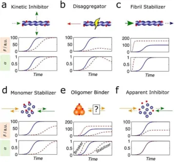
inhibitors are adopted based on mechanistic and physico-chemical considerations. Here we make a shorter selection of examples that are representative of the modes of action sum-marized in Figure 3. For the sake of simplicity each mechanism is illustrated separately, although synergistic and antagonistic effects may take place simultaneously. The selected examples are listed in Table 1.
Purely kinetic inhibitors (Figure 3a) slow down the rate of fluorescence increase without changing the final fluorescence, which is a value indicative of the amount of amyloid fibrils pro-duced. This is achieved through kinetic inhibition of any of the steps of primary nucleation, secondary nucleation or fibril elon-gation that increase the amyloid-specific signal (indicated by green arrows in Figures 1 and 3). Fluorination of the hydropho-bic residues valine or phenylalanine of the LVFFD peptide in-creases the duration of the lag phase of Ab42 aggregation without significantly changing the normalized aggregation rate v50 or the value of the total fluorescence increase F1.[28]
Therefore, the fluorinated peptides are considered kinetic in-hibitors acting upon the step of primary nucleation.[28]
The lead compound CLR01 is a molecular tweezer that spe-cifically binds to lysine residues thus disrupting important in-teractions for protein aggregation.[29] The impact of CLR01 on
ThT progress curves changes according to the amyloidogenic protein being studied; in the case of Ab40 aggregation, a ten-fold excess added during the exponential and plateau phases induced an evident disaggregating effect manifested by a pro-gressive loss of amyloid signal.[30] This trend, which has also
been observed during incubation of Ab42 with brazilin,[31]
cor-responds to the kinetic signature of fibril disaggregators (Fig-ure 3b).
By preventing fragmentation, fibril stabilization (Figure 3c) might reduce prion-like spreading of small aggregates within and between cells, as demonstrated for seeded spreading of Ab and tau using animal models of Alzheimer’s disease.[26, 32]
Tight binding fibril stabilizers might also decrease the amyloid
Figure 2. Example showing an optimized a-synuclein aggregation assay. A good separation between positive and negative controls was achieved by adding 0.2 mm of pre-formed fibrils (or “seeds”) to the reaction mixture con-sisting of 2 mm a-synuclein in 50 mm sodium phosphate pH 7.5, 200 mm NaCl and 70 mm ThT (Z. S#rk#ny and P. M. Martins, personal communication). a) Symbols: ThT fluorescence (F) measured over time in a 384-well plate, each well containing 0.2 mm of seeds in the presence (blue) and absence (gray) of soluble a-synuclein. A Z0factor of 0.66 was obtained using mean
values (m, dashed lines) and standard deviations (SD, shaded areas corre-spond to 3 > SD) calculated for the F measurements at time point 187 hours (FF) in 192 positive controls (red symbols) and 192 negative controls (green
symbols). b) Amyloid conversion calculated as the normalized fluorescence increase (a ¼ DF=DFF) taking as reference the instantaneous negative
con-trols (DF ¼ F @ m@). c) Mean values of amyloid conversion ((a, symbols), t50
(vertical dashed line) and v50(sloping dashed line) calculated from the
solubility and redirect the equilibrium from potentially more toxic, small oligomers to amyloid fibrils (large green arrow in Figure 3c).[7,8,12] This hypothesis has been explored through
the development of orange G-related compounds bound to the steric zipper Ab16-21 structure.[33]
According to the supersaturation-dependence of ka and kb,
protein-specific modulation of solubility (C*) is an attractive
approach to change the kinetics—and not only the thermody-namics—of amyloid fibril formation. This can be achieved using as target the amyloid pharmacophore of the soluble pro-tein (Figure 3c) or, alternatively, the metastable oligomers (Fig-ures 3d,e). Monomer stabilizers (Figure 3d) selectively increase the solubility of the target protein, shifting the on- and off-pathway equilibria towards the dissociation of oligomers. As the result of the higher C* values, less amyloid fibrils are pro-duced and lower fluorescence signals are expected at the end of the assay (Figure 3d, middle); because the values of super-saturation decrease, larger t50 values and lower v50 values are
also expectable (Figure 3d, bottom). Monomer stabilizers and oligomer binders (Figure 3e) are not clearly distinguishable from simple progress curves analysis if the mode of action of the latter type of inhibitor comprises the stabilization of the oligomeric assembly. This is because as the amyloidogenic pro-tein is sequestered into the oligomeric state, the pool of avail-able monomers is depleted leading to the train of events that culminates in lower DF1values and slower aggregation
kinet-ics. Mimics of the islet amyloid polypeptide (IAPP) synthesized with N-methylated amide bonds were shown to inhibit the ag-gregation of IAPP and Ab40 by a common mechanism of stabi-lization of protein monomers and nontoxic oligomers.[34]
Ac-cordingly, the IAPP mimics are included in the categories of protein and oligomer stabilizers (Figures 3d,e, respectively). The stabilization of the transthyretin (TTR) tetramer by tafami-dis (Vyndaqel; Pfizer),[35]and by the repurposed drugs
diflunis-al[36]and tolcapone[37]inhibits the formation of amyloid
depos-its by preventing the dissociation of the tetramer into aggrega-tion competent TTR monomers. Commonly referred as “kinetic stabilizers’,[26]these compounds are here classified as oligomer
stabilizers (Figure 3e) to avoid confusion with the kinetic inhib-itors having no thermodynamic effects on aggregation (Fig-ure 3a).
The new evidences associating non-amyloidogenic oligo-mers to the pathogenesis of neurodegenerative diseases allow us to anticipate a renewed interest in oligomer breakers (Fig-ure 3e) as a therapeutic hypothesis to remodel toxic assem-blies into nontoxic species. In the example of Figure 3e, the aggregation equilibrium is redirected to the formation of more amyloid fibrils (Figure 3e, top) and at faster rates (Figure 3e, bottom). Other scenarios are, however, possible in which the amyloid pathway is not favored either because protein solubili-ty is concomitantly increased or, as in the case of Ab42 aggre-gation in the presence of the N-methylated peptide inhibitor SEN304,[38]other nontoxic pathways are elicited.
Figure 3. The different types of aggregation modulators recognizable by the amyloid aggregation assay. Each panel gives the mode of action (top, car-toon symbols as in Figure 1) and the kinetic signatures (middle and bottom) of each type of modulator; the progress curves represent the fluorescence (F) increase (middle) and the amyloid conversion (bottom) in the absence (solid lines) and presence (dashed lines) of test compound. The theoretical curves were computed using Equation (2) and assuming (i) kaand kb
propor-tional to DC0=C*, and (ii) F proportional to the product DC0a.[9, 43]a) Kinetic
inhibitors (associated to lower kaand kbvalues) decrease the rates of
pri-mary nucleation, secondary nucleation or fibril elongation (dashed arrows in the cartoon). b) Disaggregators provoke the dissociation of fibrils into solu-ble protein (red arrow in the cartoon). c) Fibril stabilizers decrease the values of solubility (C*) through stabilization of amyloid fibrils (large green arrow) and might prevent fibril breakage (dashed blue arrow) d) Monomer stabiliz-ers decrease the aggregation propensity (dashed orange and green arrows) by inducing higher values of C*. e) Oligomer binders may either stabilize or inhibit the formation of off-pathway oligomers, thereby decreasing or in-creasing the effective monomer concentration and the corresponding super-saturation value. f) Apparent inhibitors non-specifically alter the solution properties, which may reduce the thermodynamic propensity for aggrega-tion (by reducing DC0) but do not affect kaand kb(as DC0=C* remains
con-stant).[42]
Table 1. Examples taken from the literature illustrating the different types of aggregation modulators presented in Figure 3.
Modulator Representative Examples
Kinetic Inhibitor - Peptide LVFFD with fluorinated valine or phenylalanine during Ab42 aggregation.[28]
Disaggregator - CLR01 during Ab40 aggregation.[30]
- Brazilin during Ab42 aggregation.[31]
Fibril Stabilizer - Orange G-related compounds bound to the steric zipper Ab16-21 structure.[33]
Monomer Stabilizer - Mimics of IAPP synthesized with N-methylated amide bonds during the aggregation of IAPP and Ab40.[34]
Oligomer Binder - Tafamidis,[35]diflunisal[36]and tolcapone[37]during the aggregation of TTR.
- N-Methylated peptide inhibitor SEN304 during the aggregation of Ab42.[38]
Changing the ionic strength of amyloidogenic solutions by the addition (or removal) of salts can have a dramatic influence on amyloid aggregation in vitro,[39] which is understood in
terms of the role of solubility in determining the rate and extent of protein self-assembly.[7,8,12,40] These modulation
ef-fects are considered “apparent” (Figure 3 f) since they are un-specific and tend to be buffered in cells and tissues media. Ap-parent inhibitors that solely alter the activity coefficient of the protein will originate superimposed progress curves if repre-sented in normalized ThT fluorescence units (Figure 3 f, bottom); this has been previously illustrated using published data of Pronchik et al.[41]on the influence of hydrophobic
inter-faces on a-synuclein aggregation.[42] It should be noted that
the superimposition of normalized curves does not necessarily imply that the test compound should be discarded. In fact, Figure 3 does not exhaustively cover all possible mechanisms of inhibition, but instead it seeks to summarize the major types of modulators that can be identified by kinetic assays.
4. Primary Screenings, Secondary Screenings,
Hit Validation
Having set up a reproducible amyloid aggregation assay does not guarantee that large numbers (> 105) of chemical
com-pounds can be efficiently assessed during HTS campaigns, es-pecially if several days are required to measure full progress curves. In these cases, a primary screening strategy can be de-vised to limit the measurement frequency to the initial, middle, and final points of the reaction (Figure 4). The corre-sponding values F0, F50 and FF (Figure 4a) can be followed in
multiple plates simultaneously without advanced robotic sys-tems being necessarily required. Positive hits stand out by showing DF50=DFF ratios significantly different from the value
of 0.5 expected in the absence of modulation effects and for apparent inhibitors (Figure 4b).
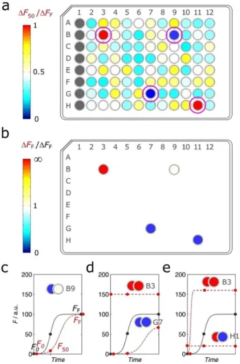
The 3-point aggregation assay can be used on the identifica-tion of positive hits as well as on the preliminary characteriza-tion of the putative inhibicharacteriza-tion mechanism. This requires the
analysis of the DF50=DFF ratio (Figure 5a) but also of DFF
values obtained in the presence and absence of the test com-pound (Figure 5b). Kinetic inhibitors, for example, are expected to originate values of DF50=DFF# 0:5 with no major impact
on the values of DFF (Figure 5c). The progress curves
charac-teristic of monomer and soluble oligomer stabilizers are not easily distinguishable (see previous representations in Figur-es 3d and 3e), and both are associated to markedly low valuFigur-es of DF50=DFF and of DFF (Figure 5d). Conversely, very high
values of DF50=DFF and of DFF may indicate the cases of fibril
stabilizers and of oligomer breakers (Figure 5e).
In the same way that Figure 3 did not cover all possible in-hibition mechanisms, Figure 5 gives a limited number of exam-ples that can be identified by the 3-point screening assay. As expectable, this initial analysis is not free from ambiguities as exemplified in Figure 5 by cell B3, whose result can be ascribed
Figure 4. A possible strategy for primary screenings based on initial, mid-point and endmid-point measurements. a) The midmid-point (t50) and endpoint (tF)
instants are defined beforehand taking into account the control progress curve (solid line) characterizing the amyloid aggregation assay. Rather than continuously measuring the full progress curve in the presence of test com-pounds (dashed line), only the fluorescence increase at time-points t50
(DF50¼ F50@ F0) and tF(DFF¼ FF@ F0), respectively, are measured. b)
Nega-tive results elicited by, e.g., fluorogenic compounds and apparent inhibitors can be identified by values of the DF50=DFFratio close to 0.5.
Figure 5. Possible readouts expected from the 3-point aggregation assay. a and b) Illustrative example of an arbitrary 96-well microplate with the assay output represented in terms of (a) the DF50=DFFratios and (b) the impact
on the value of DFF; different colors indicate different values of the readout
as indicated in the colorbars. Cells B3, B9, G7 and H11 are positive hits iden-tified in (a) and then further analyzed in (b). c to e) The combinations of DF50and DFFvalues highlighted in (a) and in (b) apply to sigmoidal
prog-ress curves (lines) obtained in the absence (solid lines) and presence (dashed lines) of (c) kinetic inhibitors, (d) soluble protein stabilizers, and (e) fibril stabilizers/oligomer breakers. Horizontal dashed lines in (d) and (e) rep-resent cases of undefined aggregation suppressors.
to fibril stabilizers, oligomer breakers (both cases represented in Figure 5e) or undefined aggregation suppressors (Fig-ure 5d). To clarify this type of doubts, the initial hits—including the undefined results—have to undergo the next round of screenings based on dose-response, full progress curve analy-sis.
In addition to the conventional representation of a over time, amyloid aggregation curves are conveniently plotted in log-linear a= 1 @ að Þ vs. time coordinates so as to detect devia-tions from the standard behavior expected by Equation (2). These deviations, which are dealt in detail elsewhere,[7]are
evi-denced by non-linear trends arising after the initial lag phase. The relative importance of off-pathway aggregation generally increases as the curvature of the ln a= 1 @ að ð ÞÞ representation gets more pronounced. Therefore, distinguishing between sim-ilar signatures of aggregation modulators such as fibril ers and oligomer breakers (Figures 3c,e), or monomer stabiliz-ers and oligomer stabilizstabiliz-ers (Figures 3d,e) is possible by repre-senting the reaction coordinates in the modified log-linear scale. Dose-response analyses of the assay measurables t50,
v50 and DFF are important to establish the potency of the
se-lected compounds by means of the half-maximal inhibitory concentration (IC50) and the half-maximal effective
concentra-tion (EC50) parameters.[44] Yet, to answer the question on
whether the right target has been reached, the relationships DFF, v50, and t50 vs. protein concentration at constant
com-pound concentration should also be looked in detail. In recent contributions we demonstrated that these scaling laws reflect the relative composition of monomers and soluble oligomers measured by direct methods.[9,43] Specifically, a direct
corre-spondence between the end-point amyloid signal and the ini-tial concentration of monomer can be expected when no off-pathway oligomers are present (Figure 6a) or when their disso-ciation is much slower than the formation of amyloid fibrils (Figure 6b). This correspondence will not be observed if the oligomerization equilibrium is reversed by the gradual vanish-ing of protein monomers provoked by the amyloidogenic pathway (Figure 6c). Examples of refractory and reversible off-pathway oligomerization were observed during the aggrega-tion of human insulin and ataxin-3, respectively.[9,43]
The oligomerization pathway can be further probed by ana-lyzing the influence of protein concentration on the half-life coordinates. The scaling laws of v50 (Figure 7a) and t50
(Fig-ure 7b) show marked deviations from linearity in the presence of slowly dissociating oligomers, with unconventional scaling exponents (g) being admissible independently of the dominant step(s) of amyloid fibrillation (Figure 7b). Within the context of HTS secondary screenings, the analysis of protein concentra-tion scaling laws in the presence and absence of test com-pounds is an effort worth making in order to discriminate which pathway is being affected and by which mechanisms.
Hit results found by indirect kinetic assays need to be vali-dated by a combination of experimental methods capable of characterizing both quantitatively and qualitatively the target aggregates. The available options can be roughly categorized into spectroscopic, chromatographic, calorimetric, light scatter-ing, microscopy and high-resolution techniques, which have
Figure 6. The scaling laws of final florescence with total protein concentra-tion (CT) provide insights into off-pathway oligomerization—Figure
repro-duced under the terms of the CC BY 4.0 license from Silva et al..[43]The initial
distributions of monomer and total protein (left side) influences the end-point scaling laws (right side). The correspondence between the initial and final stages is direct if (a) no oligomerization pathway is present or (b) oligo-merization is irreversible, and indirect if (c) oligooligo-merization is fully reversible. Green lines describe cases of protein solubility values C* ¼ 0 (solid lines) and C* > 0 (dashed lines).
Figure 7. Highly diverse kinetic scaling laws are expected depending on the relative importance of off-pathway aggregation. The scaling laws of (a) v50
and (b) t50change according to the initial degree of oligomerization and the
rate at which off-pathway oligomers dissociate (color bar). The variability of the scaling exponents (g) cannot be explained if only on-pathway mecha-nisms are considered. Figure adapted under the terms of the CC BY 4.0 li-cense from Silva et al.[43]assuming the cases of predominant secondary
been subject of updated reviews by for example, Poklar Ulrih,[45]Pryor et al.,[46] and Renaud et al..[47] The application of
these techniques may either confirm or deny the first indica-tions resulting from the amyloid aggregation assay. Illustrating this, Wobst et al.[48]employed circular dichroism (CD) to detect
changes in the secondary structure of tau protein and to con-firm that the green tea polyphenol (@)-epigallocatechin gallate (EGCG) prevents the formation of b-sheet-rich tau oligomers. On the other hand, Middleton et al.[49] used isotope labelling
and two-dimensional infrared spectroscopy to show how ag-gregation inhibitors may function by complex structural pro-cesses that are not detected by ThT fluorescence. Infrared nanospectroscopy has recently been used to characterize oli-gomeric and fibrillar species during amyloid formation of ataxin-3.[50] Nuclear Magnetic Resonance (NMR) spectroscopy
can provide high-resolution structural information as that used to unveil the ligand binding details of an anti-amyloidogenic compound (phthalocyanine tetrasulfonate) to a-synuclein. Al-ternatively, NMR spectroscopy can be utilized to follow the for-mation of intermediate and off-pathway oligomers by periodi-cally collecting the1H NMR spectra associated to monomer
de-pletion. This has been done while studying the effect of Cu2+
on a-synuclein aggregation,[51] and to demonstrate the ability
of EGCG to shift off-pathway aggregation of Ab40.[52]
Solid-state NMR,[53]together with X-ray microcrystallography,[54]X-ray
fiber diffraction[55] and cryo-electron microscopy[56]further
en-larged the resolution limits of amyloid structures,[57] and
con-tributed to reveal morphological differences between amyloid fibrils generated in vitro or in the brain.[58]With the recent
ad-vances in instrumentation, high intensity synchrotron sources, robotics/automation, speed of data processing, etc., some of the benchmark technologies are now being applied in de novo screenings of aggregation modulators. For example, in-hibitors of human IAPP and Ab40 aggregation were identified by ion mobility spectrometry-mass spectrometry using ThT fluorescence monitoring as a control method to detect possi-ble hydrophobic interactions that become lost in the gas phase.[59] The scope of light scattering techniques is also
ex-panding from proof-of-concept to high-throughput applica-tions as in the cases of dynamic light scattering (DLS) for size distribution monitoring,[60] and small angle X-ray scattering
(SAXS) for characterizing the oligomeric equilibrium of non-ag-gregated samples.[61]In another approach, a time-resolved
fluo-rescence resonance energy transfer (FRET) assay was devel-oped to screen 7:46 > 105 compounds and identify 56 hits
that markedly inhibit a-synuclein and several phenyl-benzoxa-zol compounds that promote a-synuclein aggregation (proag-gregators).[62] Important for subsequent phases of hit to lead
optimization,[5,18] cell-based assays based on phenotypic and
cytotoxicity readouts have also been used for compound screening purposes in the identification of IAAP aggregation inhibitors.[63]
5. Conclusions and Perspectives
Despite all the advances in the understanding of protein ag-gregation kinetics, the translation of this knowledge into new
practical tools for drug discovery has been limited. We believe that this scenario is about to change on account of two rea-sons both connected with the key role played by soluble oligo-mers in the process of amyloid fibril formation. First, there is a growing interest in anti-oligomerization compounds as a possi-ble therapeutic strategy to block the progression of neurode-generative disorders and amyloidoses. Second, soluble mers, and particularly non-amyloidogenic off-pathway oligo-mers leave distinctive signatures in amyloid aggregation curves and scaling laws that help distinguishing which type of protein aggregates are being targeted. In the present work we reviewed the different mechanisms of action of known anti-amyloid and anti-olgomerization compounds and showed how their effect can be identified in reaction progress curves. The systematization of this analysis led us to propose the 3-point aggregation assay for the primary screening of large libraries of chemical compounds based on ThT fluorescence readouts at the starting-point, (supposed) half-life and (supposed) end-point instants of the reaction. We also reviewed the chemical kinetic tools available for the secondary screening and hit vali-dation phases, which may involve the analysis of full progress curves represented in modified coordinates and the analysis of DFF, v50, and t50 scaling laws.
Acknowledgements
This work was financed by (i) FEDER-Fundo Europeu de Desen-volvimento Regional funds through the COMPETE 2020-Opera-cional Programme for Competitiveness and Internationalisation (POCI), Portugal 2020, and by Portuguese funds through FCT-Fundażo para a CiÞncia e a Tecnologia/Minist8rio da CiÞncia, Tecnologia e Ensino Superior in the framework of the projects 01-0145-FEDER-031173 (PTDC/BIA-BFS/31173/2017), POCI-01-0145-FEDER-007274, POCI-01-0145-FEDER-031323 (“Institute for Research and Innovation in Health Sciences”) and POCI-01-0145-FEDER-006939 (Laboratory for Process Engineering, Envi-ronment, Biotechnology and Energy-UID/EQU/00511/2013), and by (ii) FEDER through Norte Portugal Regional Operational Programme (NORTE 2020), under the PORTUGAL 2020 Partner-ship Agreement in the framework of Project Norte-01-0145-FEDER-000008 and by national funds (PIDDAC) through FCT project “LEPABE-2-ECO-INNOVATION”-NORTE-01-0145-FEDER-000005.
Conflict of interest
The authors declare no conflict of interest.
Keywords: amyloid diseases · drug discovery · nucleation · protein aggregation · soluble oligomers
[1] H.-P. Shih, X. Zhang, A. M. Aronov, Nat. Rev. Drug Discovery 2018, 17, 19. [2] a) D. M. Walsh, D. J. Selkoe, Nat. Rev. Neurosci. 2016, 17, 251; b) M. L.
Choi, S. Gandhi, FEBS J. 2018, 285, 3631. [3] D. J. Selkoe, Ann. Neurol. 2013, 74, 328.
[4] a) J. Sevigny, P. Chiao, T. BussiHre, P. H. Weinreb, L. Williams, M. Maier, R. Dunstan, S. Salloway, T. Chen, Y. Ling, Nature 2016, 537, 50; b) D. B. Schenk, M. Koller, D. K. Ness, S. G. Griffith, M. Grundman, W. Zago, J. Soto, G. Atiee, S. Ostrowitzki, G. G. Kinney, Mov. Disord. 2017, 32, 211. [5] R. Macarron, M. N. Banks, D. Bojanic, D. J. Burns, D. A. Cirovic, T. Gar-yantes, D. V. Green, R. P. Hertzberg, W. P. Janzen, J. W. Paslay, Nat. Rev. Drug Discovery 2011, 10, 188.
[6] A. Villar-Piqu8, M. Schmitz, N. Candelise, S. Ventura, F. Llorens, I. Zerr, Mol. Neurobiol. 2018, 1.
[7] R. Crespo, E. Villar-Alvarez, P. Taboada, F. A. Rocha, A. M. Damas, P. M. Martins, J. Biol. Chem. 2016, 291, 2018.
[8] R. Crespo, E. Villar-Alvarez, P. Taboada, F. A. Rocha, A. M. Damas, P. M. Martins, J. Phys. Chem. B 2017, 121, 2288.
[9] A. Silva, B. Almeida, J. S. Fraga, P. Taboada, P. M. Martins, S. Macedo-Ri-beiro, Angew. Chem. Int. Ed. 2017, 56, 14042; Angew. Chem. 2017, 129, 14230.
[10] G. Meisl, J. B. Kirkegaard, P. Arosio, T. C. Michaels, M. Vendruscolo, C. M. Dobson, S. Linse, T. P. Knowles, Nat. Protoc. 2016, 11, 252.
[11] a) N. Cremades, C. M. Dobson, Neurobiol. Dis. 2018, 109, 178; b) J. Pujols, S. PeÇa-D&az, D. F. L#zaro, F. Peccati, F. Pinheiro, D. Gonz#lez, A. Carija, S. Navarro, M. Conde-Gim8nez, J. Garc&a, S. Guardiola, E. Giralt, X. Salvatella, J. Sancho, M. Sodupe, T. F. Outeiro, E. Dalfj, S. Ventura, Proc. Natl. Acad. Sci. USA 2018, 115, 10481.
[12] R. Crespo, F. A. Rocha, A. M. Damas, P. M. Martins, J. Biol. Chem. 2012, 287, 30585.
[13] L. Bentea, M. A. Watzky, R. G. Finke, J. Phys. Chem. C 2017, 121, 5302. [14] J. N. Buxbaum, R. P. Linke, J. Mol. Biol. 2012, 421, 142.
[15] I. Dolado, J. Nieto, M. J. M. Saraiva, G. Arsequell, G. Valencia, A. Planas, J. Comb. Chem. 2005, 7, 246.
[16] D. J. Lindberg, A. Wenger, E. Sundin, E. Wes8n, F. Westerlund, E. K. Esb-jçrner, Biochemistry 2017, 56, 2170.
[17] a) C. Aldrich, C. Bertozzi, G. I. Georg, L. Kiessling, C. Lindsley, D. Liotta, K. M. Merz, A. Schepartz, S. Wang, ACS Cent. Sci. 2017, 3, 143; b) N. Thorne, D. S. Auld, J. Inglese, Curr. Opin. Chem. Biol. 2010, 14, 315. [18] A. Roy, High-Throughput 2018, 7, 4.
[19] D. Kashchiev, R. Cabriolu, S. Auer, J. Am. Chem. Soc. 2013, 135, 1531. [20] K. G. Malmos, L. M. Blancas-Mejia, B. Weber, J. Buchner, M.
Ramirez-Al-varado, H. Naiki, D. Otzen, Amyloid 2017, 24, 1.
[21] J.-H. Zhang, T. D. Chung, K. R. Oldenburg, J. Biomol. Screening 1999, 4, 67.
[22] L. Giehm, D. E. Otzen, Anal. Biochem. 2010, 400, 270. [23] J. Howitt, A. F. Hill, J. Biol. Chem. 2016, 291, 26589.
[24] a) M. Grey, C. J. Dunning, R. Gaspar, C. Grey, P. Brundin, E. Sparr, S. Linse, J. Biol. Chem. 2015, 290, 2969; b) R. Gaspar, J. Pallbo, U. Weininger, S. Linse, E. Sparr, Biochim. Biophys. Acta Proteins Proteomics 2018, 1866, 1062.
[25] A. J. Doig, P. Derreumaux, Curr. Opin. Struct. Biol. 2015, 30, 50.
[26] Y. S. Eisele, C. Monteiro, C. Fearns, S. E. Encalada, R. L. Wiseman, E. T. Powers, J. W. Kelly, Nat. Rev. Drug Discovery 2015, 14, 759.
[27] P. Velander, L. Wu, F. Henderson, S. Zhang, D. R. Bevan, B. Xu, Biochem. Pharmacol. 2017, 139, 40.
[28] J. A. Loureiro, R. Crespo, H. Bçrner, P. M. Martins, F. A. Rocha, M. Coelho, M. C. Pereira, S. Rocha, J. Mater. Chem. B 2014, 2, 2259.
[29] S. Sinha, D. H. J. Lopes, Z. Du, E. S. Pang, A. Shanmugam, A. Lomakin, P. Talbiersky, A. Tennstaedt, K. McDaniel, R. Bakshi, P.-Y. Kuo, M. Ehrmann, G. B. Benedek, J. A. Loo, F.-G. Kl-rner, T. Schrader, C. Wang, G. Bitan, J. Am. Chem. Soc. 2011, 133, 16958.
[30] T. Schrader, G. Bitan, F.-G. Kl-rner, Chem. Commun. 2016, 52, 11318. [31] W.-J. Du, J.-J. Guo, M.-T. Gao, S.-Q. Hu, X.-Y. Dong, Y.-F. Han, F.-F. Liu, S.
Jiang, Y. Sun, Sci. Rep. 2015, 5, 7992.
[32] T.-P. V. Huynh, D. M. Holtzman, Trends Neurosci. 2018, 41, 483.
[33] L. Jiang, C. Liu, D. Leibly, M. Landau, M. Zhao, M. P. Hughes, D. S. Eisen-berg, eLife 2013, 2, e00857.
[34] L.-M. Yan, A. Velkova, M. Tatarek-Nossol, G. Rammes, A. Sibaev, E. An-dreetto, M. Kracklauer, M. Bakou, E. Malideli, B. Gçke, J. Schirra, M. Storr, A. Kapurniotu, Angew. Chem. Int. Ed. 2013, 52, 10378; Angew. Chem. 2013, 125, 10569.
[35] T. Coelho, G. Merlini, C. E. Bulawa, J. A. Fleming, D. P. Judge, J. W. Kelly, M. S. Maurer, V. Plant8-Bordeneuve, R. Labaudiniere, R. Mundayat, Neurol. Ther. 2016, 5, 1.
[36] J. L. Berk, O. B. Suhr, L. Obici, Y. Sekijima, S. R. Zeldenrust, T. Yamashita, M. A. Heneghan, P. D. Gorevic, W. J. Litchy, J. F. Wiesman, JAMA J. Am. Med. Assoc. 2013, 310, 2658.
[37] R. Sant’Anna, P. Gallego, L. Z. Robinson, A. Pereira-Henriques, N. Ferreira, F. Pinheiro, S. Esperante, I. Pallares, O. Huertas, M. R. Almeida, N. Reix-ach, R. Insa, A. Velazquez-Campoy, D. Reverter, N. Reig, S. Ventura, Nat. Commun. 2016, 7, 10787.
[38] H. Amijee, C. Bate, A. Williams, J. Virdee, R. Jeggo, D. Spanswick, D. I. C. Scopes, J. M. Treherne, S. Mazzitelli, R. Chawner, C. E. Eyers, A. J. Doig, Biochemistry 2012, 51, 8338.
[39] a) A. Campos-Ram&rez, M. M#rquez, L. Quintanar, L. F. Rojas-Ochoa, Bio-phys. Chem. 2017, 228, 98; b) M. Adachi, M. Noji, M. So, K. Sasahara, J. Kardos, H. Naiki, Y. Goto, J. Biol. Chem. 2018, 293, 14775.
[40] a) Y. Goto, M. Adachi, H. Muta, M. So, Biophys. Rev. 2018, 10, 493; b) C. Ferreira, F. A. Rocha, A. M. Damas, P. M. Martins, Cryst. Growth Des. 2016, 16, 4285.
[41] J. Pronchik, X. He, J. T. Giurleo, D. S. Talaga, J. Am. Chem. Soc. 2010, 132, 9797.
[42] P. M. Martins, Prion 2013, 7, 136.
[43] A. Silva, Z. S#rk#ny, J. Fraga, P. Taboada, S. Macedo-Ribeiro, P. Martins, Biomolecules 2018, 8, 108.
[44] B. Y. Feng, B. H. Toyama, H. Wille, D. W. Colby, S. R. Collins, B. C. H. May, S. B. Prusiner, J. Weissman, B. K. Shoichet, Nat. Chem. Biol. 2008, 4, 197. [45] N. P. Ulrih, Crit. Rev. Food Sci. Nutr. 2017, 57, 2144.
[46] N. E. Pryor, M. A. Moss, C. N. Hestekin, Int. J. Mol. Sci. 2012, 13, 3038. [47] J.-P. Renaud, C.-w. Chung, U. H. Danielson, U. Egner, M. Hennig, R. E.
Hubbard, H. Nar, Nat. Rev. Drug Discovery 2016, 15, 679.
[48] H. J. Wobst, A. Sharma, M. I. Diamond, E. E. Wanker, J. Bieschke, FEBS Lett. 2015, 589, 77.
[49] C. T. Middleton, P. Marek, P. Cao, C.-C. Chiu, S. Singh, A. M. Woys, J. J. de Pablo, D. P. Raleigh, M. T. Zanni, Nat. Chem. 2012, 4, 355.
[50] F. S. Ruggeri, G. Longo, S. Faggiano, E. Lipiec, A. Pastore, G. Dietler, Nat. Commun. 2015, 6, 7831.
[51] A. Villar-Piqu8, T. L. da Fonseca, R. Sant’Anna, P. M. Szegç, L. Fonseca-Or-nelas, R. Pinho, A. Carija, E. Gerhardt, C. Masaracchia, E. A. Gonzalez, G. Rossetti, P. Carloni, C. O. Fern#ndez, D. Foguel, I. Milosevic, M. Zweck-stetter, S. Ventura, T. F. Outeiro, Proc. Natl. Acad. Sci. USA 2016, 113, E6506.
[52] J. Wang, T. Yamamoto, J. Bai, S. J. Cox, K. J. Korshavn, M. Monette, A. Ramamoorthy, Chem. Commun. 2018, 54, 2000.
[53] R. Tycko, Biophys. J. 2018, 114, 185a. [54] R. Riek, D. S. Eisenberg, Nature 2016, 539, 227.
[55] Y. K. Al-Hilaly, S. J. Pollack, D. M. Vadukul, F. Citossi, J. E. Rickard, M. Simp-son, J. M. D. Storey, C. R. Harrington, C. M. Wischik, L. C. Serpell, J. Mol. Biol. 2017, 429, 3650.
[56] B. Li, P. Ge, K. A. Murray, P. Sheth, M. Zhang, G. Nair, M. R. Sawaya, W. S. Shin, D. R. Boyer, S. Ye, D. S. Eisenberg, Z. H. Zhou, L. Jiang, Nat. Commun. 2018, 9, 3609.
[57] F. Chiti, C. M. Dobson, Annu. Rev. Biochem. 2017, 86, 27.
[58] a) J. L. Guo, V. M. Y. Lee, Nat. Med. 2014, 20, 130; b) W. Qiang, W.-M. Yau, J.-X. Lu, J. Collinge, R. Tycko, Nature 2017, 541, 217.
[59] L. M. Young, J. C. Saunders, R. A. Mahood, C. H. Revill, R. J. Foster, L.-H. Tu, D. P. Raleigh, S. E. Radford, A. E. Ashcroft, Nat. Chem. 2015, 7, 73. [60] P. Nedumpully-Govindan, A. Kakinen, E. H. Pilkington, T. P. Davis, P. C. Ke,
F. Ding, Sci. Rep. 2016, 6, 19463.
[61] M. A. Graewert, D. Franke, C. M. Jeffries, C. E. Blanchet, D. Ruskule, K. Kuhle, A. Flieger, B. Sch-fer, B. Tartsch, R. Meijers, D. I. Svergun, Sci. Rep. 2015, 5, 10734.
[62] M. Kurnik, C. Sahin, C. B. Andersen, N. Lorenzen, L. Giehm, H. Moham-mad-Beigi, C. M. Jessen, J. S. Pedersen, G. Christiansen, S. V. Petersen, R. Staal, G. Krishnamurthy, K. Pitts, P. H. Reinhart, F. A. A. Mulder, S. Mente, W. D. Hirst, D. E. Otzen, Cell Chem. Biol. 2018, 25, 1389.
[63] J. C. Saunders, L. M. Young, R. A. Mahood, M. P. Jackson, C. H. Revill, R. J. Foster, D. A. Smith, A. E. Ashcroft, D. J. Brockwell, S. E. Radford, Nat. Chem. Biol. 2016, 12, 94.
Manuscript received: November 20, 2018 Revised manuscript received: January 11, 2019 Accepted manuscript online: January 15, 2019 Version of record online: February 1, 2019