FACULDADE DE FARMÁCIA
DEPARTAMENTO DE MICROBIOLOGIA E IMUNOLOGIA
Mycobacterium tuberculosis host adaptation and
evolution reflected by defense mechanisms
against oxidative stress
Olga Maria Elviro Mestre
DOUTORAMENTO EM FARMÁCIA
MICROBIOLOGIA
FACULDADE DE FARMÁCIA
DEPARTAMENTO DE MICROBIOLOGIA E IMUNOLOGIA
Mycobacterium tuberculosis host adaptation and
evolution reflected by defense mechanisms
against oxidative stress
Olga Maria Elviro Mestre
Tese orientada pela Prof.ª Dr.ª Brigitte Gicquel e pela Prof.ª Dr.ª Madalena
Pimentel, especialmente elaborada para a obtenção do grau de doutor em
Farmácia (Microbiologia)
The studies presented in this thesis were performed at Unité de Génétique Mycobacteriénne, Institut Pasteur, Paris, under de supervision of Professor Brigitte Gicquel and Professor Madalena Pimentel.
Olga Maria Elviro Mestre was financially supported by a PhD fellowship (SFRH/BD/39079/2007) from Fundação para a Ciência e Tecnologia (FCT), Lisboa, Portugal. The work presented in this thesis was supported by the TB-ADAPT (LSHP-CT-2006-037919) project, funded by the European Commission under the Health Cooperation Work Programme of the 6th Framework Programme, as well as by TB-VIR (Grant agreement n° 200973) and NEWTBVAC (HEALTH-F3-2009-241745) projects, from the 7th Framework Programme.
De acordo com o disposto no ponto 1 do artigo nº40 do regulamento de Estudos Pós-Graduados da Universidade de Lisboa, deliberação nº 93/2006, publicado em Diário da Républica – II série nº 153 – 5 de Julho de 2003, a autora desta dissertação declara que participou na concepção e execução do trabalho experimental, interpretação dos resultados obtidos e redacção dos manuscritos.
III
ABSTRACT
Part of the success of Mycobacterium tuberculosis lies in its ability to thrive inside macrophages, where it is exposed to strong antimicrobials molecules, such as reactive oxygen species (ROS). However, the role of defense mechanisms against ROS in M. tuberculosis pathogenesis remains unclear. We used, for the first time, a functional genomic approach to investigate M. tuberculosis responses against ROS. By screening a transposon mutant library we identified a high number of mutants, with increased susceptibility to H2O2,in genes related to cell envelope functions, including
mmpL9. This revealed the importance of M. tuberculosis cell envelope against oxidative stress. However, we have also identified genes implicated in other kind of defense mechanism against ROS, such as moaD1. Infection of human macrophages has shown that both mmpL9 and moaD1 have a role in M. tuberculosis intracellular lifestyle, and previous studies suggested the same for several other genes identified during our screening. Therefore, these represent potential virulence factors useful for the development of future anti-tuberculosis strategies.
DNA repair, recombination and replication (3R) are important mechanisms in maintaining genome stability by repairing DNA damages, such as those induced by ROS, however, variations on 3R genes potentially increase genomic variability, due to an increase in mutation rates. Thus, analysis of polymorphisms in 3R genes could indicate which strains are more prone to adapt and evolve. We have analyzed polymorphisms in 3R genes in a collection of strains of a successful family of M. tuberculosis strains, the Beijing/W family. Specific 3R polymorphisms were found to characterize particular groups of Beijing/W strains for which a phylogeny was constructed. Certain groups were found to be predominant, suggesting that strains of these genotypes might have some selective advantage. Therefore, particular 3R SNPs may define pathogenic features that have contributed to the evolution of the Beijing/W family.
Keywords: Mycobacterium tuberculosis, Reactive oxygen species, macrophage, DNA repair recombination and replication, The Beijing/W family, Polymorphisms
V
RESUMO
Mycobacterium tuberculosis, a bactéria responsável pela tuberculose, infecta cerca de 1/3 da população mundial e é responsável pela morte de aproximadamente 1,5 milhões de pessoas por ano. Estes números podem ser explicados, em parte, pela capacidade desta bactéria persistir no interior de macrófagos, num ambiente extremamente agressivo, no qual é exposta a diferentes moléculas antimicrobianas tais como as espécies reactivas de oxigénio. Estas, sendo altamente reativas, causam danos em todo o tipo de moléculas incluindo lípidos, proteínas e DNA. Desta forma, os mecanismos de defesa utilizados pelo M. tuberculosis contra as espécies reativas de oxigénio são sem dúvida importantes para este, porém, o seu papel na virulência e patogénese desta bactéria ainda é pouco claro.
O trabalho desenvolvido nesta tese consistiu na investigação dos mecanismos de defesa utilizados pelo M. tuberculosis contra as espécies reactivas de oxigénio. Com este objectivo, realizou-se pela primeira vez, um screening de uma biblioteca de mutantes construída por transposição, na estirpe clinica da família Beijing/W - GC1237, de forma a selecionar mutantes sensíveis a espécies reativas de oxigénio. A análise de cerca de 6000 mutantes permitiu a selecção de 18, sensíveis a peróxido de hidrogénio, para os quais a amplificação e posterior sequenciação do sítio de inserção do transposão levaram à identificação de 11 genes. Verificou-se que grande parte dos genes apresenta funções associadas ao invólucro celular bacteriano, o que revela que o invólucro do M. tuberculosis representa a primeira barreira de defesa contra as espécies reactivas de oxigénio nesta bactéria. O gene mmpL9 foi um dos identificados, cuja proteína se encontra possivelmente envolvida no transporte de lípidos para o invólucro celular. Curiosamente, três mutantes com diferentes mutações neste gene foram seleccionados. Identificaram-se também alguns genes envolvidos noutro tipo de mecanismos tais como, moaD1, envolvido na síntese do cofactor molibdénio.
Uma vez que o M. tuberculosis tem de fazer face às espécies reativas de oxigénio no interior de macrófagos, o passo seguinte consistiu em analisar o fenótipo de alguns destes mutantes nestas células. Para o efeito, macrófagos humanos foram infectados com um dos mutantes no gene mmpL9 ou com o mutante no gene moaD1, uma vez que estudos anteriores demonstraram o papel de diferentes MmpLs na virulência desta bactéria e que outros genes envolvidos na síntese do cofactor molibdénio são importantes para a sobrevivência do M. tuberculosis no interior de macrófagos. Em ambos os casos observou-se uma diminuição na capacidade de crescimento destas
VI estirpes no interior destas células, em comparação com a estirpe selvagem. A complementação destas estirpes mutantes restaurou o fenótipo da estirpe selvagem, o que demonstra o papel destes genes no crescimento de M. tuberculosis em macrófagos humanos. Porém, a complementação restaurou apenas parcialmente a sensibilidade destes mutantes às espécies reactivas de oxigénio in vitro, em comparação com a estirpe selvagem. No entanto, mmpL9 e moaD1 deverão ter um papel na resposta às espécies reativas de oxigénio em M. tuberculosis, caso contrário, a complementação não teria restaurado de todo o fenótipo selvagem, nas condições referidas.
A comparação da sequência do gene mmpL9 da nossa estirpe selvagem, GC1237, com a estirpe de referência laboratorial, H37Rv, revelou a presença de uma deleção de um nucleótido na posição 218. Contudo, os nossos resultados obtidos a partir da infecção de macrófagos com o mutante mmpL9 e com a mesma estirpe após complementação da mutação sugerem que o gene mmpL9 deverá ser expresso apesar desta deleção. É possível que ocorra uma reniniação da tradução a partir de um codão de iniciação situado a montante da deleção, na posição 258, no gene mmpL9. Para além disso, a complementação do nosso mutante mmpL9 com o respectivo gene da estirpe H37Rv, restaurou apenas parcialmente o fenótipo da estirpe selvagem, GC1237, durante a infecção de macrófagos. Estas observações sugerem que a proteína MmpL9 na estirpe GC1237 poderá ser diferente da existente na estirpe H37Rv. A análise da sequência de todos os genes mmpL existentes no genoma de M. tuberculosis, numa coleção de estirpes que representam diferentes famílias, sugeriu que a deleção identificada poderá realmente ter um efeito na proteína. Verificou-se ainda que esta deleção é aparentemente específica para todas as estirpes modernas da família Beijing. Isto indica que a deleção do nucleótido 218 poderá ter conferido alguma vantagem selectiva a estas estirpes que contribuiu para a sua evolução.
Para além de moaD1 e mmpL9, alguns dos outros genes identificados durante o screening como importantes para a resposta de M. tuberculosis às espécies reativas de oxigénio, foram igualmente descritos, em estudos anteriores, como importantes para a capacidade desta bactéria sobreviver intracelularmente. Isto indica uma correlação entre sensibilidade ao stress oxidativo e capacidade de sobreviver ou replicar-se no interior de macrófagos. No entanto, a infecção de macrófagos tratados com inibidores da NADPH oxidase, a enzima que produz as espécies reativas de oxigénio nestas células, com os mutantes nos genes mmpL9 e moaD1, não permitiu confirmar esta hipótese. É possível que estes mutantes sejam sensíveis a outras moléculas antimicrobianas encontradas no interior do macrófago, tais como as espécies reativas de azoto, que, em conjunto com as espécies
VII reativas de oxigénio, são responsáveis pelo crescimento atenuado observado em macrófagos. Em conclusão, este trabalho permitiu a identificação de genes envolvidos na defesa do M. tuberculosis contra as espécies reactivas de oxigénio, os quais aparentam ter igualmente um papel na sobrevivência desta bactéria nas suas células hospedeiras, os macrófagos. Estes genes representam potenciais factores de virulência que poderão ser úteis para o desenvolvimento de futuros antibióticos ou vacinas.
Numa segunda parte desta tese, realizou-se uma análise dos polimorfismos existentes em genes de reparação, recombinação e replicação (3R) de DNA numa família específica de estirpes de M. tuberculosis, a família Beijing. Os mecanismos de reparação, recombinação e replicação são extremamente importantes na manutenção da estabilidade genómica, através da reparação de danos causados no DNA, tais como os induzidos pelas espécies reativas de oxigénio. Por outro lado, estes mecanismos podem ser igualmente responsáveis pela introdução de variabilidade genética. Polimorfismos nos genes que codificam para as proteínas envolvidas nestes mecanismos poderão induzir um aumento na taxa de mutação, e consequentemente, a acumulação de mutações vantajosas para as estirpes, como por exemplo, mutações responsáveis pela resistência a antibióticos. Deste modo, a análise de polimorfismos nos genes 3R poderá indicar que estirpes estão mais predispostas a adaptar-se ou evoluir. A família Beijing é uma família de M. tuberculosis que apresenta uma maior virulência e que tem sido identificada um pouco por todo o mundo como estando frequentemente associada a resistência a antibióticos. Deste modo, analisaram-se polimorfismos em genes 3R numa coleção de estirpes Beijing, isoladas em várias partes do mundo. Diferentes polimorfismos foram identificados que permitiram a discriminação de vários grupos, para os quais foi construída uma árvore filogenética. Estes resultados foram congruentes com os resultados obtidos utilizando outros marcadores genéticos, o que significa que as mesmas associações filogenéticas foram obtidas, para a colecção de estirpes utilizada neste estudo, usando polimorfismos nos genes 3R ou outro tipo de marcadores genéticos.
Uma grande percentagem das estirpes foi agrupada num dos grupos, Bmyc10, que incluiu estirpes isoladas em diferentes partes do mundo. Um estudo anterior demonstrou que estirpes com características genéticas que definem as deste grupo são igualmente predominantes numa outra região geográfica, não representada no nosso estudo. Em conjunto, estes resultados sugerem que o genótipo das estirpes do grupo Bmyc10 poderá ter-lhes conferido alguma vantagem selectiva que permitiu a sua evolução. Isto é igualmente sugerido num estudo anterior, no qual a caracterização do efeito da mutação no gene mutT2, que caracteriza todas as estirpes do
VIII grupo Bmyc10, indica que esta induz uma alteração na função da proteína que pode ser vantajosa para as estirpes que a possuem. Portanto, alguns dos polimorfismos identificados no nosso estudo poderão ter um efeito na função das respectivas proteínas, o que significa que este tipo de análise é importante para compreender o significado dos mesmos.
Em conclusão, a análise de polimorfismos em genes 3R demonstrou que diferentes grupos podem ser identificados na família Beijing caracterizados por polimorfismos específicos. Estes podem ter conferido alguma vantagem que permitiu a expansão de certos grupos e contribuiu para a evolução desta família. A análise da variabilidade genética entre estirpes é importante uma vez que estas podem reflectir diferenças patogénicas. Este estudo poderá contribuir para aprofundar os nossos conhecimentos acerca dos mecanismos que determinam o êxito das estirpes da família Beijing.
Palavras-Chave: Mycobacterium tuberculosis; Espécies reactivas de oxigénio; Macrófago; Reparação, recombinação e replicação de ADN; Família Beijing; Polimorfismos
IX
ACKNOWLEDGMENTS
I would like to thank, first of all, to my supervisor, Professor Brigitte Gicquel, for accepting me in her laboratory, and for all the support, help and guidance during the last six years. Thank you for believing in me and for giving the opportunity to learn all the things I have learnt that allowed me to evolve professionally and personally and became the person I am today. Pour tout ça, un grand Merci.
Queria agradecer igualmente à Professora Madalena Pimentel, co-orientadora desta tese. Agradeço todo o apoio que me deu durante estes cinco anos em que, esteve sempre disponível para nos reunirmos e discutirmos sobre a evolução dos trabalhos, e para me ajudar e aconselhar. Obrigada pela sua ajuda e apoio, principalmente nesta fase final, foi muito importante para mim.
Ao Tiago, por ter acreditado em mim e me ter apoiado incondicionalmente. Sem ti este trabalho nunca teria sido possível. Obrigada por todo o apoio e carinho, que tu e a Zrinka sempre me deram. Por me teres ouvido e aconselhado e por me teres orientado e guiado. Vou estar eternamente agradecida por tudo.
À Helena, a minha grande amiga, a quem dificilmente conseguirei exprimir o quanto estou agradecida. Pelo apoio incondicional e pela paciência que tiveste, sobretudo nos momentos mais difíceis. Obrigada por todos os momentos únicos e inesquecíveis que passámos juntas no laboratório e em Paris! Obrigada pela amizade, que vai para sempre durar, não tenho dúvidas.
À Mena, amiga do coração, foste um apoio imprescindível neste cinco anos. Este percurso não teria sido possível se não tivesses estado presente para me ajudares, escutares e aconselhares. Por isso, muito obrigada a ti, ao Elias, e ao Sebastian por tornarem a minha estadia em Paris mais fácil e por me proporcionarem momentos únicos e especiais.
À Soraia pela amizade e por teres estado sempre presente e disponível para me apoiares apesar da distância, e sobretudo pela ajuda e apoio nesta fase final, muito obrigada por tudo amiga.
Ao Bruno e à Elisabete, que estiveram sempre disponíveis para me apoiar, obrigada! Ao Hugo, muito obrigada pela constante preocupação e apoio. Obrigada aos três pelos momentos inesquecíveis passados juntos em Paris, nunca esquecerei!
Ao Amine, merci beaucoup pour tous les moments passés ensemble au labo, pour travailler et aussi pour décontracter et rigoler. Pour toute ton aide et soutien et aussi pour ta patience, essentiel pendant ces dernières années, je te remercie vraiment du fond du cœur.
X À Raquel, por todas as horas passadas no P3, foste uma ajuda importante, obrigada, pelo apoio e pela preocupação e carinho que tu e Marco me deram.
À tous les personnes avec qui j’ai travaillé au laboratoire à l’Institut Pasteur, en particulier à Sandrine et Véronique, pour toute votre aide, soutient et compagnie, pour les bons moments passés ensemble, je vous remercie et je m’en souviendrais toujours!
A special thanks to Alessandro for all the advices, support, discussions, and long hours spent in the P3, and off course for the Italian meals in the weekends!
A todos os meus amigos, em especial à Telma que sempre se preocupou e sempre me apoiou, obrigada pela tua amizade incondicional.
À minha família, primos, tios e avós muito obrigada pela constante preocupação e apoio!
Aos meus pais, pelo amor e apoio incondicional que sempre me deram, sem eles sem dúvida alguma que eu não teria conseguido chegar até aqui e ser o que sou hoje. Por tudo isso e pela preocupação e ajuda, não tenho palavras que possam agradecer tudo o que já fizeram por mim, a não ser dizer que vos adoro e que tenho muito orgulho em ser vossa filha.
À minha irmã e ao meu cunhado, e claro, às minhas sobrinhas lindas, pelo amor que me dão e por todo o apoio que me deram ao longo destes anos, sobretudo pela paciência nesta fase final. A vossa ajuda foi imprescindível para finalizar esta tese, muito muito obrigada, adoro-vos!
Ao meu irmão e cunhada obrigada pelo carinho, apoio e preocupação incondicional, muito obrigada!
Aos meus sogros, ao Nuno e à Inês, por me tratarem de uma forma tão especial, por se preocuparem comigo e pelo apoio e força que sempre me deram, obrigada.
Por fim, ao Rui, pelo teu amor, apoio, ajuda, compreensão e sobretudo pela enorme paciência que tiveste durante estes cinco anos. Sem ti seguramente eu não teria tido a força que tive para concluir esta tese. Obrigada por tudo e por fazeres parte da minha vida. Por tudo isto e muito mais estarás sempre no meu coração.
XI
ABREVIATIONS
3R DNA repair, recombination and replication 8-oxoG 7,8-dihydro-8-oxo-2'-deoxyguanosine
Ahp Alkyl hydroperoxide
AIDS Acquired Immunodeficiency Syndrome APsites apurinic or apyrimidinic sites
BCG Bacillus Calmette-Guérin BER Base excision repair
CGD Chronic Granulomatous Disease CFU Colony-Forming Unit
CMN Corynebacterium Mycobacterium Nocardia DAT Diacyltrehaloses
DCs Dendritic Cells
DC-SIGN Dendritic cell-specific intercellular adhesion molecule-3 grabbing nonintegrin DMSO Dimethyl Sulfoxide
DNA Deoxyribonucleic Acid DosR Dormancy regulon DPI Diphenyleneiodonium DSBs Double Strand Breaks
DTM-PCR Deletion-Targeted Multiplex PCR EEA1 Early endosome antigen 1
ESAT-6 Early secreted antigenic target protein 6 GMD GDP-D-Mannose-Dehydratase
HIV Human Immunodeficiency Virus HR Homologous Recombination iNOS inducible Nitric Oxide Synthase IFNγ Interferon gamma
LAM Lipoarabinomannan LM Lipomannan
LpdC Lipoamide dehydrogenase C LSPs Large Sequence Polymorphisms ManLAM Mannose-capped Lipoarabinomannan MDR Multi-Drug Resistant
MIRUs Mycobacterial Interspersed Repetitive Units MLST Multilocus Sequence Typing
MmpL Mycobacterial Membrane Protein Large MMR Mismatch repair
MNNG N-methyl-N’-nitro-N-nitrosoguanidine MoCo Molybdenum Cofactor
XII MR Mannose Receptor
Msr Methionine Sulfoxide Reductase NER Nucleotide Excision Repair NHEJ Non-Homologous End-Joining nsSNPs non-synonymous SNPs NTF Noise Transfer Function OD Optical Density
PCR Polymerase Chain Reaction PDIM Phthiocerol Dimycocerosate PGLs Phenolic Glycolipids
Phox Phagocyte NADPH oxidase
PI3P phosphatidylinositol-3-phosphate PIMs Phosphatidyl-myo-Inositol Mannosides
pks Polyketide biosynthesis
PMA Phagosome maturation arrest/ Phorbol 12-myristate 13-acetate (reagent) PPRs Pattern Recognition Receptors
rhM-CSF Recombinant Human Macrophage Colony Stimulating Factor RD Region of Difference
RecA-NDp RecA/LexA-independent promoter
RFLP Restriction Fragment Length Polymorphism RLUs Relative Light Units
RND Resistance, nodulation and cell division proteins RNS Reactive nitrogen species
ROS Reactive Oxygen Species SLs Sulfatides
SNPs Single Nucleotide Polymorphisms SOD Superoxide Dismutase
Spoligotyping Spacer Oligonucleotide Typing SSBs Single Strand Breaks
sSNPs synonymous SNPs TAT Triacyltrehaloses TB Tuberculosis
TDM Trehalose Dimycolate TDR Totally Drug resistant TLRs Toll-like Receptors TMM Trehalose Monomycolate Trx Thioredoxin
UV Ultraviolet
VNTR Variable Number of Tandem Repeat Vps33B Vacuolar Protein Sorting 33B
VitD3 Vitamin D3
WHO World Health Organization XDR Extensively-drug resistant
XIII
PUBLICATIONS
Most of the work presented in this thesis is included in the following published or submitted manuscripts in international peer-reviewed journals:
Mestre O, Hurtado-Ortiz R, Dos Vultos T, Namouchi A, Cimino M, Pimentel M, Neyrolles O, Gicquel B. (2012). High throughput phenotypic selection of Mycobacterium tuberculosis mutants with impaired resistance to reactive oxygen species identifies genes important for intracellular growth PLoS One; Submitted
Mestre O, Luo T, Dos Vultos T, Kremer K, Murray A, Namouchi A, Jackson C, Rauzier J, Bifani P, Warren R, Rasolofo V, Mei J, Gao Q, Gicquel B. (2011). Phylogeny of Mycobacterium tuberculosis Beijing strains constructed from polymorphisms in genes involved in DNA replication, recombination and repair. PLoS One. 20;6(1):e16020.
The following manuscripts have also been published during the Ph. D. studies:
Dos Vultos, T., Mestre, O., Rauzier, J., Golec, M., Rastogi, N., Rasolofo, V., Tonjum, T., Sola, C., Matic, I., and Gicquel, B. (2008). Evolution and diversity of clonal bacteria: the paradigm of Mycobacterium tuberculosis. PloS one 3, e1538.
Dos Vultos, T., Mestre, O., Tonjum, T., and Gicquel, B. (2009). DNA repair in Mycobacterium tuberculosis revisited. FEMS microbiology reviews 33, 471-487.
Wang, C., Peyron, P., Mestre, O., Kaplan, G., van Soolingen, D., Gao, Q., Gicquel, B., and Neyrolles, O. (2010). Innate immune response to Mycobacterium tuberculosis Beijing and other genotypes. PloS one 5, e13594.
XV
TABLE OF CONTENTS
Abstract III Resumo V Acknowledgments IX Abreviations XI Publications XIII Table of contents XVChapter 1: General introduction 1
1.1 Tuberculosis na ancient disease 3
1.2 TB today’s major issues 3
1.3 The genus Mycobacterium 6
1.3.1 The Mycobacterium tuberculosis complex 7
1.3.2 Mycobacterial cell envelope 7
1.4 Molecular evolution of mycobacteria 9
1.4.1 Molecular genetic markers that define M. tuberculosis complex genetic diversity
9
1.4.2 Phylogeny of the M. tuberculosis complex 12
1.4.3 The Beijing/W family 15
1.5 M. tuberculosis Infection 17
1.5.1 Recognition of M. tuberculosis and initiation of immune response 18
1.5.2 Granuloma formation 20
1.6 M. tuberculosis interactions with macrophages 22
1.6.1 M. tuberculosis evasion of macrophage’s antimicrobial mechanisms 23
1.6.1.1 Inhibition of phagosome maturation 24
1.6.2 Nitrosative and oxidative stress relevance in tuberculosis 26 1.6.3 M. tuberculosis responses against oxidative stress caused by ROS 27
1.6.3.1 The formation of ROS 27
1.6.3.2 ROS scavenging systems 29
1.6.3.3 Protein and lipid repair 31
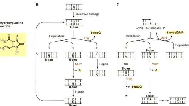
1.6.3.4 Oxidative DNA damage caused by ROS 33
1.6.3.5 M. tuberculosis DNA repair 34
1.6.3.6 Oxidative stress response regulation 44
XVI Chapter 2: Identification of M. tuberculosis virulence factors involved in defense
mechanisms against oxidative stress
49
2.1 Abstract 51
2.2 Introduction 53
2.3 Materials and Methods 55
2.4 Results 61
2.4.1 Screening for mutants with increased susceptibility to oxidative stress 61 2.4.2 Sensitive mutants to oxidative stress are affected in cell envelope
functions
62
2.4.3 Quantification of sensitivity of mutants to oxidative stress 64 2.4.4 MmpL9 and MoaD1 are important for M. tuberculosis survival in human
macrophages
66
2.4.5 The mmpL9 deletion at position 218 is conserved in Beijing/W strains 67 2.4.6 MmpL9 and MoaD1 are required to counteract other forms of stress
existent in macrophages
69
2.5 Discussion 77
Chapter 3: Evolution and diversity of M. tuberculosis Beijing/W strains reflected by polymorphisms in 3R genes
83
3.1 Abstract 85
3.2 Introduction 87
3.3 Materials and Methods 89
3.4 Results 93
3.4.1 Identification of polymorphisms in 3R genes in Beijing/W strains 93 3.4.2 Construction of a phylogeny for Beijing/W strains using 3R SNPs 96
3.5 Discussion 99
Chapter 4 : Concluding Remarks 103
References 111
XVII
FIGURES
Chapter 1Figure 1 – Estimated MDR-TB cases among notified TB patients in 2010 4
Figure 2 – Mycobacterial cell envelope constitution 8
Figure 3 – Representation of M. tuberculosis genome of the H37Rv reference strain, indicating the major genetic markers used for strain genotyping
10
Figure 4 – M. tuberculosis phylogeography 13
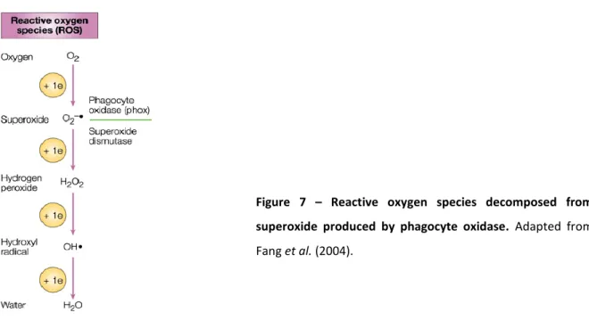
Figure 5 – M. tuberculosis infection and granuloma formation 21 Figure 6 – Assembly of the phagocyte NADPH oxidase NOX2 28 Figure 7 – Reactive oxygen species decomposed from superoxide produced by
phagocyte oxidase
29
Figure 8 – Cellular reactions leading to oxidative DNA damage via the Fenton Reaction.
34
Figure 9 – Major mechanisms repairing DNA damages in bacteria 35 Figure 10 – Enzymatic systems that protect from the mutagenic effects of
8-oxoguanine
36
Figure 11 – Early steps during the repair of double strand breaks (DSBs) 41
Chapter 2
Figure 1 – Growth curves of M. tuberculosis H37Rv, ΔmutT1, ΔmutM and ΔmutY in increasing concentrations of hydrogen peroxide.
61
Figure 2 – Growth curves of M. tuberculosis GC1237 in increasing concentrations of hydrogen peroxide.
62
Figure 3 – Susceptibility of transposon mutants to oxidative stress. 65 Figure 4 – Intracellular growth of mmpL9 and moaD1 mutants in human
macrophages.
67
Figure 5 – Graphical representation of the distribution of polymorphisms in mmpL9 in several M. tuberculosis genomes.
69
Figure 6 – Survival of mmpL9 and moaD1 mutant and complemented strains exposed to hydrogen peroxide.
70
Figure 7 – Macrophages treated with the NADPH oxidase inhibitors, apocynin and DPI. 71 Figure 8 – Intracellular growth of mmpL9 and moaD1 mutants in human macrophages
treated with Phox inhibitors.
72
Figure 9 – Macrophages activated with Vitamin D3. 73
Figure 10 – Intracellular growth of mmpL9 and moaD1 mutants in VitaminD3-activated macrophages treated with Phox inhibitors.
74
Figure 11 – Susceptibility of mmpL9 and moaD1 mutants to acidic pH and starvation. 75 Figure 12 – Representation of the mmpL9 gene in M. tuberculosis GC1237 78
XVIII Chapter 3
Figure 1. Phylogenetic network based on SNPs discovered in the collection of 58 Beijing/W isolates.
95
Figure 2. Phylogenetic network based on SNPs characterized in the entire collection of 305 Beijing/W isolates.
97
TABLES
Chapter 2Table 1. List of oligonucleotides (5’-3’) used in this study. 56 Table 2. List of identified sensitive mutants to oxidative stress using hydrogen
peroxide.
63
Table 3. Statistical analysis of SNP density in mmpL genes in a collection of M. tuberculosis genomes.
68
Chapter 3
Table 1. List of oligonucleotides used for SNP analysis. 90 Table 2. List of primers used in Multiplex-PCR for Large Sequence Polymorphisms
(LSPs) analysis.
92
Table 3. List of SNPs identified by sequencing 3R genes in Beijing/W strains, which define different sequence types (Figure 1 and 2).
94
Annex
Table A1. Description of M. tuberculosis Beijing/W strains belonging to each node found in Figure 1 and 2, and respective country of isolation.
CHAPTER 1
Tuberculosis an ancient disease
1.1
Tuberculosis (TB) is an ancient disease. Skeletal abnormalities, typical of TB, were found in Egyptian mummies, dated of several thousand years ago, and DNA analyses have later confirmed the presence of the disease-causing pathogen Mycobacterium tuberculosis. At this time TB was probably sporadic, but, as population densities increased, the disease reached epidemic proportions in Europe and North America in the 18th and 19th centuries (Daniel, 2006).
The development of a live attenuated vaccine, Bacillus Calmette-Guérin (BCG), by Albert Calmette and Camille – Guérin in 1921, and the discovery of the first antibiotic against TB, streptomycin, by Selman Waksman in 1943, led to the opinion that appropriate control measures had become available for fighting TB. Between 1950 and 1965 TB-control programs were established. However, these initiatives have had limited success in developing countries. In industrialized countries there was a pronounced reduction of infection and death rates. Consequently, TB programs became a lower priority because the disease was considered close to elimination. In the early 1980s, cases of TB began to raise due to factors such as increases in prison and homeless populations, intravenous drug use and especially Human Immunodeficiency Virus/Acquired Immunodeficiency Syndrome (HIV/AIDS) epidemics. By 1993 increasing TB could no longer be ignored so the World Health Organization (WHO) declared the disease a global emergency (Daniel, 2006) (Harries and Dye, 2006). In 2000, the World Health assembly endorsed the establishment of a Global Partnership to Stop TB with several priorities for the next 50 years (WHO at www.who.int/topic/tuberculosis/en/).
TB today’s major issues
1.2
Despite the efforts to combat the disease, TB remains today a major public-health problem. Every 15 seconds someone dies from TB, which means that about 1.5 million people die each year from this treatable disease. The current TB epidemic is being sustained and fuelled by two important factors: the HIV infection, and its association with active TB disease, and increasing resistance of M. tuberculosis strains to anti-TB drugs (WHO at www.who.int/topic/tuberculosis/en/).
Despite the availability of an effective antibiotherapy for decades, tuberculosis control has been seriously challenged due to the appearance of drug-resistant strains. Drug resistance in M. tuberculosis arises mainly from spontaneous mutations in the chromosome (Heym et al., 1994), and not from mobile genetic elements such as plasmids or transposons, acquired by horizontal
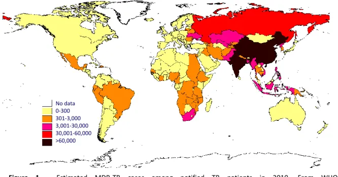
gene transfer, as observed in other bacteria. Then, irregular drug supply, inappropriate prescription and poor patient adherence to treatment allow selection of strains carrying drug-resistance mutations. These strains can be transmitted and other mutations involved in resistance to individual drugs can be accumulated resulting in the appearance of multidrug-resistant (MDR) strains, defined as resistant to at least rifampin and isoniazid, the most effective first-line anti-TB drugs (WHO at www.who.int/topic/tuberculosis/en/). The WHO estimates that there were 290 000 MDR-TB cases among notified TB cases in 2010 (Figure 1). The estimated number of prevalent MDR-TB cases seems to reach 650 000 among the 12 million total prevalent cases of TB (WHO, 2011). The number of extensively-drug resistant strains (XDR), defined as MDR plus resistance to any fluoroquinolone and to one of the three injectable second line drugs (capreomycin, kanamycin, or amikacin), has been increasing every year, and, by the end of 2010, 77 countries had reported at least one case of XDR TB to WHO (WHO at http://www.who.int/tb/challenges/mdr/en/index.html). More recently, the appearance of totally drug-resistant tuberculosis (TDR), resistant to all antituberculosis drugs, seriously threatens TB control (Velayati et al., 2009).
Figure 1 - Estimated MDR-TB cases among notified TB patients in 2010. From WHO at
http://www.who.int/tb/challenges/mdr/en/index.html.
More than one in ten TB cases occur among HIV-infected people, and almost one in four deaths among people with HIV is due to TB. In 2010 350 000 people died of HIV-associated TB (WHO, 2011)(WHO at http://www.who.int/tb/publications/TBHIV_Facts_for_2011.pdf). In fact, immunodeficiency caused by HIV infection greatly increases susceptibility to infection and the risk of developing TB, which is 21-34 times higher in HIV co-infected individuals than in non-infected
No data 0-300 301-3,000 3,001-30,000 30,001-60,000 >60,000
(WHO, 2011). On the other hand, TB may accelerate the course of HIV infection, increasing the viral load in some patients (Day et al., 2004; Whalen et al., 1995). In addition to contribute to an increase in TB burden in general, HIV might also be contributing to the increase of MDR-TB prevalence (Wells et al., 2007). Co-infection of TB in HIV patients further complicates treatment because it has to be combined with antiretroviral therapy. This leads to an increased pill burden, drug-drug interactions that may lead to suboptimal concentrations of anti-TB drugs, and finally overlapping side effects (Swaminathan et al., 2010; Wells et al., 2007). Moreover, TB patients co-infected with HIV are more likely to progress to disease, and thus potentially accelerate the propagation and spread of these MDR strains (Gandhi et al., 2010). The intersection of drug-resistance and HIV epidemics might represent a lethal combination and further threaten TB control.
The development of an effective vaccine against TB could also have a significant impact in the control of the disease. The only available vaccine today is BCG, a live vaccine derived from an attenuated Mycobacterium bovis strain, obtained after serial passages (Calmette, 1931). It has been used since 1921, yet, it has given little contribution to contain the current TB pandemic. It is effective in protection against disseminated TB in children, like meningitis and miliary TB (Trunz et al., 2006), but, it seems to have variable efficacy against adult pulmonary TB – the most prevalent form of disease – being particularly limited in endemic areas (Colditz et al., 1994; Fine, 1995). TB vaccine research has made huge progress and as a result twelve vaccine candidates have been developed and entered clinical trials (Kaufmann, 2011). Nevertheless, we do not know with certainty whether they will confer robust protection against TB in humans.
Finally, enormous progress has been made in the last decade resulting in the appearance of much promising tools for diagnosing TB (Palomino, 2012). Still, no tests are available that are universally applicable to all patients including HIV-coinfected people and children with TB, for which diagnosis is usually more difficult. Additionally, an important gap in this field is the development of tools to determine progression to disease in people infected with M. tuberculosis without active disease (latent TB). This would be extremely important given that one-third of the world’s population seem to be latently infected with M. tuberculosis (WHO at www.who.int/topic/tuberculosis/en/).
Although the number of incident cases has decreased substantially during the last twenty-years, Portugal has still an elevated rate of TB incidence, being the highest among western European countries ((WHO, 2011) - Annex 3). There is also a high prevalence of MDR-TB and alarming rates
of XDR-TB, particularly in the Lisbon Health Region (Direcção Geral de Saúde (DGS) at http://www.dgs.pt/upload/membro.id/ficheiros/i012626.pdf) (Perdigao et al., 2008).
The genus Mycobacterium
1.3
The first notion that TB could be an infectious disease was described by Benjamin Martens in 1790, followed by Jean-Antoine Villemin who demonstrated, in 1865, the transmissible and infectious nature of TB in animals (Daniel, 2006). However, it was Robert Koch, in 1882, who first described the tubercle bacillus, M. tuberculosis (Koch, 1882; Koch, 1982). Nevertheless, the identification of mycobacteria, however, occurred earlier with the discovery of the leprosy bacillus in 1873 (Hansen, 1874(Mange, 1992).
Mycobacteria are aerobic non-motile rod-shaped actinobacteria with a high G+C content in their DNA (61-71%). These bacteria are characterized by an unusual cell envelope composed by mycolic acids with long branched chains (60 to 90 carbons), which confer alcohol and acid-fast staining properties (Levy-Frebault and Portaels, 1992; Shinnick and Good, 1994). Along with Corynebacterium and Nocardia, they form a well-defined sub-group (CMN) of actinobacteria, which share properties such as the production of mycolic acids (Embley and Stackebrandt, 1994; Ventura et al., 2007). Other mycolic acid-containing genera include Gordonia, Rhodococcus, Tsukamurella and Dietzia among others (Gurtler et al., 2004) being thus considered to be closely related to mycobacteria.
The genus Mycobacterium is highly diverse and comprises more than 100 species (Dworkin et al., 2006). It can be divided into two major groups’ – fast and slow growers, a traditional division was found to be congruent by 16S rRNA analyses (Stahl and Urbance, 1990). This definition is based on the time required for visible colonies to appear on a solid medium, after plating suspensions diluted enough to give well separated colonies (Shinnick and Good, 1994):
I. Fast growers (colonies in less than seven days) – usually non-pathogenic mycobacteria and free-living saprophytes that include M. smegmatis, commonly used as a non-pathogenic model in the laboratory, to study essentially genetics or physiology of mycobacteria (O'Toole, 2010). An exception would be M. abssecus, a fast grower that is pathogenic.
II. Slow growers (colonies in more than seven days) – usually associated with human or animal disease and include important pathogens such as M. leprae, M. ulcerans, and M. tuberculosis complex members. There are, however, slow growers that are not pathogenic, such as M. gordonae.
1.3.1 The Mycobacterium tuberculosis complex
The M. tuberculosis complex includes several closely related species, responsible for tuberculosis, with different phenotypic properties and host preferences. M. tuberculosis and M. africanum are almost exclusively human pathogens. M. microti causes disease in voles and M. pinnipedii causes TB in seals and sea lions. M. caprae infects both goats and deer and, finally, M. bovis has the largest host range although it can be mainly isolated from cattle (Brosch et al., 2002; Mostowy et al., 2005; Smith et al., 2006). Some very rare isolates of tubercle bacilli, with the ability to cause disease in humans, were found to belong to the same phylogenetic lineage as M. tuberculosis complex members as shown by 16sRNA analyses (van Soolingen et al., 1997). These bacteria, referred to as M. canetti, have shown unusual smooth colony morphology, in contrast to the rough colonies produced by the other M. tuberculosis complex members.
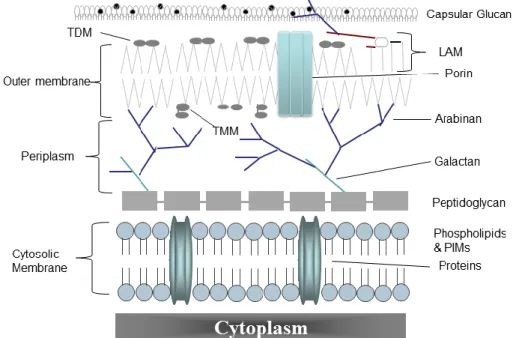
1.3.2 Mycobacterial cell envelope
Mycobacteria possess a remarkably complex cell envelope consisting of three principal layers: the cytoplasmic membrane, the cell wall core and an outer membrane (Figure 2). Cell walls are constituted by a peptidoglycan – arabinogalactan polymer core with covalently bound mycolic acids (the mycolyl-AG-peptidoglycan complex) that, along with non-covalently bound, extractable lipids, form an outermost bilayer structure (Hoffmann et al., 2008; Zuber et al., 2008). Thus, despite the classification of mycobacteria as Gram-positive bacteria it seems that the mycobacterial cell envelope contains an outer membrane containing pore-forming proteins (Niederweis et al., 2010), and, by inference, a periplasmic space (Hoffmann et al., 2008; Zuber et al., 2008) (Figure 2). This complex structure represents a permeability barrier that contributes to the intrinsic resistance of mycobacteria to antibiotics, dehydration and chemical damage (Brennan and Nikaido, 1995; Nguyen and Thompson, 2006). An outer layer (also called capsule) surrounding the outer membrane (Figure 2), mainly composed of polysaccharides, proteins and only minor amounts of lipids (Ortalo-Magne et al., 1995), also seems to exist in various mycobacterial species (Sani et al., 2010). Most of these compounds were found released in the culture fluids of in vitro-grown bacteria but seem to coat intracellularly vitro-grown bacteria (Daffe and Draper, 1998).
Numerous extractable lipids are present in mycobacterial cell walls, being many of these suspected to be present in the outermembrane. However, their exact arrangement and distribution remains poorly understood. These include: phthiocerol dimycocerosate (PDIM), a major lipid of the cell wall, important for virulence, architecture and permeability (Camacho et al., 1999; Cox et al., 1999); phenolic glycolipids (PGLs), that share a common lipid core with PDIMs and
are also associated with virulence, but are produced by only a few groups of M. tuberculosis (Reed et al., 2004; Tsenova et al., 2005); as well as glycolipids such as diacyltrehaloses (DAT), triacyltrehaloses (TAT), sulfatides (SLs), and the mycolic acid derivatives trehalose monomycolate (TMM) and trehalose dimycolate (TDM) (also known as cord factor) (Kaur et al., 2009).
Other important molecules in the mycobacterial cell envelope are mannosylated molecules such as lipoarabinomannan (LAM), the related lipomannan (LM), and phosphatidyl-myo-inositol mannosides (PIMs) (Kaur et al., 2009), that seem to be anchored not only to the outer membrane but also to the cytoplasmic membrane (Pitarque et al., 2008) (Figure 2). In M. tuberculosis and other pathogenic mycobacteria LAM is mannose-capped (ManLAM) (Brennan and Nikaido, 1995).
Figure 2 – Mycobacterial cell envelope constitution. LAM: lipoarabinomannan, PIMs:
phosphatidyl-myo-inositol mannosides, TDM: trehalose dimycolate, TMM: trehalose monomycolate. Adapted from Catalão et al. (2012), with authors’ permission. (Catalao et al., 2012)
As most of these molecules are the first to contact with host cellular constituents, the importance of these in facilitating host cell entry and modulating the host cell response has been well documented (discussed in more detail in section 1.5.1).
A large proportion of the M. tuberculosis genome is devoted to fatty acid and lipid metabolism (Cole et al., 1998). The colocalization of some mmpL (mycobacterial membrane protein Large) genes with genes involved in polyketide biosynthesis (pks) and lipid metabolism suggested that MmpLs are generally involved in lipid synthesis and/or transport (Tekaia et al., 1999). This was found to be indeed the case for MmpL7 (Camacho et al., 2001; Cox et al., 1999) as well as MmpL8 (Converse et al., 2003; Domenech et al., 2004), involved in the transport of PDIM and SL-1,
respectively. However, certain MmpLs do not colocalize with genes involved in lipid synthesis (Cole et al., 1998) and it seems that MmpL substrate specificity is dictated, in part, by their interaction with particular cytosolic enzymes (Jain and Cox, 2005).
Molecular evolution of mycobacteria
1.4
It is important to investigate diversity of bacterial pathogens for both epidemiological, to elucidate prevalence of strains, and biological reasons, because specific strains or groups of strains (lineages/families) might have specific relevant phenotypes (e.g. differences in virulence or drug susceptibility).
M. tuberculosis complex members are genetically extremely closely related when compared to other bacteria, being thus considered as monomorphic (Achtman, 2008). Therefore, some of the genotyping tools applied to other pathogens can be uninformative in M. tuberculosis (Achtman, 2008). For example, early attempts at using multilocus sequence typing (MLST) to study strain diversity in M. tuberculosis complex have shown that these bacteria are characterized by more than 99.9% similarity at the DNA level and identical 16S rRNA sequences (Sreevatsan et al., 1997).
Despite this apparent genetic homogeneity, M. tuberculosis complex members display diverse phenotypic characteristics and host ranges and several polymorphic molecular genetic markers (Figure 3) have proved to be useful to identify and discriminate M. tuberculosis complex isolates.
1.4.1 Molecular genetic markers that define M. tuberculosis complex genetic diversity
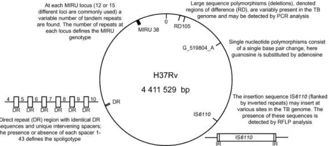
One of the most widely applied molecular methods to type M. tuberculosis strains has been the IS6110 Restriction Fragment Length Polymorphism (RFLP) typing. Individual strains vary in the number of copies and in the sites into which IS6110 is inserted. A method using a restriction enzyme has been developed and IS6110 Southern blots of the digested DNA separated by electrophoresis on agarose gels provide a RFLP pattern (van Embden et al., 1993). Unfortunately, this method has some limitations and disadvantages such as poor discrimination between isolates with low copy numbers (<5) of IS6110 and it is even absent from certain strains. In addition, it is a technically demanding and expensive technique and requires high amounts of DNA (Kato-Maeda et al., 2011). This has led to the development of other techniques, frequently based in amplification of DNA, being spacer oligonucleotide typing (Spoligotyping) and variable number of tandem repeat (VNTR) typing (Frothingham and Meeker-O'Connell, 1998) the most frequently used.
Figure 3 - Representation of M. tuberculosis genome of the H37Rv reference strain, indicating the major genetic markers used for strain genotyping. As an example one specific mycobacterial interspersed repetitive
unit (MIRU) locus, insertion sequence (IS6110), large sequence polymorphism (region of difference - RD) and single nucleotide polymorphism are shown. Adapted from Nicol et al. (2008). (Nicol and Wilkinson, 2008)
Spoligotyping involves PCR amplification of the DR locus, a single region of M. tuberculosis complex strains chromosome that contains multiple copies of a 36-bp direct repeat, separated by a DNA spacer with various sequences (Kamerbeek et al., 1997). Strains have the same overall arrangement of spacers but differ in terms of presence or absence of specific spacers. Comparison with the 43 spacers identified in M. tuberculosis H37Rv, the laboratory reference strain, and M. bovis BCG, allows discrimination and classification of strains (Van Soolingen, 2001). Forty-one VNTRs of the mycobacterial interspersed repetitive units (MIRUs) have been identified in M. tuberculosis genome (Supply et al., 2000). PCR amplification of each locus will reflect the number of copies of the repeat unit and is the basis of VNTR typing. Several number and combinations of loci have been used to genotype M. tuberculosis strains which might have similar or higher discriminatory power than IS6110-RFLP and may also vary according to certain characteristics of the sample that is being analyzed, such as geographic origin. Thus, the set of loci might have to be adapted to the type of collection of isolates to analyze. Nevertheless, MIRU-VNTR is considered by some authors as the gold standard for M. tuberculosis genotyping (Kato-Maeda et al., 2011). These methods are, however, not reliable for evolutionary and phylogenetic studies (Comas et al., 2009) because intragenomic recombination events could occur independently at the level of repetitive sequences (Achtman, 2008).
Recently, systematic comparative genomic analyses have identified Single Nucleotide Polymorphisms (SNPs) and Large Sequence Polymorphisms (LSPs) as robust markers that generate
portable and comparable data for studying population variation. This kind of markers have been especially used to infer phylogenetic relations in M. tuberculosis (Baker et al., 2004; Dos Vultos et al., 2008; Filliol et al., 2002; Fleischmann et al., 2002; Gagneux et al., 2006a; Gutacker et al., 2006; Gutacker et al., 2002; Hershberg et al., 2008). LSPs correspond to genomic deletions that have been suggested as an important mechanism for genetic variation in M. tuberculosis. Affected sequences might have functional relevance in pathogenesis and immunity since deleted sequences can include putative open reading frames as well as intergenic regions and housekeeping genes (Alland et al., 2007). LSPs may be informative markers for discrimination of strains and have been, therefore, proposed for genotyping and, as these are irreversible mutations, have also been used for constructing phylogenies and to investigate M. tuberculosis complex evolution (Alland et al., 2007; Brosch et al., 2002; Gagneux et al., 2006a; Tsolaki et al., 2005). SNPs also contribute to strain variability and may have a functional effect. For example, drug-resistance is mainly caused by SNPs in M. tuberculosis (Zhang and Yew, 2009). SNPs have also proved to be useful markers to differentiate strains and to study the phylogenetic relatedness of M. tuberculosis strains (Dos Vultos et al., 2008; Hershberg et al., 2008). A high number of such polymorphisms can be identified by performing whole-genome sequencing which can be extremely informative, particularly for genetically monomorphic bacteria like M. tuberculosis (Achtman, 2008).
Choosing the appropriate genetic marker will depend on the purpose of the study. For epidemiologic analyses it is important to have markers with a high discriminatory power that change relatively rapidly, but, are stable enough to make connections between related isolates. For example, recognition of the regional specificity of circulating M. tuberculosis strains in Portugal dates back to 1999 when early RFLP-IS6110 genotyping studies of an MDR cluster in the Lisbon Health Region identified a prominent family, designated family Lisboa (Portugal et al., 1999). In subsequent years RFLP-IS6110 and 12 loci MIRU–VNTR genotyping identified clusters of circulating strains with similar genotypic characteristics (Perdigao et al., 2008; Perdigao et al., 2011), several of which, including the Lisboa family, were associated with XDR–TB. The results show that active transmission of MDR- and XDR-TB is taking place and that the high prevalence of observed XDR-TB is due to the continued transmission of particular genetic clusters such as the Lisboa family, identified in 1999 as strongly associated with MDR-TB (Portugal et al., 1999) and now associated with XDR-TB (Perdigao et al., 2008; Perdigao et al., 2011).
To define phylogenetic associations unambiguously, genetic markers, need to be unique and, ideally, irreversible. Robust phylogenetic markers will exhibit low rates of convergent evolution
(Kato-Maeda et al., 2011) providing solid phylogenetic frameworks in which specific strains can be positioned and biologically meaningful groups defined.
1.4.2 Phylogeny of the M. tuberculosis complex
Although many molecular methods, such as the referred in the previous section, have been used to categorize M. tuberculosis complex strains, the phylogenetic relationships among this group have only recently started to be detailed. Analyses of genomic deletions (LSPs) have shown that the animal-adapted organisms of the M. tuberculosis complex are part of the same evolutionary lineage, defined by deletion of region of difference (RD) 9 (Brosch et al., 2002). Furthermore, M. bovis, which was initially thought to cause disease in a variety of animals, can be divided in different subspecies characterized by additional specific deletions, which include M. caprae, M. microti and M. pinnipedii (Brosch et al., 2002; Mostowy et al., 2005). It was suggested that the M. tuberculosis complex consists of distinct and genetically defined groups with specific host-adapted groups called ecotypes (Smith et al., 2006). M. africanum shares the RD9 deletion with these animal-adapted members of the M. tuberculosis complex which has led to the speculation that it is not primarily adapted to humans but has an animal reservoir (Smith et al., 2006). However, this remains to be confirmed and meanwhile, given that M. africanum clearly infects and transmits among humans being responsible for up to 40% of human TB in west Africa (de Jong et al., 2006; Kallenius et al., 1999), it is considered as human-adapted. The same analysis of genomic regions also showed that M. canetti had almost all the RD regions intact (Brosch et al., 2002). Although these strains share the same 16S rRNA sequence with M. tuberculosis complex members as referred previously, they present high levels of nucleotide variation in housekeeping genes and clear evidence of horizontal gene transfer (Gutierrez et al., 2005). This suggests that M. canetti strains are relatives of a highly diverse progenitor of the M. tuberculosis complex, M. prototuberculosis, that might have underwent an extreme bottleneck in a relatively recent past. Consequently, much of the original diversity disappeared leaving only M. canetti strains among the M. tuberculosis complex members (Gutierrez et al., 2005).
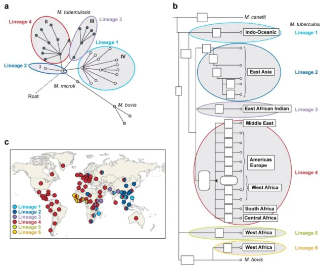
Other studies have defined the phylogeny of M. tuberculosis and M. africanum, the human-adapted species of the M. tuberculosis complex. Gagneux et al. (2006) also used LSPs to describe six major lineages, two of which included M. africanum. Each lineage seems to be associated with particular human populations (Figure 4). Several studies have used SNPs for the same purpose. Baker et al. (2004) analysed polymorphisms in seven housekeeping genes identifying 36 synonymous SNPs (sSNPs) that allowed discrimination of 4 main lineages in M. tuberculosis. These
four main lineages correspond to the same identified after by Gagneux et al. (2006) using LSPs (Figure 4).
Figure 4 – M. tuberculosis phylogeography. Current major lineage assignments (lineage 1 to 6) are indicated
by coloured lines. (a) Tree originated from the study of Baker et al. (2004) based on 36 synonymous SNPs identified by sequencing seven housekeeping genes in a collection of 225 strains. Roman designations correspond to lineage assignments in the study. (b) Tree based on large sequence polymorphisms (LSPs) discovered by Gagneux et al. (2006). (c) Association of M. tuberculosis lineages with specific geographic regions according to data from Gagneux et al. (2006) and Filliol et al. (2006). Adapted from Achtman et al. (2008). (Filliol et al., 2006; Gutacker et al., 2006)
Gutacker et al. (2006) have screened 5069 isolates for 36 sSNPs that have been identified by comparison of two M. tuberculosis genomes and defined nine main lineages. Three of these lineages corresponded to the ones described by Gagneux et al. (2006) and Baker et al. (2004). Filliol et al. (2006) have used a similar approach to identify 159 sSNPs and screened 219 isolates of M. tuberculosis and M. bovis. Ten major groups were identified, one of them being specific to M. bovis and three corresponding to the same previously described (Baker et al., 2004; Gagneux et al., 2006a). These studies have identified thus, four main M. tuberculosis lineages and two M. africanum lineages, associated with particular geographic locations (Figure 4).
The fact that both LSP and SNP-based analyses of large strain collections resulted in single trees without homoplasy indicated that horizontal gene transfer is extremely rare in M. tuberculosis complex and it has a highly clonal population structure (Supply et al., 2003).
Although these studies have provided a picture of M. tuberculosis and M. africanum evolution, some limitations didn’t allow a better elucidation. The number of SNPs was either too limited (Baker et al., 2004) or resulted from comparison of a limited number of genomes which can lead to branch collapsing (Filliol et al., 2006; Gutacker et al., 2006) and biased phylogenies. As for the phylogeny inferred from LSPs (Gagneux et al., 2006a), genetic distances based on genomic deletions are difficult to interpret. In conclusion, genotyping approaches that rely on LSPs or a limited numbers of sSNPs can define deep phylogenetic population structures, but they are still characterized by low discriminatory power.
More recently, two studies have contributed to a better knowledge of M. tuberculosis evolution by overcoming the limitations of the previous studies (Dos Vultos et al., 2008; Hershberg et al., 2008), providing a more detailed picture of M. tuberculosis phylogeny. These studies reveal that the human-adapted members of M. tuberculosis complex are much more genetically diverse than generally recognized and some of this genetic diversity is likely to have functional consequences (Hershberg et al., 2008). The genetic diversity found among the human-adapted members of M. tuberculosis complex was just as pronounced as that among animal-adapted strains, even though the latter are adapted to distinct animal host species (Hershberg et al., 2008). In the study of Vultos et al. (2008) a specific group of genes was analysed: genes involved in DNA repair, recombination and replication (3R) genes. In M. tuberculosis the main source of genetic variation should arise from random mutations, although it was recently suggested that recent recombination might also occur in M. tuberculosis (Namouchi et al., 2012). Defective 3R systems can potentially increase genomic variability due to higher mutation rates (Denamur and Matic, 2006; Tonjum and Seeberg, 2001). Thus, polymorphisms in 3R genes seems to be an important mechanism in evolution and adaptation of microorganisms (Taddei et al., 1997; Tonjum and Seeberg, 2001) as strains with higher mutation rates (mutators) may, under certain conditions, have a selective advantage. Analysis of polymorphisms in such genes in a collection of strains representative of global M. tuberculosis complex diversity has identified high levels of polymorphisms, suggesting that these genes may have a role in M. tuberculosis complex evolution (Dos Vultos et al., 2008). In 2010, Comas et al. (2010) used 21 whole genome sequences to generate the first whole genome – based phylogeny of human-adapted M. tuberculosis, and confirmed the existence of six main phylogenetic lineages. More recently whole genome analysis of M. tuberculosis complex strains
from major lineages has also confirmed previous phylogenetic analysis using finished and partial genome data (Namouchi et al., 2012). It was also shown that purifying selection is acting on M. tuberculosis genetic diversity (Namouchi et al., 2012), in contrast to what has been previously suggested (Hershberg et al., 2008).(Comas et al., 2010)
There is increasing evidence that the observed M. tuberculosis strain variation is biologically significant and that specific genotypes are associated with specific phenotypes. Indeed, different M. tuberculosis genotypes show differences in terms of virulence and pathogenesis in animal models (Lopez et al., 2003; Palanisamy et al., 2009) as well as survival and transcriptional profiles within macrophages (Homolka et al., 2010). Although this genetic diversity seems to influence clinical presentation of the disease in humans (Caws et al., 2008; Thwaites et al., 2008) this is still not so clear (Coscolla and Gagneux, 2010) as several factors have a role in the development of TB. One of the most studied M. tuberculosis genotypes due to its association with increased virulence, as well as other characteristics that suggest that these strains have a selective advantage and are more adapted than strains from other genotypes, is the Beijing/W family (East-Asian lineage according to Gagneux et al. (2006)).
1.4.3 The Beijing/W family
The Beijing family was first described in 1995. A large proportion of M. tuberculosis strains in the Beijing area of China had highly similar multi-banded IS6110 RFLP patterns and identical spoligotyping patterns (van Soolingen et al., 1995). In the second half of the 1990’s the multidrug resistant M. tuberculosis strain identified in New York, “W” strain (Bifani et al., 1996), was recognized as a member of the Beijing genotype family which became the Beijing/W family (Bifani et al., 2002; Kurepina et al., 1998). Since then, the Beijing/W genotype was found to be the most frequently observed genotype of M. tuberculosis to cause disease globally (Glynn et al., 2002). According to the fourth international spoligotyping database, strains from this genotype were found to be present in the largest number of countries globally and represented 13% of global isolates (Brudey et al., 2006). In some regions including Russia, Bangladesh, South Africa, and Western Europe, the incidence of Beijing/W strains seems even to be increasing (Tuberculosis, 2006). This contrasts to what seems to happen with other genotypes that appear to be predominantly restricted to defined geographical regions, and suggests that Beijing/W strains might have some selective advantage that have allowed their global spread.
When compared to other genotypes, Beijing/W strains were found to grow significantly faster in human macrophages (Li et al., 2002; Theus et al., 2004) and have shown increased virulence in
different animal models of infection (Lopez et al., 2003; Tsenova et al., 2005). However, it is not possible to conclusively demonstrate an association between the Beijing/W strain genotype and clinical presentation in humans (Coscolla and Gagneux, 2010; Parwati et al., 2010). In addition, Beijing/W strains are often associated with drug-resistance (Tuberculosis, 2006).
Beijing/W strains share several genetic characteristics (Bifani et al., 2002) which differentiate them from other all M. tuberculosis genotypes, including: a spoligotyping pattern containing at least three of the nine spacers, 35 to 43, with an absence of hybridization to spacers 1 to 34, which classically define and identify Beijing/W strains (Kremer et al., 2004); similar IS6110 RFLP patterns (15-26 copies); IS6110 insertion between dnaA and dnaN genes encoding DNA replication proteins and insertion of variable numbers of IS6110 in the noise transfer function (NTF) chromosomal region (Kurepina et al., 1998) that define typical/modern (one or several IS6110 insertions) and atypical/ancient (no IS6110 insertions) Beijing/W strains (Mokrousov et al., 2006). VNTR typing has been the most frequently used marker to discriminate Beijing/W strains. However, special consideration is given to VNTR typing of Beijing/W isolates once the standardized set of 15 or 24 loci used to type M. tuberculosis strains are not always sufficient to discriminate among Beijing/W isolates (Iwamoto et al., 2007). In addition VNTR loci might have different discriminatory powers among Beijing/W strains depending on strain collection characteristics such as geographical origin (Kremer et al., 2005).
By comparative whole-genome hybridization of Beijing/W strains Tsolaki et al. (2005) have shown that there are LSPs associated with this family. These correspond to genomic regions variably deleted in Beijing/W strains in relation to M. tuberculosis H37Rv (RDs). RD105, RD181, RD150, RD142, were found to, not only define but also subdivide this family into four different subgroups (Tsolaki et al., 2005) that might be associated with different phenotypes. Hanekom et al. (2007) have shown that the Beijing/W family can be divided into seven different lineages and that, by analysis of epidemiological data, differences between these lineages in their ability to spread and cause disease might exist. Thus, even closely related strains from the same lineage can accumulate genomic diversity, which can lead to different pathogenic properties. Indeed, an earlier study has shown that not all Beijing/W strains are hypervirulent in mice (Dormans et al., 2004). In subsequent studies this difference among Beijing/W strains was shown in different animal models (Aguilar et al., 2010; Palanisamy et al., 2009) as well as in human macrophages (Theus et al., 2007). A very recent study has shown, in guinea pigs, that the LSP-defined Beijing/W sublineages have indeed differences in pathogenicity (Kato-Maeda et al., 2012).