Universidade de Aveiro
2014/2015
Departamento de Química
João Paulo Duarte
Calixto
Concentração de biomarcadores tumorais utilizando
sistemas aquosos bifásicos
Concentration of tumor biomarkers using aqueous
biphasic systems
I Universidade de Aveiro
2014/2015
Departamento de Química
João Paulo Duarte
Calixto
Concentração de biomarcadores tumorais utilizando
sistemas aquosos bifásicos
Concentration of tumor biomarkers using aqueous
biphasic systems
Dissertação apresentada à Universidade de Aveiro para cumprimento dos requisitos necessários à obtenção do grau de Mestre em Bioquímica, ramo de Bioquímica Clínica, realizada sob a orientação científica da Doutora Mara Guadalupe Freire Martins, Investigadora Coordenadora do Departamento de Química, CICECO, da Universidade de Aveiro, e co-orientação do Professor Doutor João Manuel da Costa Araújo e Pereira Coutinho, Professor Catedrático do Departamento de Química da Universidade de Aveiro.
III
Aos meus pais, o suporte do meu bem-estar e ao meu irmão, a minha maior referência…
V
O júri
presidente
Prof. Dr. Pedro Miguel Dimas Neves DominguesProfessor Auxiliar com Agregação do Departamento de Química da Universidade de Aveiro
Drª. Mara Guadalupe Freire Martins
Investigadora Coordenadora do Departamento de Química, CICECO, da Universidade de Aveiro
Drª. Ana Catarina Almeida Sousa
Estagiária de Pós-Doutoramento do Centro de Investigação em Ciências da Saúde, CICS-UBI, da Universidade da Beira Interior
VII Agradecimentos
Gostaria de começar esta fase de agradecimentos por agradecer aos meus orientadores, a Drª Mara Freire e o Professor João Coutinho, pelo excelente acompanhamento e pela força, otimismo e entusiasmo que me incutiram desde o início.
Gostaria igualmente de agradecer ao Path e a todos os seus elementos por tudo o que me ensinaram e por terem feito de mim um de vós. Obrigado, em especial a ti, Matheus Pereira, por tudo o que de fantástico fizeste por mim. Pela paciência, pelo apoio, pelos conselhos ou mesmo por não me dares trabalho às sextas para eu poder sair às quintas à noite, muito obrigado. Se esta tese existe, isso deve-se muito a ti.
Obrigado a todos os professores, não só da Universidade de Aveiro, mas também do Colégio Dr. Luís Pereira da Costa, por todos os conhecimentos que me transmitiram e por serem um suporte base da nossa sociedade que nem sempre é devidamente reconhecido. Obrigado a todos os colegas da equipa de Bioquímica e da AAUAv e ao meu treinador.
Obrigado a todos os amigos e colegas de curso que comigo trilharam e que fizeram destes 5 anos, os 5 melhores anos da minha vida. Torna-se difícil e injusto individualizar, no entanto, Henrique, Rafa, Daniel, Joel, Diogo, Rafa, Tiago, André, João Pedro, David, Mauro, Diana e Margarida, muito obrigado!
Obrigado a todos os meus familiares, por se preocuparem e serem atenciosos e por me irem perguntando, ao longo destes 5 anos, “Então como está Aveiro?” ou “Isso da Bioquímica serve para o quê?”. Um agradecimento especial aos meus avós, às minhas tias Isabel e Alcinda Calixto e aos meus primos Alexandre Calixto dos Santos e Diana Calixto Gil.
Por fim, muito obrigado às 3 pessoas mais importantes da minha vida: o meu pai, a minha mãe e o meu irmão. Ao meu pai, Antero Calixto, obrigado por seres o meu maior fã, obrigado por todos os conselhos, por me dares, com a mãe, todas as condições para concluir o meu percurso académico e por fazeres de mim o homem que sou hoje. À minha mãe, Fernanda Calixto, obrigado por todo o carinho e atenção nos momentos mais difíceis, obrigado por me acordares devagarinho ao Domingo quando fico a dormir até à hora do almoço, obrigado pelas remessas semanais de tupperwares e por me teres ensinado todas aquelas pequenas coisas “da casa” que nestes 5 anos me deram muito jeito. Ao meu irmão, Cláudio Calixto, obrigado acima de tudo por seres a minha maior referência e a pessoa mais importante da minha vida. Obrigado por me teres ajudado a crescer, por tudo que fazes por mim e por estares sempre do meu lado. Todos temos um ídolo, alguém que tentamos seguir e para o qual olhamos “para cima” em sinal de respeito e admiração. O meu ídolo, desde a minha infância, és tu!
Neste momento, a dor da nostalgia acaba por me atraiçoar o coração, no entanto, o que de fantástico vivi, e a sensação de que, se tivesse a oportunidade de voltar atrás eu faria exatamente tudo do mesmo modo, acaba por ser superior e dá-me força para continuar.
Obrigado, do fundo do coração, a todos os que comigo percorreram este caminho!
IX
Palavras-chave Cancro da próstata, marcadores tumorais, antigénio prostático específico, sistemas aquosos bifásicos, técnica de concentração, líquidos iónicos
Resumo De acordo com dados disponibilizados pela Organização Mundial de Saúde, cerca de 8,2 milhões de pessoas morrem anualmente com cancro. A elevada taxa de mortalidade associada ao cancro resulta da maioria dos pacientes não efetuar exames de rotina e porque a manifestação dos sintomas, na maioria dos casos, acontece quando o paciente já se encontra numa fase avançada da doença. Atualmente, o cancro da próstata representa a segunda maior causa de morte entre indivíduos do sexo masculino em todo o mundo. Tendo em conta que não existe cura para casos avançados de cancro da próstata, a estratégia passa por um diagnóstico precoce que permita aumentar a taxa de sucesso dos tratamentos. O antigénio prostático específico (PSA) é um biomarcador importante do cancro da próstata que pode ser detetado em fluidos biológicos, nomeadamente sangue, urina e sémen. No entanto, os kits comerciais disponíveis utilizam amostras de sangue e os métodos analíticos normalmente utilizados na sua deteção e quantificação requerem pessoal especializado, equipamento específico e um processamento extensivo das amostras, resultando em processos com um elevado custo associado. Assim, o objetivo deste mestrado passou por desenvolver um método simples, eficiente e menos dispendioso para a extração e concentração de PSA a partir de amostras de urina utilizando sistemas aquosos bifásicos (SAB) constituídos por líquidos iónicos.
Numa fase inicial, determinaram-se os diagramas de fases de um conjunto de sistemas aquosos bifásicos constituídos por um sal orgânico e por líquidos iónicos. Em seguida, avaliou-se a capacidade dos mesmos para a extração do PSA. Os resultados obtidos demonstram que, nos sistemas em estudo, o antigénio prostático específico é totalmente extraído para a fase rica em líquido iónico num único passo. Por fim, averiguou-se a aplicabilidade dos SAB estudados para a concentração do PSA a partir de soluções aquosas e de urina. A baixa concentração deste biomarcador na urina (clinicamente significativo abaixo de 150 ng/mL) dificulta a sua deteção através de técnicas analíticas convencionais. Os resultados obtidos demonstraram que é possível extrair e concentrar PSA até 250 vezes, numa única etapa, sendo este detetável através de técnicas menos dispendiosas.
XI
keywords Prostate cancer, cancer biomarkers, prostate specific antigen, aqueous biphasic systems, concentration technique, ionic liquids
abstract According to the World Health Organization, around 8.2 million people die each year with cancer. Most patients do not perform routine diagnoses and the symptoms, in most situations, occur when the patient is already at an advanced stage of the disease, consequently resulting in a high cancer mortality. Currently, prostate cancer is the second leading cause of death among males worldwide. In Portugal, this is the most diagnosed type of cancer and the third that causes more deaths. Taking into account that there is no cure for advanced stages of prostate cancer, the main strategy comprises an early diagnosis to increase the successful rate of the treatment. The prostate specific antigen (PSA) is an important biomarker of prostate cancer that can be detected in biological fluids, including blood, urine and semen. However, the commercial kits available are addressed for blood samples and the commonly used analytical methods for their detection and quantification requires specialized staff, specific equipment and extensive sample processing, resulting in an expensive process. Thus, the aim of this MSc thesis consisted on the development of a simple, efficient and less expensive method for the extraction and concentration of PSA from urine samples using aqueous biphasic systems (ABS) composed of ionic liquids.
Initially, the phase diagrams of a set of aqueous biphasic systems composed of an organic salt and ionic liquids were determined. Then, their ability to extract PSA was ascertained. The obtained results reveal that in the tested systems the prostate specific antigen is completely extracted to the ionic-liquid-rich phase in a single step. Subsequently, the applicability of the investigated ABS for the concentration of PSA was addressed, either from aqueous solutions or urine samples. The low concentration of this biomarker in urine (clinically significant below 150 ng/mL) usually hinders its detection by conventional analytical techniques. The obtained results showed that it is possible to extract and concentrate PSA, up to 250 times in a single-step, so that it can be identified and quantified using less expensive techniques.
XIII
Contents
1. General introduction... 1
1.1. Scopes and objectives ... 3
1.2. Cancer update overview ... 5
1.2.1. Epidemiology ... 5
1.2.2. Carcinogenesis: molecular changes and risk factors ... 6
1.3. Cancer biomarkers ... 11
1.4. Prostate cancer ... 16
1.4.1. Etiology ... 16
1.4.2. Signals, symptoms and diagnosis ... 16
1.5. Prostate-specific antigen as a prostate cancer biomarker ... 17
1.5.1. Prostate cancer screening, stage and grade ... 18
1.5.2. Monitoring therapy and disease recurrence ... 20
1.5.3. Molecular characteristics ... 20
1.5.3.1. Biosynthesis structure ... 20
1.5.3.2. Structure and physicochemical properties ... 21
1.5.3.3. Physiological role of PSA in prostate and in external tissues... 22
1.5.3.4. Molecular derivatives of PSA and their role in diagnosis ... 23
1.5.3.5. Stability of total and free PSA ... 24
1.6. Analytical methods for the quantification of PSA ... 25
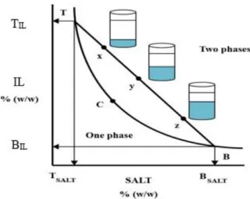
1.7. Aqueous biphasic systems (ABS) for the extraction and concentration of proteins 28 1.7.1. Phase diagrams ... 28
1.7.2. Ionic liquids as phase-forming components of ABS... 29
2. Extraction of PSA using phosphonium-based ILs + salt ABS ... 35
2.1. Introduction ... 36
2.2. Experimental Section ... 38
2.2.1. Chemicals ... 38
2.2.2. Experimental Procedure ... 38
2.2.2.1. Synthesis and characterization of the Good’s buffers ionic liquids ... 38
2.2.2.2. Phase diagrams and tie-lines (TLs) ... 39
2.2.2.3. Partition of PSA ... 41
XIV
2.3.1. Characterization of the synthetized ionic liquids ... 42
2.3.2. Phase Diagrams and Tie-lines ... 45
2.3.3. Partition of PSA ... 50
2.4. Conclusions ... 52
3. Concentration of PSA using model IL-based ABS ... 55
3.1. Introduction ... 56
3.2. Experimental section ... 56
3.2.1. Chemicals ... 56
3.2.2. Experimental procedure ... 57
3.2.2.1. Lever-arm Rule ... 57
3.2.2.2. Concentration factors of PSA ... 57
3.2.2.3. Size-exclusion HPLC (SE-HPLC) ... 58
3.3. Results and discussion ... 58
3.3.1. Concentration factors of PSA... 58
3.4. Conclusions ... 61
4. Concentration of PSA from human fluids using optimized IL-based ABS ... 64
4.1. Introduction ... 65
4.2. Experimental Section ... 66
4.2.1. Chemicals ... 66
4.2.2. Experimental procedure ... 66
4.2.2.1. Size-exclusion HPLC (SE-HPLC) ... 66
4.2.2.2. Sodium dodecyl sulphate polyacrylamide gel electrophoresis (SDS-PAGE) ... 67
4.3. Results and discussion ... 67
4.3.1. Concentration of PSA from human urine samples ... 67
4.4. Conclusions ... 70
5. Final remarks and future work ... 73
6. References ... 76
Appendix A ... 87
BLItz® Pro system calibration curve ... 87
Appendix B ... 89
Experimental binodal data ... 89
Appendix C ... 98
XVI
List of tables
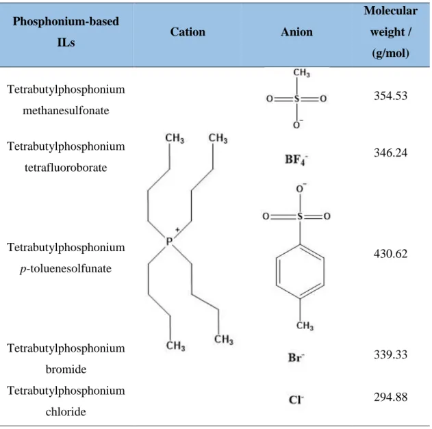
Table 1.1. Categories of cancer types according to their source (15). ... 7 Table 1.2. Cancer biomarkers identified in biological fluids or tissues in cancer diagnosis, stage, response to treatment and prognosis (47). ... 13 Table 1.3. PSA values of reference for men with 40-79 years old in blood (66). ... 19 Table 1.4. Risk stratification of CaP patients according to PSA levels, Gleason grade and Clinical stage (69). ... 19 Table 1.5. Comparison of literature methods for the purification/concentration/quantification of PSA isoforms from different human matrices... 26 Table 1.6. Chemical structures of phosphonium-based ionic liquids (114). ... 31 Table 1.7. Chemical structures of GBs: MES: 2-(N-morpholino)ethanesulfonic acid; TES: 2-[[1,3-dihydroxy-2-(hydroxymethyl)propan-2-yl]amino]ethanesulfonic acid; HEPES: 4-(2-hydroxyethyl)-1-piperazineethanesulfonic acid; CHES: N-cyclohexyl-2-aminoethanesulfonic acid; Tricine: N-2(2-Hydroxy-1,1-bis(hydroxymethyl)ethyl)glycine. ... 33 Table 2.1. Chemical structures and molecular weight (g/mol) of the synthesized GB-ILs ([P4444][MES], [P4444][TES], [P4444][HEPES], [P4444][CHES], [P4444][Tricine]). ... 44
Table 2.2. Correlation parameters used to describe the experimental binodal data by Eq. 1 and respective standard deviations (σ) and correlation coefficients (R2). ... 47
Table 2.3. Data for the tie-lines (TLs) and tie-line lengths (TLLs). Initial mixture compositions are represented as [Salt]M and [IL]M, whereas [Salt]Salt and [IL]Salt are the
compositions of IL and salt at the IL-rich phase, respectively, and vice-versa. ... 48 Table 2.4. Extraction efficiency of PSA (EEPSA%) at 25º C in the ABS composed of ILs and
K3C6H5O7. ... 50
Table 3.1. Concentration factors (initial compositions of the mixtures in Appendix C) in the ABS composed of [P4444][GB]+ K3C6H5O7 + water. ✓: achieved CF; ✕: not achieved CF.
... 60 Table 3.2. Concentration factor of 250-fold achieved for PSA at the IL-rich phase for the systems composed of GB-IL + K3C6H5O7+ PSA. ✓: achieved CF; ✕: not achieved CF. . 60
XVII
Table B.1.1. Experimental weight fraction data for the binodal curve of the systems composed of [P4444][GB] (1) + K3C6H5O7 (2) at (25 ± 1) °C……….90
Table B.1.2 Experimental weight fraction data for the binodal curve of the systems composed of [P4444][GB] (1) + K3C6H5O7 (2) at (25 ± 1) °C………95
Table C.1.2. Experimental data for CF of the systems composed of [P4444][GB] + K3C6H5O7
XIX
List of figures
Figure 1.1. Bars-plot of new cancer cases and deaths of worldwide population in 2012 (numbers in millions of new cancer cases/deaths) (12). ... 5 Figure 1.2. Number of new cancer cases in Portuguese population (males) in 2012 (dark-blue), and predictions to 2015 (blue) and 2030 (light-blue). Numbers for the 5 most common types of cancer in Portugal (13). ... 6 Figure 1.3. Schematic representation of PSA processing in epithelial cells of the prostate. Adapted from (75). ... 21 Figure 1.4. 3D representation of PSA using light-assisted molecular immobilization technology (78). ... 22 Figure 1.5. Different fPSA percursor forms (bPSA, proPSA and intact unclipped PSA) and PSA complexes (PSA-ACT, PSA-API and PSA-A2M) (5). ... 24 Figure 1.6. Phase diagram of an ABS: TCB = binodal curve; C = critical point; TB = tie-line; T = composition of the top phase; B = composition of the bottom phase; X, Y and Z = mixture compositions at the biphasic region. Adapted from (108). ... 29 Figure 2.1. GB-ILs synthesis. An equimolar excess of the aqueous solution of buffer was added to [P4444]OH solution (A). The mixture was dried at 50-60 °C under reduced pressure
in a Carrousel 6 plus reaction station (B). A mixture of acetonitrile and methanol (1:1, v:v) was added to the viscous liquid and then vigorously stirred at room temperature for 1 h (Fig. 2.1. C). The solvent mixture was evaporated and the GB-IL was dried in vacuum (10 Pa) for 3 days at room temperature (D). ... 39 Figure 2.2. Blitz® PRO System and Super Streptavidin Biosensors. ... 41 Figure 2.3. Macroscopic appearance of the synthesized Good's Buffer ionic liquids at 25ºC. From the left to the right: [P4444][MES], [P4444][TES], [P4444][CHES], [P4444][HEPES],
[P4444][Tricine]. ... 43
Figure 2.4. Phase diagrams for the systems composed of IL + K3C6H5O7 + H2O at 25ºC and
atmospheric pressure (in wt%) with the corresponding binodal data adjusted by Eq. 1: [P4444][Tricine] (); [P4444][HEPES] (); [P4444][TES] (); [P4444][MES]
();[P4444][CHES] (); ... 45
Figure 2.5. Phase diagrams and TLs (■) and adjusted binodal data using Eq. 1 (-), at 25ºC and atmospheric pressure, for the ternary systems composed of K3C6H5O7 + water +
XX
[P4444][MES] (); [P4444][CHES] (); [P4444][HEPES] (); [P4444][Tricine] ();
[P4444][TES] ()... 49
Figure 2.6 PSA secondary structure (1) and their active site (2) (using the Discovery Studio software). ... 51 Figure 3.1. Different compositions along the same TL obtained by applying the lever-arm rule which allow to obtain different CF for the ABS composed of K3C6H5O7 + [P4444][CHES]
+ water. ... 59 Figure 3.2. HPLC spectra of bottom (left) and top (right) phases for the systems composed of GB-IL + K3C6H5O7 + water/PSA. A spectrum of pure PSA in water (C= 50 ng/L) is also
provided as an insert for terms of comparison. ... 61 Figure 4.1. ABS composed of [P4444][CHES] + salt + human urine (150 ng/mL of PSA
added). ABS after being vigorously stirred (on the left) and after being centrifuged for 10 min and left in equilibrium for 15 min (on the right). ... 68 Figure 4.2. SE-HPLC profile of pure PSA in aqueous solution, human urine and in the top and bottom phases of an ABS composed of [P4444][CHES] + salt + human urine (150 ng/mL
of PSA added). ... 69 Figure 4.3. SDS-PAGE analyses. A: Molecular Wweight marker; B: Pure human serum; C: Pure human urine. ... 70 Figure A.1. Calibration curve for PSA in aqueous solution………..88 Figure C.1.3. Experimental data for CF of the systems composed of [P4444][GB] + K3C6H5O7
XXII
List of symbols
wt% – weight fraction percentage;
σ – standard deviation;
Abs – absorbance (dimensionless);
Mw – molecular weight (g·mol-1);
R2 – correlation coefficient (dimensionless);
α – ratio between the top weight and the total weight of the mixture (dimensionless);
[𝐼𝐿] – concentration of ionic liquid (wt% or mol·kg-1);
[𝐼𝐿]IL – concentration of ionic liquid in the ionic-liquid-rich phase (wt%); [𝐼𝐿]Salt – concentration of ionic liquid in the salt-rich phase (wt%); [𝐼𝐿]M – concentration of ionic liquid in the initial mixture (wt%); [𝑆𝑎𝑙𝑡] – concentration of salt (wt% or mol·kg-1);
[𝑆𝑎𝑙𝑡]IL – concentration of salt in the ionic-liquid-rich phase (wt%);
[𝑆𝑎𝑙𝑡]Salt – concentration of salt in the salt-rich phase (wt%);
[𝑆𝑎𝑙𝑡]M – concentration of salt in the initial mixture (wt%); [𝑆𝑎𝑙𝑡] – concentration of salt (wt% or mol·kg-1);
EEPSA% – percentage extraction efficiency of PSA (%);
CFPSA – PSA concentration factor;
wIL – weight of the IL-rich phase;
XXIV
ACRONYMS
A2M – alpha-2-macroglobulin; ABS – aqueous biphasic systems; ACT – alpha-1-antichymotrypsin; API – alpha-1-protease inhibitor B – bottom phase;
BPH – benign prostate hyperplasia; BPSA – benign PSA;
BSA – bovine serum albumin C – critical point;
CaP – prostate cancer;
CHES – N-cyclohexyl-2-aminoethanesulfonic acid; CML – chronic myelogenous leukemia;
DNA – deoxyribonucleic acid; DRE – digital rectal examination;
ELISA – enzyme-linked immunosorbent assay; ER – endoplasmic reticulum;
FDA – Food and Drug Administration; fPSA – free PSA;
GBs – Good’s buffers;
HEPES – N-cyclohexyl-2-aminoethanesulfonic acid; HPLC – High-performance liquid chromatography; IGF – insulin-like growth factor;
IGFBP – insulin-like growth factor binding protein; iPSA – intact PSA;
K3C6H5O7 – potassium citrate;
MES – 2-(N-morpholino)ethanesulfonic acid; PAP – prostatic acid phosphatase.
XXV pI – isoelectric point;
proPSA – precursor isoforms of PSA; PSA – prostate specific antigen; RNA – ribonucleic acid;
ROS – reactive oxygen species; SPE – solid-phase extraction;
SPR – surface plasmon resonance technology; T – top phase;
TL – tie-line; tPSA – total PSA;
TRIFA – immunochemiluminescent; TRUS – transrectal ultrasound; TS – tumor suppressor;
[P4444][CHES] – tetrabutylphosphonium 2-( cyxlohexylamino)ethanesulfonate;
[P4444][HEPES] – tetrabutylphosphonium
2-[4-(2-hydroxyethyl)piperazin-1-yl]ethanesulfonate;
[P4444][MES] – tetrabutylphosphonium 2-(N-morpholino)ethanesulfonate;
[P4444][TES] – tetrabutylphosphonium
2-[[1,3-dihydroxy-2-(hydroxymethyl)propan-2-yl]amino]ethanesulfonate;
[P4444][Tricine] – tetrabutylphosphonium
1
3 1.1. Scopes and objectives
Prostate cancer is the second more common type of cancer, and represents 15% of the cancer-related diseases in males (1). Despite the efforts in the search of new and effective therapies, up to now, none of the known therapies is entirely effective when the diagnosis is carried out at an advanced stage, emphasizing thus the importance of early detection as a powerful way to increase the curative successful rate (2). Prostate cancer is usually identified through biopsy, a procedure that is still known as the diagnostic “gold strand”. However, associated with this procedure, there are several and serious side effects, like febrile urinary tract infection (UTI), urosepsis, bleeding and hematuria. In addition, this procedure is also connected to 15%-20% of false negative results (3). Therefore, it is of crucial importance to find non-invasive diagnostic tools that could replace the commonly used biopsy. The identification and quantification of cancer biomarkers in human serum and urine is a potential alternative and has received a significant attention in the past few years (4). For instance, the identification and quantification of the prostate specific antigen (PSA) is the most widely used assay to detect prostate cancer at earlier stages bypassing unnecessary biopsies and reducing the number of patients where metastatic disease is found (5).
Currently, there are diverse companies (6) that offer several commercial kits for the PSA quantification in blood samples, but some of them require the use of immunoassay-qualified antibodies, specific and expensive equipment and highly skilled technical operators – time-consuming and expensive immunoassays (7,8). Thus, the main objective of this work comprises the development of an alternative technique for the extraction and concentration of PSA from human urine using a liquid-liquid extraction procedure. This procedure should allow the concentration of PSA up to levels capable of being detected by more accessible and less expensive equipment, such as high-performance liquid chromatography (HPLC). To achieve this goal, aqueous biphasic systems (ABS) composed of water, ionic liquids (ILs) and organic salts were investigated. In the past decade, a large effort has been dedicated to IL-based ABS since these systems have demonstrated superior extraction efficiencies and selectivities when compared to the more traditional polymer-based ABS (9). Moreover, moderately toxic imidazolium-based ILs with no buffering capacity have been the most studied class of phase-forming components of IL-based ABS (10). The control of the pH of the aqueous medium is highly
4
relevant when working with the extraction and concentration of proteins (such as PSA) since they can denaturate and precipitate, leading therefore to non-accurate results. To overcome these drawbacks, ILs comprising anions derived from Good’s buffers, which have shown to have self-buffering characteristics (11), in combination with biodegradable organic salts, were investigated in this work.
This work starts with a brief statistical analysis on the cancer incidence and mortality, followed by a small introduction to carcinogenesis, where the role of mutations, tumor suppressor genes and oncogenes are highlighted with special emphasis on prostate cancer (CaP). The molecular characteristics of PSA (a prostate cancer biomarker) and its derivatives are then presented and reviewed. Further, the common methods for PSA quantification from human fluids, and IL-based ABS as a liquid-liquid extraction technique, are introduced and described. Finally, this work ends with the experimental results achieved using IL-based ABS for the extraction and concentration of PSA from human urine samples and respective discussion. Outstanding complete extractions and concentration factors up to 250-fold were obtained in a single-step, allowing thus the use of HPLC as a less expensive technique for the quantification of PSA levels in human fluids.
5 1.2. Cancer update overview
1.2.1. Epidemiology
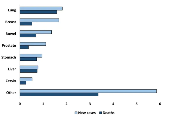
Cancer is the major death cause in the world with an estimated 14.1 million new cases and 8.2 million deaths in 2012 and, according to projections for 2030, the number of new cases will increase 68% (up to 23.6 million) (12). More than 80 billion € are spent each year in USA to deal with cancer healthcare (13). Therefore, more investment in cancer early detection assays and in new therapies in order to increase the lifespan of patients, as well as to decrease medical costs of advanced cases of disease, are urgent requirements. Lung, female breast, bowel and prostate cancers are the most common solid neoplasms (Fig. 1.1.). Portugal has a rate of 246.21 per 100,000 individuals which means an incidence of 49,174 cancer new cases per year, resulting in an overall number of 25,758 deaths in 2012 (14).
Figure 1.1. Bars-plot of new cancer cases and deaths of worldwide population in 2012 (numbers
in millions of new cancer cases/deaths) (12).
Prostate cancer (CaP) is the second most common diagnosed cancer in males with more than 1.11 million new cases worldwide and almost 40% of them – 417,000 – were
0 1 2 3 4 5 6 7 Other Cervix Liver Stomach Prostate Bowel Breast Lung
6
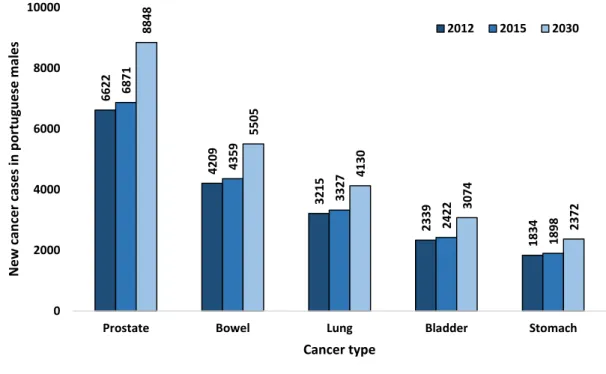
diagnosed in Europe, meaning that the CaP incidence is not equally distributed around the world (1). However, CaP mortality is equally distributed around the world (15).Thus, cancer incidence is not equally distributed but mortality is. In Portugal, for instance, CaP is the third most common cancer-related cause of death (1582 deaths) and the most diagnosed cancer (6622 cases). The number of new cases has been increasing in the past few years and there are some alarming reports that predict a rise of more than 30% in new CaP cases to 2030 (Fig. 1.2.) (1,14).
Figure 1.2. Number of new cancer cases in Portuguese population (males) in 2012 (dark-blue),
and predictions to 2015 (blue) and 2030 (light-blue). Numbers for the 5 most common types of cancer in Portugal (13).
1.2.2. Carcinogenesis: molecular changes and risk factors
Cancer is the word used to describe a group of diseases in which abnormal cells tend to divide. In this multistep process, cells can suffer severe behavioral and metabolic alterations and ultimately invade distant tissues and form metastases via blood and lymphatic systems (16). Cancer types can be divided into 5 categories according to their source (Table 1.1.). These include carcinoma, sarcoma, leukemia, lymphoma, myeloma and central nervous system cancers.
6622 4209 3215 2339 1834 6871 4359 3327 2422 1898 8848 5505 4130 3074 2372 0 2000 4000 6000 8000 10000
Prostate Bowel Lung Bladder Stomach
N e w can ce r case s i n p o rtu gu e se m al e s Cancer type 2012 2015 2030
7
Table 1.1. Categories of cancer types according to their source ((17)15).
Cancer type Source
Carcinoma
Cancer begins in the skin or in tissues around internal organs. Carcinoma is subdivided into adenocarcinoma (starts in glandular cells), basal cell carcinoma (begins in the lower part of the epidermis), squamous cell carcinoma (begins in squamous cells) and transitional cell carcinoma (occurs mainly in the organs of the urinary system).
Sarcoma Cancer begins in connective or supportive tissues, like cartilage, fat, bone and blood vessels.
Leukemia Bone barrow produces a large number of abnormal blood cells that enter into the bloodstream.
Lymphoma Develops in glands or nodes of the lymphatic system. Lymphomas are classified into Hodgkin lymphoma and non-Hodgkin lymphoma.
Myeloma Cancer that originates in plasma cells of the bone marrow. The plasma cells produce some of the proteins found in blood.
Mixed types The type components may be within one category or from different categories, such as adenosquasmous carcinoma, mixed mesodermal tumor and teratocarcinoma.
The process by which cells lead to cancer is called carcinogenesis and is a multifactorial process that involves 3 major steps (18). The first step comprises the initiation, in which due to genotypical and phenotypical changes that arise spontaneously or are induced by exposure to a carcinogen, normal cells become more likely to malign evolution. A cell that suffers such changes is called an initiated cell and suffers mutations that induce proliferation but not differentiation. Thereby, the proliferating initiated cells have less time to be repaired by the DNA repair system and the injury becomes permanent and irreversible; so, the cellular genome undergoes mutations that favor the neoplastic development of the affected cells (19). The cellular genes involved in this step are the proto-oncogenes. They are groups of genes that when mutated cause normal cells to become cancerous. However, they are not discarded by evolutionary processes because they have important roles, such as the encoding of proteins that stimulate cellular division, inhibition of cellular differentiation and are involved in signal transduction. Proto-oncogenes have, as usual in all other genes, a regulatory region that regulates the gene expression, and a structural region that encodes the amino acid sequence of a protein.
8
Mutations in any of these regions can activate a proto-oncogene, creating an oncogene
(20). There are several types of known oncogenes, such as RAS, MYC, ERK, TRK or
WNT. Another oncogene type is the Bcr-Abl gene, found on the Philadelphia chromosome (21). This oncogene is a known example of an oncogenic chromosomal translocation wherein a piece of chromosome 22 end with the BCR geneis broken, and then suffers fusion with a fragment of chromosome 9 that carries the ABL-1 gene, yielding thus the Philadelphia chromosome with the BCR-ABL fused gene. This oncogene is translated into tyrosine kinase receptors that are involved in cell cycle regulation and stimulation of cell division, leading to uncontrolled cell division which can cause chronic myelogenous leukemia (CML) and other types of leukemia (21).
The second step of carcinogenesis is promotion. After initiation, a cell can remain latent for weeks, months or years until and unless it is stimulated to undergo further proliferation, disturbing the cellular balance. Promoter compounds act at this stage only in the activated cells, inducing cell proliferation of susceptible tissues, helping to fix mutations, increasing alterations in genetic expression and causing changes in cellular growth control. This causes the onset of new oncogenes and disruption of the tumor suppressor genes action, leading to neoplastic transformation and development (22,23). Tumor Suppressor (TS) genes are a group of genes that regulate cell proliferation by checking the cell cycle progression, are involved in DNA repair processes preventing the accumulation of mutations in cancer-related genes and stimulate cell death when an injury is detected, and so, they are known as negative growth regulators (24). One of the most studied TS genes is p53, which is a protein encoded by the TP53 gene and has an important role in cellular and genomic equilibrium – reason for “The guardian of the genome” label. Mutations in this gene were found in at least 50% of human cancers. This TS gene regulates the cells cycle in the G1-S checkpoint, induces cell differentiation and promotes apoptosis. In a normal cell, when DNA damage is detected, p53 binds to DNA and stops the cell cycle. This happens because p21, a protein whose transcription is activated by p53, binds to cdk-cyclin complex inhibiting its kinase activity which causes a delay in G1 checkpoint and enabling the DNA repair mechanisms to act. If DNA is repaired, p53 promotes its differentiation and multiplication. If the cell cannot be repaired, p53 gives information to be eliminated by apoptotic processes. Therefore, when p53 is mutated, it can lose its function causing suppression of apoptosis, promoting cell division and hindering the differentiation of the cells. There are another important types of TS genes, like RB1, APC, BRCA1/2, among others (25).
9
The last step in carcinogenesis is the progression, a step where pre-neoplastic lesions and/or benign neoplasms turn into malign lesions. Progression is characterized by irreversibility, because once in this condition, cells are compromised into the malign development. The acquired genetic instability causes undifferentiated cells to proliferate faster while the well differentiated and slow growth cells die. There are also some biochemical, metabolic and morphological changes, as well as the appearance of new cell clones with genetic heterogeneity with a high capacity to proliferate and metastasize, which will induce the formation of a tumor mass (26,27).
There are few theories regarding the explanation of how cancer starts and what causes its development; the most widely accepted is the “two hit” hypothesis, based on the mechanisms of retinoblastoma, proposed by Alfred Knudson in 1971 (28). In an initial stage of retinal cancer, retinoblasts do not differentiate into retinal photoreceptors cells and nerve cells, and continue to proliferate forming tumors in retina able to metastasize to other parts of the body. Knudson (28) studied 48 patients with retinoblastoma for 20 years, and tabulated the patient age, family history, gender, and if the tumor occurs in one or in both eyes. With the obtained data and knowing that retinoblastoma is caused by a recessive mutation, the researcher verified that bilateral hereditary retinoblastoma occurred at a younger age while unilateral nonhereditary retinoblastoma is somehow delayed (28). Based on these findings, the researcher concluded that two mutations or “two hits” are required, one in each copy of a TS gene (today called RB1) (28). In hereditary cases, patients get one mutation and need to acquire only one to develop retinoblastoma in a process known as loss of heterozygosity. In what concerns to non-hereditary cases, patients have to acquire 2 mutations during their life to develop this type of cancer, what explains why nonhereditary retinoblastoma is developed latter. This is one of the most accepted theories for cancer-related researches; however, it is not 100% correct because it only takes into account mutations in TS genes (28,29).
Family history seems to be one of the most important factors which can lead to cancer (28). In general, 5-10% of all cancers are hereditary (30). One of the most common syndromes is the hereditary non-polyposis colorectal cancer (HNPCC) or Lynch syndrome. This syndrome is mostly related with endometrial cancer but it is also associated with ovary, stomach, pancreas, kidney and brain cancers. In HNPCC, genes involved in the DNA repair mechanism are mutated leading to cancer development (31).
Aging is another predisposing factor in carcinogenesis. During aging, DNA strand-break occurs in tissues and there is the accumulation of DNA adducts, assumed to
10
be caused by an imbalance between cellular pro-oxidants and antioxidants. In cellular metabolic processes, free radicals and reactive species of oxygen (ROS) are produced. These cellular antioxidants are accumulated over the years and damage macromolecules and organelle functions due to their high reactivity (32). Inflammation is also an important factor in oncogenesis. With aging, cellular damage increases, and in order to fix this damage, cells tend to produce cytokines, such as the tumor necrosis factor (TNF)-α, interleukin (IL)-1, IL-6,interferon (IF)-γ and C-reactive protein (CRP), resulting in a state known as low-grade chronical inflammation. The rising in cytokines concentration affect angiogenesis what may enhance cell proliferation (33). Finally, epigenetic alterations are also connected to cancer. These are genome modifications during cell division that do not involve changes in the DNA sequence, have a hereditary character, are able to modify gene expression and affect all tissues and cells throughout life (34). DNA methylation at CpG islands is a common epigenetic alteration. CpG islands are short regions of 0.5-4 kb in length rich in CpG and are located in promoter regions of half the genes in mammalian genome, including TS genes (35). Usually, they are un-methylated; yet, in cancer, hipermethylation occurs blocking the action of the genes involved in cells cycle regulation, resulting in their uncontrollable proliferation and cancer development (36). Histone modifications, such as ubiquitylation, methylation, acetylation and phosphorylation are other epigenetic alterations more usual with aging. They affect the chromatin structure leading to deregulated expression of important genes in cancer progression (34).
The human’s diet also affects the probability of cancer development. Some studies indicated that a longer and planed low-fat dietary pattern can decrease the probability of developing cancer (37,38). A fiber-rich diet is also associated with a lower risk of colon cancer (39). Excessive consumption of alcohol has been proved to be associated with the development of head, neck, esophageal, liver, breast and colorectal cancers. For instance, ethanol in alcoholic drinks is metabolized to acetaldehyde that damages genetic material and proteins (40). There are still some carcinogenic compounds added to food or produced during its preparation. One of them is acrylamide, present in foods heated above 120 ºC, such as potato chips, and that was found to have a positive association with renal cancer (41). Heterocyclic amines (HCs) and polycyclic aromatic hydrocarbons (PAHs), produced when amino acids, sugars and creatinine of the meat are directly exposed to fire, particularly when they are grilled or barbecued, were also found to be mutagenic
11
bioaccumulate in the body, and thus be responsible for some type of cancer, and in particular of pancreatic and breast cancers (42).
People lifestyle is another important factor in the probability of developing cancer. Overexposure to UV sunlight radiations leads to gene mutations, oxidative stress and inflammatory responses. In addition, UV radiations cause direct mutations in the TS gene p53, and are responsible for cancers like melanoma and basal cell carcinoma (44). Cigarette smoke, either as active or as a passive smoker, is also a carcinogenic inductor. More than 7000 chemicals are found in cigarretes and at least 69 of them were identified as carcinogenic, including nitrosamines, radioactive elements and PAHs, and whose action is responsible for the oral, esophageal, lung and pancreatic cancers (45). In summary, a balanced diet combined with frequent physical activities are fundamental in cancer prevention, particularly for breast, endometrial and colon cancers (46).
Viral infections caused by Helicobacter pylori and Human papillomaviruses can also promote carcinogenesis. Helicobacter pylori is a food and water contaminant capable of suppressing immune responses and causing gastric cancer (47). Human
papillomaviruses are a group of sexually transmitted viruses responsible for the largest
majority of cervical, vulvar and vaginal cancers (48). 1.3. Cancer biomarkers
One of the most revolutionary advances in oncologic diagnoses and related therapies was the detection and role of cancer biomarkers. According to the National Cancer Institute, a biomarker is a “biological molecule found in blood, other body fluids or tissues that is a sign of a normal or abnormal process, or of a condition or disease” and “may be to see how well the body responds to a treatment for a disease or condition” (49). Biomarkers can be produced by cancer cells or produced by the human body in response to cancer (Table 1.2.). Usually, most biomarkers are proteins but, more recently, additional biomarkers such as DNA, metabolites or RNA transcripts have been identified
(4).
The “ideal” tumorous biomarker does not exist. In fact, it is difficult to find a 100% reliable biomarker especially in early detection and diagnosis. Their lack of specificity, and the fact that some markers are normally produced by the body and their action as cancer biomarkers is associated to concentration levels in body fluids, leads to some controversy in what regards the cut-off values established for biomarkers as an indication of cancer (50). Thus, an ideal biomarker, should be easy to identify in recurrent
12
screenings, should be able to diagnosis cancer in an early-stage, and should allow the evaluation of the prognosis status. Moreover, their identification and quantification should be consistent and accurate, and by non-invasive, cheap, and easily accessible methods (4,51). In general, the PSA quantification by blood tests is still the most reliable screening of prostate cancer (5).
13
Table 1.2. Cancer biomarkers identified in biological fluids or tissues in cancer diagnosis, stage, response to treatment and prognosis (51).
Tumorous biomarker Biological fluids or tissues Cancer type Utility
Alpha-Fetoprotein (AFP) Blood Liver, germ cells tumors
Liver cancer diagnosis and response to treatment;
Stage, prognosis and response of germ cell tumors.
Beta-2-microglobulina (B2M) Blood, urine or cerebrospinal fluid
Multiple myeloma, chronic lymphocytic leukemia
Prognosis and to follow the individual response to treatment.
Beta-human chorionic gonadotrofin
(Beta-hCG) Urine or blood
Choriocarcinoma, testicular cancer
Identification of stage, prognosis, and response to treatment.
BCR-ABL fusion gene Blood and/or bone marrow Chronic myeloid leukemia Diagnosis and to monitor the disease status.
Bladder tumor antigen (BTA) Urine Bladder
Confirm diagnosis; used with NMP22 to test cancer.
14
CA15-3/CA27.29 Blood Breast cancer To assess therapy results and
disease recurrence.
CA19-9 Blood Pancreas, gallbladder, bille
duct, stomach To assess therapy results.
CA-125 Blood Ovaries
Diagnosis, assessment of response to treatment, and evaluation of recurrence.
Calcitonin Blood Medullary thyroid
Diagnosis, assessment of response to treatment, and evaluation of recurrence.
Carcinoembryonic antigen (CEA) Blood Colorectal cancer and breast cancer
To check if cancer has spread; Assessment of response to treatment and evaluation of recurrence.
CD20 Blood Non-Hodgkin lymphoma To check if a targeted therapy
is appropriate.
Chromogranin A (CgA) Blood Neuroendocrine tumors
Diagnosis, assessment of response to treatment, and evaluation of recurrence.
15 Immunoglobulins Blood and urine
Multiple myeloma and Waldenström macroglobulinemia
Diagnosis, assessment of response to treatment, and evaluation of recurrence.
Lactate dehydrogenase Blood Germ cell tumors
To assess the cancer stage, prognosis, and response to treatment.
Prostate-specific antigen (PSA) Blood and urine Prostate
Diagnosis, assessment of response to treatment, and evaluation of recurrence. Prostatic acid phosphatase (PAP) Blood Prostate, lung, multiple
16 1.4. Prostate cancer
1.4.1. Etiology
Prostate cancer (CaP) is one of the most common males’ cancers in the world (1). The vast majority of prostatic tumors are adenocarcinomas with origin in prostatic epithelium. CaP is a multifactorial disease, where there is a relation between aging and environmental, physiological and molecular factors. Advanced age and its associated oxidative stress, low grade-chronical inflammation and epigenetic alterations are the most significant risk factors with an important role in tumor initiation (52). Around 60% of tumors are diagnosed in men with more than 65 years old, and 97% in men with more than 50 years old (1). Exposure to UV radiation, diet, alcohol consumption, family history, ethnicity and hormones are additional factors involved in the development of CaP. African or American men with more than 65 years and with a first-line familiar who had this type of cancer are those with a higher risk in CaP development (52).
In what concerns the molecular changes, some important mechanisms should be highlighted. The Wnt signaling pathway is a major signaling pathway involved in invasiveness of prostate tumor cells. Her-2/neu (ERBB2) is a proto-oncogene involved in, among others, prostate cancer. When active it is important in the differentiation and cells growth of tumor wherein high levels of Her-2/neu are related with metastatic prostate cancer. Phosohoinositide-3 Kinase/AKT (PI3K) is a mediator of several oncogenic signaling pathways and it is activated by receptor tyrosine kinases yielding secondary messengers promoters of cellular proliferation and survival. Due to somatic mutations, PI3K can be hyperactivated promoting the prostate cancer progression (53). Nowadays, there are no effective therapies for CaP if the tumor already spread the limits of the organ. Therefore, efforts and investments in the CaP early detection are critical requirements to increase the curative successful rate (14).
1.4.2. Signals, symptoms and diagnosis
Prostate cancer, as many other cancer types, can be effectively treated with surgery and radiation if detected at an early stage. However, a relevant percentage of patients is only diagnosed after demonstrating symptoms like haematuria, urinary obstruction, polyuria, among others. In more severe cases, when the tumor reaches the lymph nodes or bones, oedema and pain are major symptoms (54). At this stage, the treatments typically used are no longer efficient and the 5-year relative survival rate
17
decreases (2). This fact puts on evidence the screening importance attempting an early detection of CaP and the need to perform diagnostic tests even in the absence of signals and symptoms of the disease, which is in fact the biggest challenge in CaP diagnosis.
Digital rectal examination (DRE), measurement of the serum total PSA and transrectal ultrasound (TRUS) guided biopsies are the usual methods to detect CaP. If DRE and PSA as initial tests indicate the presence of the disease, TRUS guided biopsy is further carried out to confirm the diagnosis. Despite being widely used, these tests display however some disadvantages. The PSA cut-off test is not consensual because a serum PSA concentration above the threshold limit is not necessarily a result of CaP. Recent ejaculations, benign prostate hyperplasia (BPH) and urinary or prostatic infections can also increase PSA concentration. So, in the case of such over-diagnostics, unnecessary treatments, including prostatectomy and radiation therapy, will be conducted. DRE is a qualitative test and it is not possible to reach/evaluate the entire prostatic gland, especially in small tumors that have not reached yet the prostatic capsule. Finally, the TRUS guided biopsy is an invasive technique and does not allow distinguishing between CaP and BPH. Furthermore, there is a significant risk of infection and related consequences (fever, pain, hematuria and sepsis) (50,55).
1.5. Prostate-specific antigen as a prostate cancer biomarker
The first references to prostate cancer date from 1649 when Riolan described the enlargement of prostate as a clinical manifestation of a possible tumor and related it with the bladder obstruction (56). At this time, the diagnosis was done based only on the family history and physical examination (57). Only in 1938, PAP (prostatic acid phosphatase), the first prostate tumor biomarker, was proposed by Gutman (58). However, despite being one enzyme secreted by columnar epithelium secretory cells, it can be produced by other organs, blood components and several non-prostatic malignancies (Table 1.2.). Therefore, it is a non-specific biomarker. In addition, its concentration only significantly rises when the adenocarcinoma of prostate is metastasized, what makes of PAP an useless biomarker in CaP early detection (59,60).
Between 1960 and 1970, several researches were dedicated to the search of tumor specific antigens that could be useful in clinical diagnosis, with special focus in oncology
(57). In 1960, Rubin Flocks reported some features of prostate antigens (61). The
researcher showed that antibodies created against human prostate tissue were species- and tissues-specific; the antigens in benign and malignant tissue were similar and no specific
18
antigens could be isolated (62). In 1966, Mitsuwo Hara characterized a protein, “gamma-seminoprotein” (63), and in 1970, Tien Shun Li and Carl Beling reported some antigens in human semen (64). It is believed that both discoveries were related with PSA; nonetheless, the real “discovery” of PSA is attributed to Ming C. Wang and co-workers when they purified and characterized PSA from prostatic tissue (65). The research group revealed that PSA is only produced by prostatic tissue and is present in BPH and CaP
(65). Once discovered its potential, the applications and use of PSA have been amplified.
PSA was approved in 1987 by the Food and Drug Administration (FDA) to monitor the disease status in patients with prostate cancer (66). Latter, in 1994, the same organization approved the PSA blood test as an easy and inexpensive screening tool to be used in association with DRE to detect prostate cancer (67). A serum PSA measurement of ≥ 4.0 ng/mL is considered as a clinically relevant value where 25% of patients with values above this cut-off value had CaP (57), while urinary PSA has a cut-off value of 150 ng/mL
(68). This cut-off value in urine demonstrated an impressive sensitivity of 92.5% to CaP
diagnosis; however, due to the small number of cases analyzed, more studies have to be conducted to corroborate these results (69). The analysis of PSA in urine samples is also an important tool in distinguish amongst CaP and BPH, particularly when serum PSA is between 2.5 ng/mL and 10 ng/mL (68).
1.5.1. Prostate cancer screening, stage and grade
The PSA test is believed to be the best approach in the CaP early detection and probably the most used cancer blood test in medicine. A PSA value of ≥ 4.0 ng/mL in serum has been defined as abnormal and can indicate the presence of a prostatic tumor
(70). FDA recommends an annual screening of CaP for men with 50 or more years old,
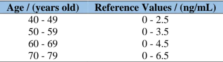
and in African American men with more than 40 years old because ethnicity is a relevant factor. More than in other cancer types, age is a significant factor that influences the CaP development and PSA baseline values. Thus, urologists have to adjust the PSA cut-off levels according to age. Table 1.3. presents the reference PSA values in blood for men with 40-79 years old (1,71).
19
Table 1.3. PSA values of reference for men with 40-79 years old in blood (66).
Age / (years old) Reference Values / (ng/mL)
40 - 49 0 - 2.5
50 - 59 0 - 3.5
60 - 69 0 - 4.5
70 - 79 0 - 6.5
Values of PSA between 4.0 and 10.0 ng/mL in blood are considered suspicious and the test needs to be repeated. Also the patient needs to be supervised to check whether these values continue to rise, and if DRE also suggests the presence of a tumor, a TRUS guided biopsy has to be done to confirm diagnosis. For values above 10 ng/mL the test is repeated after several days for confirmation because there are some factors that can cause fluctuations in PSA concentrations, and a TRUS guided biopsy has to be carried out because for these values a patient already has a chance of 67% of CaP (72).
The knowledge of the PSA levels also allows to evaluate the grade of the prostatic tumor: the higher the PSA levels, the more advanced is the disease and the larger is the tumor (70). PSA levels when combined with the Gleason sum score and the clinical stage can be used as predictors of the patient prognosis (Table 1.4.).
The Gleason classification is a system used in the evaluation of men with CaP prognosis. This classification is made according to histological and cellular characteristics of the tumor and is the sum of histological patterns (from 1-well differentiated- to 5- poorly differentiated) from the two predominant cellular types in tumor giving a score that can vary from 2 to 10. Scores from 0-6 have a good prognosis, 7 have moderated prognosis and scores between 8 and 10 correspond to undifferentiated tumor and the ones with worst prognosis (73). In what concerns the clinical stage, this is done to primarily evaluate the physical characteristics of the tumor and varies from T0 (no evidence of tumor) to T4 (tumor has invaded nearby tissues).
Table 1.4. Risk stratification of CaP patients according to PSA levels, Gleason grade and Clinical
stage (69).
Risk PSA levels / (ng/mL) Gleason grade Clinical stage
Low < 10 ng/mL ≤ 6 T1-T2a
Intermediate 10 - 20 ng/mL 7 T2b-T2c
20
1.5.2. Monitoring therapy and disease recurrence
The risk stratification according to PSA levels is extremely important in the identification of CaP patients. For suspicious PSA levels, the first action consists on an active surveillance. 3-monthly PSA measurements and a re-biopsy should be done at 1 year, and whether clinical manifestations continue to suggest the presence of a tumor, new clinical actions should be implemented. External beam radiotherapy (EBRT) is often performed, but radical prostatectomy is the most common procedure when PSA levels suggest that the tumor is only localized within the prostate (74).
After radical prostatectomy, the PSA blood test is also important to check whether treatment was successful or whether there are signals of disease recurrence. After a few weeks, PSA values should decrease to undetectable concentrations (half time PSA in serum is approximately 2.2-3.2 days (75)) due to the removal of prostate. If the PSA concentration increases, there are evidences that the prostate was not totally removed or that the biomarker is being produced by metastases of the original tumor (74).
The PSA blood test has been extremely useful in early diagnosis of CaP leading to a significant increase of tumors detection at local stages, thus resulting in a decline of the mortality rate. However, there are some drawbacks associated to the PSA blood test. PSA can also be produced by other types of tumors, by prostate enlargement and inflammation or even by recent sexual activity leading to false positive results and unnecessary surgeries (50,55). In order to overcome these drawbacks, new tests with PSA have been developed to increase the diagnostic accuracy, including PSA density, free total PSA ratio and other molecular forms, such as free PSA, proPSA and BPSA (5).
1.5.3. Molecular characteristics
PSA is a serine protease which is part of kallikrein related peptidases (KLKs). KLKs are the largest cluster of peptidases in human genome and are located in a locus of chromosome 19q13.3-13.4 being responsible for the expression of 15 homologous serine proteases with an important role in several biological processes, such as reproduction, inflammation blood clothing and cancer. PSA is encoded by one of these genes, the KLK3 gene, whose expression is regulated by androgens (76).
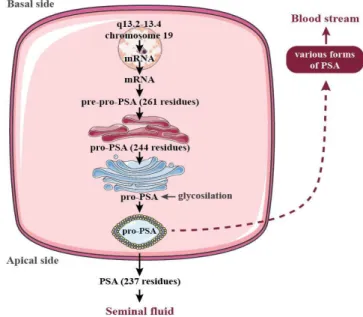
1.5.3.1. Biosynthesis structure
PSA transcription and biosynthesis occur mainly in epithelial cells and periurethral glands in a process regulated by androgens. The KLK3 gene transcription
21
and translation yield an inactive proenzyme (pre-pro-PSA) with 261 residues. 17 of these are an amino acid leader sequence that is cleaved and works as a signal that directs pre-pro-PSA to the membrane on endoplasmic reticulum (ER). In ER, the originated pre-pro-PSA (244 residues) has also a pro-peptide with 7 amino acids that is cleaved in N-terminals between the arginine at position 7 and isoleucine at position 8 in a process of zymogen activation that can be autocatalytic or catalyzed by trypsin like-peptidases (76). PSA is its active form (237 residues) and has 5 intrachain disulfide bounds, a single asparagine-linked oligosaccharide, a weight of 28-33 kDa, and is secreted into semen, being the most abundant protein in seminal fluid with a concentration between 0.5 and 2.0 mg/mL (Fig. 1.3.) (77). Once active, PSA can diffuse into circulation and be inactivated by the binding of protease inhibitors or by divalent inhibitors, particularly zinc. However, PSA can also became inactive due to proteolytic inactivation of the carboxy-terminals of the amino acid residues Lysine (Lys) 145 and/or Lys 182 and circulates in an unbounded state known as free PSA f(PSA) (78).
Figure 1.3. Schematic representation of PSA processing in epithelial cells of the prostate.
Adapted from (75).
1.5.3.2. Structure and physicochemical properties
PSA is a serine protease glycoprotein with 28-33 kDa. PSA consists of a single polypeptide with 237 amino acids and a single N-linked sugar moiety at asparagine (Asn) 61 with two fucose linkages in the inner most of N-acetylglucosamine (GlcNAc) (79). PSA also has in its mature form a single carbohydrate unit at Asn45 with 5 dissulfide
22
bonds which increases the total mass in 2-3 kDa. It was demonstrated that 92% of PSA is composed of peptides and 8% of carbohydrates, whose 4.84% is hexose, 2.87% is hexosamine and 0.25% is sialic acid (80). The protein consists of two 6-strand antiparallel β-barrels and three α-helices as depicted in Fig. 1.4..
Figure 1.4. 3D representation of PSA using light-assisted molecular immobilization technology
(78).
The catalytic site (Serine (Ser) 195, Histidine (His) 57, and Aspartate (Asp) 102) is conserved and located in a cleft between two β-barrels. The sequence also contains the GWG motif, which is a typical pattern present in many proteins with proteolytic activity and the presence of the His-Asp-Ser triad and a catalytic domain similar to KLKs make it a serine protease. The GWG motif contains a Trp located in the β-barrel nearby the disulphide bond Cysteine (Cys)157-Cys 22 (81).
PSA has 5 isoenzymes, one with 5 antibody binding sites and an isoelectric point ranging between 6.7-7.2 (81). Trauma, stress or a tumor can rise the PSA serum levels but they are usually below 0.1 ng/L, depending on its production, distribution and elimination. fPSA forms are cleared from blood primarily in kidneys (half time of 12 hours). Complexed forms of PSA take more time to be eliminated (2.2-3.2 days) because they are larger and cannot be subjected to renal clearance (78).
1.5.3.3. Physiological role of PSA in prostate and in external tissues
PSA is a chymotrypsin-like protease with some properties that differentiate PSA from chymotrypsin and other serine proteases. The physiological function of PSA consists on liquefying seminal fluid after ejaculation in order to facilitate the sperm
23
transport in the vagina. Seminal fluid has gelified vesicles in coagulum shape and they are composed of seminogelins (SEMG1 and SEMG2), fibronectin, laminin and gelatin which are proteolytic degraded to liquefy sperm in a zinc-mediated process. In prostate, Zn2+ concentrations are high and the pH is alkaline, and the major role of zinc ions consists on inhibiting the proteolytic effect of PSA (78).
Typically, PSA biological effects are only confined to the interior of prostate glands where is predominantly in an active form. In blood, PSA is an intact and non-catalytic form due to internal cleavages or is covalently attached to α1-anthichymotrypsin (ACT) in levels from < 0.1 to 104 ng/mL. Levels above 102 ng/mL are found essentially in men with advanced prostate cancer (82). In CaP, the layer of basal cells and the basement membrane that surrounds prostate glands is disrupted and PSA can reach bloodstream and can influence neoplastic growth and metastases. PSA in bloodstream cleaves the insulin-like growth factor binding protein-3 (IGFBP-3), one of the major serum binding proteins for insulin-like growth factor-1 (IGF-1). IGF-1 is a risk factor for CaP and IGFBP-3 cleavages increases the IGF-1 availability increasing the proliferative effect of tumor cells (83). As mentioned, it has the capacity to cleave extracellular matrix glycoproteins, such as laminin and fibronectin, and also the urokinase-type plasminogen activator, that are involved in tumor invasion and metastases. Other studies suggested that PSA has an antiangiogenic effect and mitogen activity for osteoblasts transforming the growth factor-beta by proteolytic modulation of osteoblasts surface receptors (78).
1.5.3.4. Molecular derivatives of PSA and their role in diagnosis
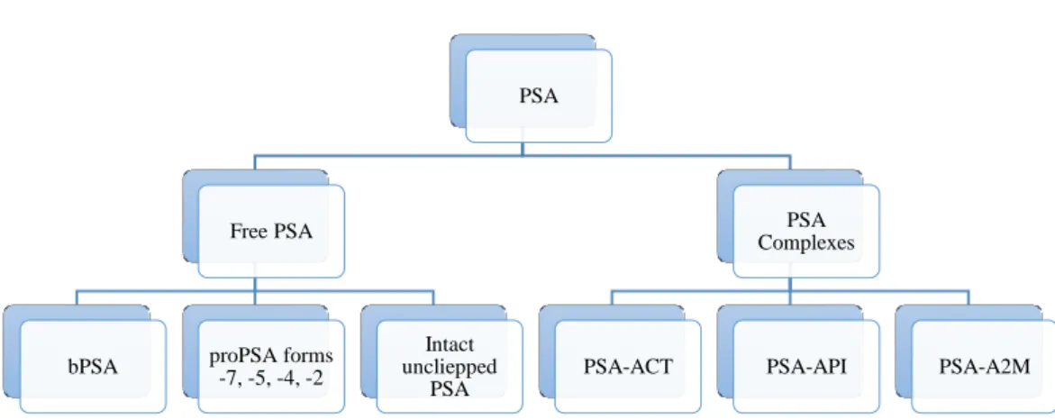
Contrarily to initial findings, where PSA was considered a protein composed of 237 amino acids, it is now known that serum PSA can occur in two forms: (i) in a “complexed form” with protease inhibitors; or (ii) in a free unbounded form (fPSA), and as shown in Fig. 5. 65%-95% of PSA occurs linked to a protease inhibitor, ACT, in a PSA-ACT complex that is higher in CaP patients probably because of the loss of the prostate integrity (5,84).
In respect to fPSA, there are 3 distinct forms of inactive PSA. One of them, benign PSA (bPSA), is a clipped subform with 2 internal peptide bond cleavages at Lys 182 and Lys 145 and is associated with the transition zone of patients with nodular BPH (85). The second subform, inactive PSA, is “intact”, unclipped PSA (iPSA), that is similar to native PSA but with some structural changes that make it inactive. The third PSA form is proPSA (pPSA). pPSA comprises a 7 amino acid N-terminal pro leader peptide, [-7]
24
proPSA, that can be cleaved to form enzymatically active PSA. [-7]proPSA can also be truncated by proteolytic cleavage to 4]proPSA, 5]proPSA and 2]proPSA. [-2]proPSA is of particular interest because it is not cleaved further and it accumulates in cancer tissues so it can have a diagnostic utility especially in the 2.5-4 ng/mL range (5). [-2]proPSA is also important in the prediction of CaP aggressiveness because it is a parameter of prostate health index, phi (𝑝ℎ𝑖 =[−2]𝑝𝑟𝑜𝑃𝑆𝐴
𝑓𝑃𝑆𝐴 × √𝑃𝑆𝐴), which is three times
more specific than PSA (86).
Figure 1.5. Different fPSA percursor forms (bPSA, proPSA and intact unclipped PSA) and PSA
complexes (PSA-ACT, PSA-API and PSA-A2M) (5).
Some efforts have been done to refine and to improve the measurements of PSA levels aiming at gathering a more accurate diagnosis. The fPSA to tPSA ratio, usually known as %fPSA, has shown clinical relevance, and with a better performance of PSA and tPSA while differentiating between CaP and BPH when PSA values are in the grey zone (4-10 ng/mL) and DRE results are negative (82,87).
1.5.3.5. Stability of total and free PSA
The stability of PSA, important in clinical tests, is influenced by pH and temperature, particularly for fPSA isoforms. Biological samples containing PSA should be stored at 4°C and analyzed within 8 hours after collection or at -70°C and at pH of 5.5 for latter analysis. At these conditions, PSA can be stored for 9 months with only 5% loss
(88). tPSa is more stable and studies have shown that it can be stored at 4°C for a week,
at -20°C in a domestic freezer for a month and even at -80°C for 7 years (89,90).
PSA
Free PSA
bPSA proPSA forms-7, -5, -4, -2
Intact uncliepped
PSA
PSA Complexes
25
1.6. Analytical methods for the quantification of PSA
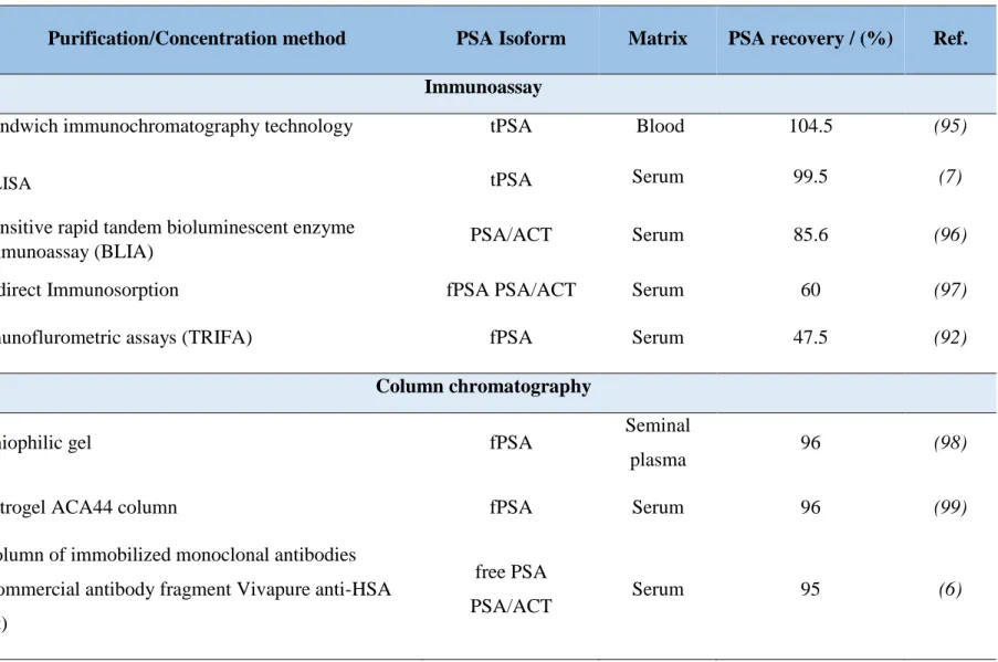
Since the PSA discovery and purification by Ming C. Wang in 1979 (65) and after the knowledge of the PSA importance in the CaP early detection, diagnosis and monitoring, several tests to extract, concentrate and quantify PSA, fPSA, PSA-ACT and tPSA were developed and are now commercially available (7). The PSA purification/fractionation method with ammonium sulfate is usually followed by other separation methods, such as gel-filtration chromatography, affinity chromatography, high performance liquid chromatography (HPLC) and polyacrylamide gel electrophoresis
(91). However, for clinical purposes, immunoassays are recommended (by FDA). These
immunoassays require anti-PSA monoclonal antibodies that will specifically bound to PSA (91). There are various kinds of PSA immunoassays although the most widely used are the enzyme-linked immunosorbent assay (ELISA) (7), time-resolved immunofluorometric assay (TRIFA) (92), fluorescence microscopy (93), surface plasmon resonance technology (SPR) (94), and immunochromatography (95). Table 1.5. represents a review of literature of the immunoassays and column chromatography methods used for the extraction and quantification of different PSA isoforms.
Albeit these methods have some benefits and lead to high purification factors they still present major disadvantages. In particular, specific and highly expensive material and equipment are required, as well as highly qualified and trained technicians. Moreover, they are usually time-consuming. In fluorescence and electrochemical-based methods, the analyte to be quantified has to be dyed in order to amplify the respective signal and the sample can be lost during these preparation steps. In addition, the prolonged exposure of the fluorescence dye to excitation light sources can cause photo bleaching and quenching of signals leading to false negative results (96). ELISA is an expensive technique, because of the cost of antibodies required. It also has low reproducibility and the requirement of signal amplification using a biochemical reaction (96). Thus, it is clear that it is necessary a new method, more economically viable and simpler to concentrate and quantify PSA, and where aqueous biphasic systems (ABS) fit.
![Table 1.7. Chemical structures of GBs: MES: 2-(N-morpholino)ethanesulfonic acid; TES: 2- 2-[[1,3-dihydroxy-2-(hydroxymethyl)propan-2-yl]amino]ethanesulfonic acid; HEPES: 4-(2-hydroxyethyl)-1-piperazineethanesulfonic acid; CHES: N-cyclohexyl-2-aminoethanes](https://thumb-eu.123doks.com/thumbv2/123dok_br/15956392.1098484/60.892.149.745.188.874/structures-morpholino-ethanesulfonic-hydroxymethyl-ethanesulfonic-hydroxyethyl-piperazineethanesulfonic-aminoethanes.webp)
![Figure 2.1. GB-ILs synthesis. An equimolar excess of the aqueous solution of buffer was added to [P 4444 ]OH solution (A)](https://thumb-eu.123doks.com/thumbv2/123dok_br/15956392.1098484/66.892.131.757.234.438/figure-synthesis-equimolar-excess-aqueous-solution-buffer-solution.webp)
