i
UNIVERSIDADE DE LISBOA
Faculdade de Medicina Veterinária
THE ROLE OF MOB PROTEINS IN PROTOZOAN CELL
CYCLE REGULATION
Alexandra Jorge Tavares
TESE DE DOUTORAMENTO EM CIÊNCIAS VETERINÁRIAS ESPECIALIDADE DE CIÊNCIAS BIOLÓGICAS e BIOMÉDICAS
CONSTITUIÇÃO DO JÚRI
Doutora Maria Helena Antunes Soares Doutora Ana Maria Luís Ramos Tomás Doutor António José Freitas Duarte Doutora Maria Luísa Santos Sousa Cyrne Doutora Mónica Bettencourt Dias
Doutor José Alexandre da Costa Perdigão e Cameira Leitão
ORIENTADORA
Doutora Maria Helena Antunes Soares
CO-ORIENTADOR
Doutor José Alexandre da Costa Perdigão e Cameira Leitão
2015 LISBOA
iii
A
GRADECIMENTOSÉ com enorme felicidade que, por fim, escrevo estas linhas para agradecer aos que de uma ou outra forma contribuíram para que este trabalho fosse possível.
Em primeiro lugar tenho de agradecer à minha orientadora Professora Doutora Helena Soares. Obrigada Helena por todo o apoio e incentivo que ao longo dos anos me deu. Por ter sempre acreditado que este dia ia chegar e por me ter ensinado tanto. Obrigada pelas discussões científicas e menos científicas, pelas ideias, pela partilha. O percurso foi sinuoso mas chegámos cá! Obrigada acima de tudo pela amizade. Porque sem dúvida não teria sido possível sem si!
Quero também agradecer muito ao meu co-orientador Doutor Alexandre Leitão. Obrigada por sempre ter apoiado a minha “causa”. Obrigada pelas várias discussões, pelo incentivo e por me ter apresentado, com infinita paciência, ao mundo dos parasitas. Sei que não foi fácil! Obrigada pela amizade e por ter feito que este dia fosse possível.
Um agradecimento à Faculdade de Medicina Veterinária, CIISA, nomeadamente ao Professor Doutor Luís Tavares, por ter aprovado a realização deste projecto nas infraestruturas da faculdade.
Agradeço também ao Instituto Gulbenkian de Ciência e ao Centro de Química e Bioquímica da FCUL pelas infraestruturas disponibilizadas para a realização de parte do trabalho aqui apresentado. Um agradecimento especial à Imaging Facility do IGC.
À Sofia Nolasco quero agradecer toda a amizade, cumplicidade e ajuda. Sofia, muito do que hoje sei no laboratório devo-o a ti. Obrigada por estares sempre disposta a ajudar e a ensinar e obrigada pela verdadeira amizade que construímos ao longo destes anos. Obrigada ainda pelo teu excelente sentido prático! Partilhámos muito neste tempo, momentos bons e menos bons mas sempre estiveste lá e ajudaste no que pudeste. Independentemente do que o futuro nos reserve, a amizade está garantida.
Ao João Gonçalves tenho também muito a agradecer. Obrigada por me teres transmitido tanto do que sabias e por sempre teres contribuído com boas ideias para o projecto. Obrigada acima de tudo pela amizade, pela disponibilidade, pelas gargalhadas,
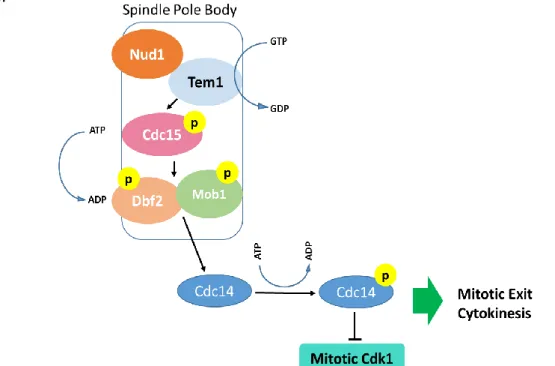
Figure 1 - The Mitotic Exit Network. Schematic representation of the signaling cascade of MEN as described in the budding yeast. The GTPase Tem1, the kinase Cdc15 and the complex Dbf2-Mob1 are localized at the spindle pole body. A cascade of phosphorylation events culminates with the activation of the phosphatase Cdc14 that is released into the cytoplasm where it counteracts Cdk1 activity promoting mitotic exit and cytokinesis.
iv
pelos bons momentos que partilhámos e pelo apoio nos menos bons. Obrigada pelo sentido de humor inconfundível que tanta falta faz a um laboratório.
Ao João Coelho quero também agradecer toda a ajuda e amizade. Obrigada pelas conversas, pela partilha e pelo entendimento mútuo Obrigada pela preciosa ajuda no mundo da filogenia. Também eu fico à espera de um agradecimento para breve!
À Dulce Santos quero também agradecer a amizade. Dulce, foi muito bom ter-te conhecido pois tens sempre uma palavra amiga e sensata a dizer. Muitas vezes me deste força e me fizeste ver diferentes perspectivas. Obrigada pela boa disposição e pelo humor sagaz que tantas vezes animou aquele gabinete.
A todos os colegas e amigos do laboratório, Samuel, Afonso, Eduardo, Rita, Sara, um muito obrigada pelo excelente ambiente que se vivia na bancada. É sem dúvida muito bom quando nos levantamos de manhã e pensamos que é um privilégio poder trabalhar com pessoas disponíveis e bem-dispostas Obrigada pela amizade ao longo deste tempo.
Quero ainda agradecer aos membros do Grupo da Biologia Redox, FCUL, com quem tive o prazer de trabalhar. De uma maneira ou de outra contribuíram para este percurso.
Ao Gonçalo Costa, pela ajuda preciosa que me deu na produção da proteína Mob1 de Toxoplasma gondii. Obrigada também por todas conversas e partilha de conhecimento.
Deixo também um agradecimento ao Professor Doutor Álvaro Tavares pelas discussões e ideias. Tudo começou também devido a si e por isso agradeço a oportunidade.
I would also like to thank to my colleagues and friends of the Chromosome Dynamics Lab at IGC. Thank you Raquel for all the times that you gave me time to work on the thesis or to solve PhD related things! Your understanding and support were very important. I am also thankful to Lina, Ewa, Cíntia and Mihailo, you’ve become good friends and it is pleasant to work with you all.
Uma palavra muito especial para a Mariana Santos. Sem ti, acho que ainda estaria agora a fazer formatações! Obrigada pela amizade e disponibilidade.
v
Por fim, tenho que agradecer por ter a sorte de ter uma família cujo apoio e incentivo têm sido incondicionais. Obrigada mãe e pai por sempre acreditarem em mim, por perceberem as dificuldades e apoiarem as minhas decisões de fazer aquilo que gosto realmente de fazer. Vocês são em muito responsáveis pelo que sou hoje. Obrigada por tudo! À mana Mariana, pela amizade, cumplicidade e entendimento.
A ti Pedro, por me apoiares, por acreditares, por perceberes a dedicação que implica a escrita duma tese. Sempre me transmitiste que eu sou capaz, principalmente nos momentos em que a motivação enfraquece. Todos os dias me mostras o quanto vale lutar pelo que queremos. Obrigada pelo amor e por me fazeres feliz.
vi
F
INANCIALS
UPPORTThis work was supported by a Fellowship from the IEFP (Instituto de Emprego e Formação Profissional) and by a Fellowship in the FCT project PTDC/SAU-OBD/105234/2008 entitled "The role of Mob1 and Unc119 proteins in cytokinesis and tumorgenesis”
vii
The role of Mob proteins in protozoan cell cycle regulation
ABSTRACT
Proper cell division and control of cell proliferation are critical aspects in cell biology, with implications during embryonic development and in the maintenance of organisms’ homeostasis. Mob1 is a core protein of the Mitotic Exit Network and of the Hippo pathway, fundamental signaling cascades for the correct metaphase to anaphase transition and for the proper balance between cell proliferation and death. In this work we took advantage of two protozoan organisms to investigate the role of Mob1, the most ancient protein of the Hippo pathway. In the ciliate Tetrahymena thermophila we demonstrated that Mob1 has a polarized subcellular distribution, concentrating in the basal bodies of the cell posterior pole. During cell division, the protein also localizes in the region where the division plane is formed and its absence in this specific place leads to the mispositioning of division axis and cytokinesis impairment. These results revealed that Mob1 directly links proper cell polarity to correct cell division. Our studies of Mob1 in the apicomplexan parasite Toxoplasma gondii, also a permanent polarized unicellular organism, contributed to a better understanding of how parasites may regulate cell proliferation inside the host cell, a critical aspect for the course of infection. In T. gondii, Mob1 also localizes preferentially in the posterior pole of the cell, where the basal complex, which is essential for cytokinesis, is localized. Interestingly, in agreement with a role for Mob1 in proliferation control in T. gondii, we observed that mob1 mRNA levels are dramatically diminished when parasites are actively replicating inside the cell and that Mob1 overexpression leads to a delay in the parasite replication rate. Altogether, the work presented clearly positions Mob1 as an ancestral molecule playing a critical role in the cross-road of cell polarity establishment, correct cell division and proliferation control.
Keywords: Mob1 proteins, cell polarity, cell division/proliferation, Tetrahymena
ix
O papel das proteínas Mob1 na regulação da divisão celular em
protozoários
RESUMO
A divisão celular e o controlo da proliferação são aspectos fundamentais em biologia celular com implicações no desenvolvimento embrionário e na manutenção da homeostasia nos organismos. A proteína Mob1 é uma componente de duas vias de sinalização celular, a Mitotic Exit Network e a via de sinalização Hippo, cascatas de fosforilação essenciais para a correcta transição entre a metáfase a e a anáfase e para o balanço entre a proliferação/morte celular. Neste trabalho, utilizámos dois protozoários modelo para investigar a função da proteína Mob1, a mais ancestral das proteínas nas vias de sinalização referidas. No ciliado Tetrahymena thermophila, demonstrámos que a proteína Mob1 apresenta uma localização polarizada, estando principalmente concentrada nos corpos basais do polo posterior das células. Aquando da divisão celular, a Mob1 também é observada na região da célula onde se forma o eixo de divisão. Esta localização é essencial visto a ausência de Mob1 no local conduzir ao deslocamento do eixo e impedir a citocinese. O nosso estudo no parasita apicomplexa Toxoplasma gondii, um organismo também permanentemente polarizado, contribuiu para compreender melhor, o possível mecanismo de regulação da proliferação dos parasitas dentro da célula hospedeira, um aspecto essencial no desenvolvimento da infecção. Em T. gondii, a proteína Mob1 também se concentra no polo posterior da célula onde se localiza o complexo basal, uma estrutura envolvida na citocinese. Claramente suportando a nossa hipótese que a Mob1 desempenha um papel no controlo da proliferação, observámos que os níveis de RNA mensageiro do gene mob1 são drasticamente diminuídos quando os parasitas estão no período de replicação activa dentro das células hospedeiras. Adicionalmente, a acumulação da proteína no citoplasma dos parasitas provoca um atraso significativo na sua taxa de replicação. Em conjunto, o trabalho apresentado posiciona a proteína Mob1 como uma molécula ancestral envolvida na conexão entre o estabelecimento da polaridade, a correcta divisão e o controlo da proliferação celular.
Palavras-chave: Proteínas Mob1; Polaridade Celular; Controlo da Divisão/Proliferação; T. thermophila; T. gondii
xi
G
ENERALI
NDEX AGRADECIMENTOS ... III ABSTRACT ... VII RESUMO ... IX LIST OF FIGURES ...XVLIST OF TABLES ... XVII
ABBREVIATIONS ... XIX
CHAPTER I:GENERAL INTRODUCTION ... 1
1. Cell Cycle: Brief Description ... 3
1.1 The importance of regulation in cell division ... 6
1.2 Regulatory molecular mechanisms of cell cycle – a brief overview ... 7
1.3 The Metaphase to anaphase transition ... 8
1.4 Cytokinesis: the final step of cell division ... 9
2. The Mob1 Proteins ... 10
2.1 The Mob1 in the eukaryotic lineage – conserved functions ... 11
2.2 Mob1 proteins – subcellular localization ... 14
3. The Mitotic Exit Network ... 15
4. The Septation Initiation Network ... 17
5. The Hippo pathway ... 18
OBJECTIVES ... 23
CHAPTER II:MOB1 IN THE BIOLOGICAL MODEL PROTOZOA Tetrahymenathermophila ... 27
PART 1:INTRODUCTION ... 29
1. Introduction: Cell polarity ... 29
1.1 Intrinsic cell polarity ... 29
1.2 Influence of cell polarity in cell division ... 33
1.3 The influence of cell polarity on Mitotic Exit Network and Hippo pathways ... 34
2. Cell polarity in unicellular protozoans – the case of the ciliate Tetrahymena thermophila ………..…36
xii
4. Tetrahymena thermophila as a model organism ... 40
PART 2:MOB1: DEFINING CELL POLARITY FOR PROPER CELL DIVISION ... 43
1. Abstract ... 43
2. Introduction ... 44
3. Material and Methods... 46
3.1. Plasmid construction ... 46
3.2. Tetrahymena thermophila strains ... 47
3.3. Nocodazole treatment ... 47
3.4. Immunofluorescence assays ... 47
3.5. Western blot and electrophoresis analysis ... 48
3.6. Deciliation assay ... 49
3.7. Gene expression analysis by RT-PCR ... 49
4. Results ... 50
4.1. Tetrahymena thermophila Mob1 downregulation leads to incorrect cell division planes and impaired cytokinesis ... 50
4.2. Tetrahymena thermophila Mob1 accumulates at basal bodies with a polarized gradient distribution towards the posterior pole ... 55
4.3. Tetrahymena thermophila Mob1 depletion leads to a delay in cilia recovery ... 59
4.4. Tetrahymena thermophila Mob1 recruitment to the cell midzone defines the division plane ...61
5. Discussion ... 68
CHAPTER III:MOB1 IN THE APICOMPLEXAN PARASITE Toxoplasmagondii ... 71
PART 1:INTRODUCTION ... 73
1. The Toxoplasma gondii – an apicomplexan parasite ... 73
1.1. The life cycle of Toxoplasma gondii ... 75
1.2. The single cell organism Toxoplasma gondii – the complexity of a permanent polarized cell strucuture ... 77
2. Toxoplasma gondii inside the host cell ... 80
xiii
2.2. Toxoplasma gondii establishment and replication inside the host cell ... 82
3. Proliferation control in Toxoplasma gondii – the ability of stage conversion ... 86
PART2:MOB1 IS A CRITICAL PLAYER IN TOXOPLASMA GONDII REPLICATION ... 89
1. Abstract ... 89
2. Introduction ... 90
3. Material and Methods... 92
3.1. Sequence alignment and phylogenetic analysis ... 92
3.2. DNA Constructs ... 93
3.3. Cell and parasite cultivation ... 94
3.4. Quantitative Real-time PCR ... 94
3.5. Toxoplasma gondii Mob1 Antibody Production ... 94
3.6. Immunofluorescence Assays ... 95
3.7. Invasion Assays ... 95
3.8. Replication Assay ... 96
4. Results ... 97
4.1. Phylogenetic analysis of Mob1 protein in Toxoplasma gondii ... 97
4.2. Upon host cell entrance, mob1 expression in Toxoplasma gondii is greatly diminished ………..…99
4.3. Mob1 presents a polarized localization in Toxoplasma gondii and is associated with key structures involved in cell division ... 100
4.4. Mob1 increased levels lead to a delay in Toxoplasma gondii replication ... 105
5. Discussion ... 108
CHAPTER IV:CONCLUDING REMARKS AND FUTURE PERSPECTIVES ... 113
1. Concluding Remarks ... 115
2. Future Perspectives ... 118
xv
L
IST OFF
IGURESFigure 1 - Eukaryotic cell cycle.. ... 5
Figure 2 - The Mitotic Exit Network.. ... iii
Figure 3 - The Septation Initiation Network.. ... 17
Figure 4 - The Hippo pathway in Drosophila and Mammals.. ... 20
Figure 5 - The main protein complexes involved in epithelial polarity establishment. ... 31
Figure 6 - The ciliate Tetrahymena thermophila. ... 38
Figure 7 – Schematic representation of basal body structure and formation pattern. ... 38
Figure 8 - Sequential steps of oral apparatus formation in Tetrahymena thermophila during cell division. ... 39
Figure 9 – Strategy used for obtaining the Mob1 knockdown strain of Tetrahymena thermophila ... 51
Figure 10 – Validation of the Tetrahymena thermophila knockdown strain. ... 52
Figure 11 - Mob1 depletion in Tetrahymena thermophila causes incorrect positioning of the cell division plan and impaired cytokinesis. ... 53
Figure 12 - Graphic representation of the angle (h) established between the DP and the anterior–posterior axis ... 54
Figure 13 - Schematic representation of the DNA construct used to create the TtMob1 GFP strain.. ... 55
Figure 14 - Tetrahymena thermophila Mob1 localizes preferentially at the posterior pole basal bodies. ... 57
Figure 15 - Tetrahymena thermophila Mob1 basal body localization does not depend on intracytoplasmic microtubules. ... 58
Figure 16 - Tetrahymena thermophila Mob1 depletion leads to a delay in cilia recovery.. ... 60
Figure 17 - Tetrahymena thermophila Mob1 localizes at the division plane basal bodies and new oral apparatus in dividing cells. ... 64
Figure 18 - TtMob1-GFP cells were biolistically transformed with the DNA construct to disrupt the endogenous Ttmob1 locus.. ... 65
Figure 19 - Tetrahymena thermophila Mob1-KD phenotype recovery in a Mob1–GFP ShuttON/OFF strain. ... 66
Figure 20 - The Alveolata monophyletic group is composed by several different species of single-celled eukaryotes that present very diverse and distinct life styles. ... 73
Figure 21 - Toxoplasma gondii life cycle. ... 76
Figure 22 - Toxoplasma gondii cell ultra-structure ... 78
xvi
Figure 24 - Toxoplasma gondii division and budding.. ... 85 Figure 25 - Phylogenetic analysis of Mob1 proteins from different parasitic organisms.. ... 98 Figure 26 - Quantitative Real-Time PCR analysis of mob1 expression in Toxoplasma gondii . 99 Figure 27 - Toxoplasma gondii anti-Mob1Tg polyclonal serum. ... 101 Figure 28 - Analysis of Mob1 subcellular localization and distribution in Toxoplasma gondii in host cell. ... 102 Figure 29 - Mob1 is not a centrosomal protein in Toxoplasma gondii. ... 103 Figure 30 - Schematic representation of Mob1 localization in Toxoplasma gondii. ... 104 Figure 31 - Representative images of those used to quantify the ability of Toxoplasma gondii parasites to replicate in a Mob1 overexpression condition. ... 105 Figure 32 - Overexpression of Mob1 leads to a replication delay in the parasite Toxoplasma
gondii... 106
Figure 33 - Overexpression of Mob1 does not affect invasion ability in the parasite Toxoplasma
xvii
L
IST OFT
ABLESTable 1 - Nucleotide sequences of the primers used in the study of Mob1 in Tetrahymena
thermophila. ... 46 Table 2 – List of the accession numbers of the Mob proteins of the organisms used in the phylogenetic analysis ... 92 Table 3 – Nucleotide Sequences of the primers used in the study of Mob1 in Toxoplasma gondii ... 96
xix
A
BBREVIATIONSAPC/C – anaphase promoting complex/cyclosome aPKC – atypical protein kinase
ATCC - American Type Culture Collection BAG1 – bradyzoite antigen 1
BB – basal body
BIRC5 - Baculoviral IAP (inhibitor of apoptosis protein) Repeat-Containing 5 BLAST - Basic Local Alignment Search Tool
BSA – bovine serum albumin
Bub1/2 - budding uninhibited by benzimidazole-1 1/2 cAMP - Cyclic adenosine monophosphate
Cdc14 – cell division cycle 14
Cdc42 - Cell division cycle protein 42 CDKs – Cyclin dependente kinases cDNA – coding DNA
CENP-E/A – cetromeric protein E/A CEP55 – centrosomal protein 55
cGMP – cyclic guanosine monophosphate CPC - chromosomal passenger complex CPC – chromosome passenger complex CVPS – contractile vacuole pores DAPI - 4', 6-Diamidino-2-Phenylindole dd- destabilization domain
xx DIAP1- - Drosophila Inhibitor of Apoptosis Protein 1 DLG – discs-large
DMEM - Dulbecco’s modified Eagle’s médium DNA - Deoxyribonucleic acid
DP – division plane
eIF2 - eukaryotic initiation factor-2
ESCRT - endosomal sorting complex required for transport G0; G1; G2 – gap 0; 1; 2
GAP – GTP activating protein
Gapdh - glyceraldehyde-3-phosphate dehydrogenase GFP – green fluorescent protein
GST - Glutathione S Transferase GTP - Guanosine triphosphate HFF - Human foreskin fibroblast IF – immunofluorescence IMC – inner complex membrane
IPTG - isopropyl-β-D-thiogalactopyranoside KD – knock down
kDa - 103 Dalton
LATS1/2 – large tumor suppressor ½ LGL – lethal giant larvae
M –Mitosis
MAC – macronucleus
xxi MATS – Mob as tumor suppressor
MEN – mitotic exit network
MIC – (Chapter III) microneme proteins MIC – micronucleus
ml – milliliter
Mob1 – Mps one binder 1 Mps1 – multipolar spindle 1
mRNA - messenger ribonucleic acid MTT1 - metallothionein gene 1
NCBI - National Center for Biotechnology Information NDR – nuclear dbf2-related
Neo – neomycin
NIMA – never in mitosis protein NOA – new oral apparatus
NuMa1 - Nuclear mitotic apparatus protein 1 OA – oral apparatus
ORF – open reading frame
PAR proteins - partitioning-defective proteins PBS – phosphate-buffer saline
PCR – polymerase chain reaction PFA – paraformaldehyde
Plk1 – Polo-like kinase 1 RNA - Ribonucleic acid RON – rhoptry neck protein
xxii ROPs – rhoptries proteins
RT-PCR – reverse transcriptase PCR S – Synthesis
SAC – spindle assembly checkpoint Shd – shiled
SIN – Septation initiation network
siRNA – short interference Ribonucleic acid SPB – spindle pole body
SPOC – Spindle positioning checkpoint SPP – super proteose peptone
Tg ADF – T. gondii actin depolymerizing factor
TgMorn1 – T. gondii Membrane Occupation and Recognition Nexus 1 UTR – untranslated region
WB – western blot wt – wyild type
YAP1 – Yes associated protein 1 μg – microgram
μl – microliter μm – micrometer μM – micromolar
1
3
1. Cell Cycle: Brief Description
From the more simple unicellular organisms, to higher eukaryotes as mammals, the cell is the basic element whose main function is to maintain its specific genetic information and to allow its propagation to future generations. In the case of multicellular organisms, the adult arises from a more or less extended developmental period in which a unique original cell, the zygote, originates the several specialized tissues and organs, through multiple cell divisions. After the end of this developmental process, cell division process allows, in the adult, for cell renovation and, being so, the maintenance of organism homeostasis.
Regarding the unicellular organisms, they regulate their cell cycle according to the specific environment in which they live. For example, free living unicellular can control their cell proliferation relatively to the nutrient abundance, and physical stresses such as temperature. A very particular case is the one of parasitic unicellular. Here, cell cycle regulation is a crosstalk between the mechanisms of parasite survival and the host’s defense mechanisms.
Globally, the cell cycle is divided into two distinct periods: the interphase, a longer phase that concerns most of the cell’s life and mitosis, a faster phase were cells undergo division (Fig. 1). Briefly, the interphase can be divided into three distinct and consecutive stages: G1, S and G2. In G1, the cell presents a very active metabolism and growth and there is synthesis of molecules and organelles, needed for the different cell functions. At this time, cells can receive external signals that dictate their future: entering G0, a non-proliferative phase or continuing cycling and enter S phase (Alberts et al., 2007; Cooper & Hausman, 2007).
Being so, if the environment is favorable, cells go to S phase where DNA replication takes place. At this point, each chromosome is duplicated which allows that the future daughter cells receive the same DNA content initially present in the mother cell. After DNA replication the cell is almost ready for division. Right before mitosis, cells undergo a G2 period where the molecules necessary for division are synthetized. Therefore, G2, as G1, is a growth period where cell activities enable cell to divide.
Mitosis occurs in a short period of time in the life of the cell. It has, as main function, nuclear division and the equal distribution to the daughter cells of DNA molecules replicated in the S phase. Formally, mitosis is divided in four distinct stages: prophase, metaphase, anaphase and telophase. At the end of these sequentially occurring phases, cytokinesis, the individualization of the two daughter cells, takes place.
4
At prophase, the duplicated chromosomes start to condense, being this condensed state of DNA a critical point for its correct segregation. In fact, longer interphase chromosomes would be very difficult to equally segregate, and cytokinesis would not occur properly if DNA portions were still in the cleavage furrow region.
In a typical mitosis, during prophase, besides this chromatin reorganization, the duplication cycle of the centrosome (the main microtubule organizing center in animal cells) also occurs. Thus, each of the newly duplicated centrosomes migrates towards opposite poles of the cell while the mitotic spindle, which results from the reorganization of interphase microtubules, begins to be assembled. At the end of prophase nuclear envelope breakdown can be observed. The sister chromatids are connected at the specific region of the centromere, a heterochromatin rich region where the proteins of the kinetochore are recruited and assembled. In fact, it is on this specific transient structure that spindle microtubules are going to be connected.
Metaphase follows prophase and this phase is characterized by the equatorial positioning of the mitotic chromosomes in the middle region of the spindle, forming the metaphasic plate. Depending if it is a symmetric or an asymmetric mitosis, the spindle is positioned in different regions of the cytoplasm. Cells arrest in metaphase until all the kinetochores are stably connected to the spindle microtubules. Upon the fulfillment of this connection, cells can go through the anaphase. At this point, the sister chromatids of each chromosome are segregated to opposite poles of the cell.
The mitotic spindle is elongated and the centrosomes are now localized at the distal regions of the cell. At the end of mitosis, after the segregation of sister chromatids, telophase takes place. The DNA, equally distributed to daughter cells, starts to decondense and the new nuclear envelopes are assembled around it. Simultaneously, the cytoplasm of the mother cell is divided into two, in a process known as cytokinesis, which will be later described in more detail (Alberts et al., 2007; Cooper & Hausman, 2007).
5
Although the steps briefly described above are observed in a typical cell cycle, namely in animal cells, there are several organisms which present striking differences in this process. For example, the centrosome is not present in all organisms and being so there are alternative strategies to build the mitotic spindle. This is the case for Saccharomyces cerevisiae cells, which possess a structure homologue to centrosome, the spindle pole body. Also the ciliate Tetrahymena thermophila builds the mitotic spindle possessing neither centrosomes nor a spindle pole body. Other remarkable difference is the case of organisms that present closed mitosis. In this particular situation, the nuclear envelope breakdown does not take place. Examples of organisms with closed mitosis are the two models used in the present study: the ciliate T. thermophila and the apicomplexa T. gondii.
This analysis of the different details in the cell cycle/mitotic process is surely simplistic and merely illustrative. Nevertheless, for the purpose of this work it matters to be stressed that, regardless of the different strategies, in all cases, the final objective is to properly segregate the DNA content and to distribute the other cell components, namely cytoplasmic organelles.
Taking into account the importance of correctly transmitting the genetic information to the daughter cells, cell division is a tightly controlled process, existing
Figure 2 - Eukaryotic cell cycle. The cycle begins in G1, the first period after last cell division. At this time, there is a very active growth and metabolism. During S phase, replication of DNA molecules, which will be equally distributed to daughter cells, takes place. In G2 cells produce all the components that will be needed for mitosis. At this point, protein synthesis is very active. Finally, mitosis occurs and nuclear and cytoplasmic division takes place. (From (BD Biosciences)).
6
several mechanisms that work throughout the cell cycle and that guarantee, in normal conditions, its success.
1.1 The importance of regulation in cell division
During the cell cycle, either in interphase as in mitosis, there are several checkpoints that verify the precision of some of the phases described before. These checkpoints, a total of three in all cell cycles are: the G1 checkpoint, the DNA replication checkpoint (S to G2) and the Spindle Assembly checkpoint (Mitosis). The G1 checkpoint ensures that in G1 to S transition the cellular environment is suitable to initiate DNA replication and verifies if the DNA has lesions that can compromise the genetic information transmitted to next generation. In this situation, if the cell machinery does not ensure that DNA molecules are intact and without errors before the restriction point (after which cells are irreversibly committed to enter S phase), DNA will be replicated with the eventual errors. This fact implies that undetected point mutations, insertions or deletions will be transmitted to daughter cells. DNA errors accumulation, due to G1 checkpoint deregulation, is intimately connected to cancer development (Alberts et al., 2007; Cooper & Hausman, 2007).
The DNA replication checkpoint occurs at the end of S phase in transition to G2. The checkpoint machinery evaluates if the DNA was correctly and completely replicated. The consequences of G2 checkpoint failure are very similar to the previous one. Even though DNA molecules can be intact before replication, the replication machinery may itself compromise genetic information by the insertion of errors in the newly synthetized DNA molecules (Alberts et al., 2007; Cooper & Hausman, 2007).
Finally, the spindle assembly checkpoint (SAC) ensures that all the chromosomes are correctly bound to spindle microtubules to allow an even distribution to the daughter cells. Indeed, it is crucial to ensure that homologous chromatids are correctly segregated to opposite sides of the cell. For that, cells present a mechanism that in metaphase, verifies if all the chromosomes are connected, by the kinetochore, to the spindle microtubules, arresting cells in this phase until this condition is achieved. In this particular case, the deregulation of SAC may originate daughter cells with abnormal DNA content, i.e. aneuploid cells, and consequently, genomic instability (Alberts et al., 2007; Cooper & Hausman, 2007).
7
The malfunctioning of any of these control mechanisms has potentially the formation of daughter cells with severe genetic defects.
1.2 Regulatory molecular mechanisms of cell cycle – a brief overview
After presenting a framework of the problem, it now matters to address the molecular mechanisms at the basis of cell cycle dynamics. Actually, these different control mechanisms acting during the cell cycle can be interpreted as a set of biochemical switches that are negatively regulated. The molecular complexity of this process is massive, but, in a basic view, all the steps involve a protein kinase (from the serine/threonine kinases family) broadly known as CDKs (cyclin dependent kinases), whose activity oscillates during the cell cycle. These proteins are responsible for the phosphorylation and activation of the molecular players of processes such as DNA replication, mitosis and cytokinesis and are in turn regulated by another family of proteins, known as cyclins. Each cyclin binds to the respective CDK protein which activates its catalytic function. In summary, different cyclin-CDK pairs act in different cell cycle stages (Alberts et al., 2007; Cooper & Hausman, 2007). This is a negatively regulated action since, for cells to proceed in the cycle, the specific cyclin at each step has to be targeted for degradation in an ubiquitination dependent mechanism. As a consequence, the interacting CDKs are inactivated and cell cycle progresses (Alberts et al., 2007; Cooper & Hausman, 2007).
CDKs may be classified into two groups: interphase CDKs and mitotic CDKs. In G1 phase, CDK4 and CDK6 bind to cyclin-D type cyclins as a response to growth factor stimuli, typical of this point in cell cycle (for review see (Enders, 2012)). Later in the end of G1, transition to S phase, the complex CDK2-Cyclin E is formed. This complex is present at the centrosome and is involved both in DNA replication and the centrosome duplication cycle, which evidences the coordination of these two processes (Hinchcliffe, Li, Thompson, Maller, & Sluder, 1999; Matsumoto, Hayashi, & Nishida, 1999; Matsumoto & Maller, 2004; H. Zhao, Chen, Gurian-West, & Roberts, 2012). In G2 to M transition, a CDK2 is activated by the connection to cyclin A. In mitosis, the degradation of this CDK2-cyclin A complex allows the establishment of the main complex for cell division completion, the CDK1-cyclin B complex. Just before anaphase, cyclin B is targeted for degradation, which inactivates CDK1 and cells undergo DNA segregation (for review (Malumbres & Barbacid, 2009)).
8
Given the complexity of the mechanism responsible for metaphase to anaphase transition, this specific point of the cell cycle will be addressed in more detail.
Interestingly, although cell cycle transitions are the ones described above, studies in mice have shown that alone the CDK1-cyclin B complex is capable of promoting all cell cycle transitions. Thus, CDK1 emerge as the unique kinase essential for cell cycle to proceed (Santamaria et al., 2007).
.
1.3 The Metaphase to anaphase transition
One of the most critical steps for mitotic precision is the transition from metaphase to anaphase. In fact, the unbalanced distribution of chromosomes to the new cells has severe consequences as aneuploidy and genomic instability. The SAC is the mechanism in the cells that is responsible for the control of this specific step. As described, SAC machinery ensures that all the chromosomes are stably connected to spindle microtubules by the kinetochore, a transient protein scaffold assembled during mitosis. A single unattached kinetochore is capable of emitting enough signaling to maintain SAC active, which implies that the signal must be amplified (Rieder, Cole, Khodjakov, & Sluder, 1995) (Jia, Kim, & Yu, 2013). The two sister chromatids are maintained together by a protein named cohesin which persists, at least in the centromeric region, until just before anaphase onset. This physical connection of sister chromatids is critical to chromosome biorientation and consequently to chromosome segregation (for review see (Peters & Nishiyama, 2012)). Cell division proceeds until metaphase, time at which SAC activation is catalyzed by the presence of unattached kinetochores. SAC is formed by several proteins, Mad1, Mad2, Bub1, Bub2 and Mps1, firstly identified by genetic screening done in Saccharomyces cerevisiae (Li & Murray, 1991; Roberts, Farr, & Hoyt, 1994; Weiss & Winey, 1996), which are highly conserved throughout all the eukaryotic lineage (for review see (Jia, Kim, & Yu, 2013).
SAC activation’s main consequence is the inhibition of another protein complex, the APC/C (Anaphase promoting complex/ cyclosome). This complex is composed by more than 15 proteins which function is the ubiquitination of several substrates, as for example securin and cyclin B (for review see (Kim & Yu, 2011; Lara-Gonzalez & Taylor, 2012)). After all the chromosomes are properly attached, SAC signaling is extinguished and cells can undergo anaphase. Although object of intense study, the mechanism by which SAC is inactivated is not yet fully understood. Nevertheless, it has been shown that microtubules attachment to the kinetochore and chromosome
9
biorientation towards opposite sides of the cell generate tension at the kinetochore which in turn inactivates SAC (Maresca & Salmon, 2009; Uchida et al., 2009). Once SAC is silenced, the APC/C is no longer inhibited and becomes active. This complex promotes anaphase by targeting for degradation core proteins that maintain cells in metaphase. Cyclin B degradation implies the inactivation of CDK1 which allows metaphase to anaphase transition.
1.4 Cytokinesis: the final step of cell division
The last step of cell division is the physical separation of the two daughter cells by a process known as cytokinesis. The abscission of cell membrane and so, the individualization of the newly formed cells, is a process intimately associated with the corrected segregation of genetic material and is coordinated in time and space with anaphase progression.
In animal cells, after chromosome segregation, the microtubules of the anaphase spindle, in the central spindle region, serve as a platform to accumulate key regulatory and structural proteins in the place where division plane is to be formed. The major protein complex involved in the process is the chromosomal passenger complex (CPC) which is formed by the kinase Aurora B, the inner centromere protein INCENP and other two molecules, Survivin (BIRC5) and Borealin (CDCA8) (for review see (van der Horst & Lens, 2014)). Also Polo-like kinase 1 accumulates at the spindle midzone which leads to the activation of specific proteins in this region. Aurora B activates the central spindilin complex and the activity of these regulatory proteins culminates with the activation of the in locus of the small GTPase Rho (A) which in turn associates to the cell membrane in the equatorial region of the cortex, where the cleavage furrow will be formed (for review see (Green, Paluch, & Oegema, 2012; Lacroix & Maddox, 2012)).
Basically, the presence of Rho A in an active form in cell´s equator allows the establishment of a multiproteic contractile ring, which constriction based on a actomyosin-based force will lead to alterations in cell shape and simultaneously the maturation, by post-translational modifications, of central spindle microtubules. Even though contractile ring function is based on actomyosin forces, septins are also present in the structure via recruitment made by anillins that works as a linker protein to bind the components of the ring (for review see (Green et al., 2012)). Recently, using Drosophila melanogaster, it has been shown that septins are responsible by the ring
10
curvature of F-actin filaments, demonstrating its critical role in the constriction process (Mavrakis et al., 2014).
At the end of cytokinesis, it can be observed the formation of other ring-like structure, the midbody, which is formed in the region where the central spindle microtubules plus ends overlap (for review see (Schiel, Childs, & Prekeris, 2013)). This protein structure is composed by several molecules that concentrate in the intercellular bridge, which can maintain the two daughter cells connected by several hours, until abscission occurs. The specific function of most of the proteins that accumulate in midbody is currently unknown, nevertheless midbody is seen as a platform for the abscission machinery (for review see (Fededa & Gerlich, 2012)). The final step of cytokinesis, the abscission of the intercellular bridge resulting in the total individualization of the two cells, is far from only being a simple cut. In fact, this process is controlled by several protein complexes that involve both lipid and cytoskeleton dynamics remodeling and occurs asymmetrically in the intercellular bridge. Microtubules severing and actin depolymerization are observed in one or both sides of the midbody, which originates a secondary ingression in the region, placing the abscission local. This fact implies that either one of the daughter cells inherits the midbody and the molecules concentrated there or that it is discarded (for review see (Agromayor & Martin-Serrano, 2013; Schiel et al., 2013)). The biological relevance of this is still unknown.
The abscission process is initiated by the localization of the centrosomal protein CEP55 at the midbody. This localization is tightly time controlled since during anaphase PLK1 phosphorylates CEP55 which inhibits out of proper time recruitment to anaphase spindle. Later, when Plk1 is degraded, CEP55 is dephosphorylated and can relocalize in the midbody to promote abscission (Bastos & Barr, 2010). Finally, the endosomal sorting complex required for transport (ESCRT) machinery is recruited to Cep55 already present in the midbody, and mediates abscission in a mechanism that is still elusive.
2. The Mob1 Proteins
As previously mentioned, the control of cell division is a major issue during the life of the cell and several molecules and signaling pathways are implicated in this process. This regulation has consequences in all living organisms. From asexual reproduction in unicellular protozoan to the most complex metazoan where cells have to live in
11
organized tissues, cell division is tightly regulated. To this regulation, both cell external and internal factors play a critical role. In fact, new cells are only formed when nutrients and space conditions are favorable. In any case, cells that undergo division, intrinsically regulate genetic material segregation in proper quality and quantity.
Mob proteins are a small family of kinase activators whose role in cell division control has been emerging in the past years. Most of the eukaryotes analyzed to date have more than one Mob protein and the presence and function of these proteins is highly conserved. The first described member of the family, S. cerevisiae Mob1, is implicated in mitotic exit control being part of the signaling cascade known as Mitotic Exit Network (MEN) that controls anaphase to interphase transition. In multicellular organisms, Mob1 is also part of the Hippo pathway, a molecular cascade that controls cell proliferation versus cell death. Both pathways are going to be described in detail later in the present work, but for now it is worth to explore what is known about Mob1 proteins in terms of their distribution throughout eukaryotic lineage, their cellular localization in different organisms and finally their molecular functions in activation of specific NDR (nuclear dbf2-related) kinase family members.
2.1 The Mob1 in the eukaryotic lineage – conserved functions
Mob1 proteins are conserved and present in all eukaryotes. The first identified protein of the family was described as a binder of Mps1 protein in S. cerevisiae (Mob1p). This protein was implicated in spindle pole body duplication and mitotic checkpoint signaling (Luca & Winey, 1998). In this first study it was shown that yeast Mob1p is essential, required to maintain ploidy levels and for successful mitosis. Moreover, the authors also identified another Mob protein, Mob2p that contrary to Mob1p is not essential for yeast survival. This second protein of the family is involved in asymmetric budding by inducing a daughter-specific gene expression as a consequence of Cbk1/Mob2-dependent activation and localization of the Ace2 transcription factor to the daughter nucleus (Colman-Lerner, Chin, & Brent, 2001; Weiss et al., 2002). Later, the role of Mob1 in mitosis completion was dissected when Mob1 was implicated in MEN signaling control. Shortly, after the study in budding yeast, data from a study using the fission yeast Schizosaccharomyces pombe supported a role for Mob1 in septum formation, as spores in mob1 mutants failed to divide which led to highly elongated cells (Salimova, Sohrmann, Fournier, & Simanis, 2000).
12
Further studies then showed a conservation of Mob1 proteins’ role in cell division also in metazoans. The first study of Mob1 in D. melanogaster was performed by Lai, ZC and co-workers (2005). These authors demonstrated that Mob1 works as a tumor suppressor protein since mutations in the protein led to cell over proliferation and tumor development in many fly organs. Due to this phenotype, in the fly, Mob1 was named Mats, which stands for Mob as tumor suppressor. The wide variety of tumors formed indicated that this role in tissue proliferation control is not tissue specific but a general effect.
A parallel issue in cell proliferation control is apoptosis. In fact, the balance between cell proliferation and cell death is crucial to maintain tissue and organism homeostasis. Interestingly, in the same study involving D. melanogaster, it was also demonstrated that Mats facilitates cell death by negatively regulating DIAP1, a caspase inhibitor essential for cell survival. Several posterior studies showed that this role of Mats in cell proliferation control is due to its core role in the Hippo signaling pathway that will be address in detail later. This function in proliferation control is also conserved in mammals. In fact, the fly Mats and its ortholog in humans, Mob1A, share 87% identity of the aminoacid sequence and Mob1A could rescue pupal lethality in Mats mutants (Lai et al., 2005).
As stated before, Mob proteins are a small family of kinase activators, more exactly, Mob proteins are involved in the activation of NDR kinases, proteins highly conserved from yeast to humans. Human cells express four related NDR kinases – NDR1, NDR2, LATS1 (Large tumor suppressor 1) and LATS2, all involved in cell division control (for a review see (A. Hergovich, 2013)). In fact, the first study to address the function of human Mob1 (Mob1A) was about the activation of NDR kinases and the authors demonstrated that human Mob1 stimulates NDR kinases activity both in vitro and in vivo (Bichsel, Tamaskovic, Stegert & Hemmings, 2004). The activation of LATS1 kinase by Mob1 was reported later and the data suggested that the interaction between these two proteins was required for proper cytokinesis as LATS1 or Mob1A depletion by siRNAs increased telophase duration (Bothos, Tuttle, Ottey, Luca, & Halazonetis, 2005). The functional relevance of these observations was addressed later in a study also using human cell lines where it was observed that Mob1A/B depletion disrupts the normal localization of CPC at spindle midzone during early anaphase which seems to be related to the larger telophase duration (Wilmeth, Shrestha, Montaño, Rashe & Shuster, 2010). More recently, it was shown that the cytokinesis failure observed in Mob1 depleted cells is related to an over stabilization of microtubules at the end of
13
telophase, in the midbody region. In fact, Mob1A depleted cells show higher levels of acetylated microtubules (a post translational modification present in more stable microtubules) and also microtubules with higher resistance to cold and nocadozole treatment. In addition to this, Mob1 depleted cells also showed increased motility. It was observed that the resulting daughter cells were kept together by ultrafine bridges. At a first glance, one could think that the increased mobility could be attributed to a response to the intercellular cytoplasm bridge formed due to cytokinesis failure in these cells. In this case, cells would exert a mechanical pulling force to promote abscission. However, the authors observed that the increased motility was also present after abscission (Florindo et al., 2012). These findings support the notion that Mob1A and/or the kinases regulated by it have biological roles beyond cell division, such as in microtubule dynamics and cell polarization, important for cell movement.
Even though Mob1 has been studied in mammalian systems, the role of this protein is still poorly understood. The role of Mob1 in MEN and Hippo pathways, which components are conserved from yeast to humans has been a matter of great interest. In metazoans Mob1 was described as a tumor suppressor protein, mainly because of its involvement in the Hippo signaling cascade. Supporting this fact, there are some more clinical studies showing that mob1 expression is reduced in non-small-cell lung and colorectal cancers (Kosaka et al., 2007; Sasaki et al., 2007). Finally, a study performed on Mob1A/B double mutant mice demonstrated that Mob1 is needed for embryogenesis and also that mice without Mob1A/B protein are prone to tumor formation, demonstrating that its activity depends on development stage (Nishio et al., 2012).
Apart from the described studies in model organisms and human cells, the role of Mob1 proteins was also addressed in protozoans. Actually, taking into account Mob1 involvement in cell division control and the fact that this protein is deeply conserved among eukaryotes, it is an excellent molecule to explore also in protozoans. Protozoans are ancient organisms, mostly unicellular, which present a plethora of different cell shapes and different life styles. Some of these organisms are excellent models for cell biology studies since they present complex cell structures without the complexity of tissue environment. In the particular case of Mob1 proteins studies, our knowledge about its mechanism of action is still sparse. The use of more simple organisms, which nevertheless present the same basic cell characteristics of metazoan cells, will allow us to address better the fundamental role of Mob1 proteins. Besides that, the study of Mob1 in parasitic protozoa, such T. gondii will allow us to address the cell proliferation
14
control in a unicellular organism that forms multicellular associations inside the host cell. This particular aspect of T. gondii life style will allow the study of Mob1’s function in a very different cell organization context when compared to free living unicellular or multicellular organisms.
The first study of Mob1 in a protozoan was done in the parasite Trypanosoma brucei, the African trypanosome that presents two different Mob1 proteins: Mob1-A and Mob1-B. In this study, the authors showed that both in the bloodstream and procyclic forms of the parasite life cycle, Mob1 depletion by siRNA causes cytokinesis defects. In the bloodstream form, the infective form for mammalians, cytokinesis was delayed in the early stage of furrow ingression. In contrary, parasites in the procyclic form also presented defective cytokinesis due to an incorrect positioning of the cleavage furrow (Hammarton et al., 2005). More recently, in T. brucei, it was also demonstrated that NDR-like kinases (PK50 and PK53) are critical for cytokinesis. Interestingly, in this parasite NDR kinases are active even in Mob1 depletion scenario and the authors could not co-precipitate Mob-GST and PK50 and PK53. Nevertheless, it is possible that Mob1 interacts with PK50 and PK53 but does so in a transient manner incompatible with co-precipitation (Ma et al., 2010).
Except for these studies in T. brucei, the investigation of Mob1 proteins in protozoa is inexistent. In this context, in the present work, it will be presented and discussed the results obtained from the study of Mob1 proteins in the two different protozoa: the free living ciliate Tetrahymena thermophila (Chapter II) and the apicomplexa parasite T. gondii (Chapter III). This is a significant contribute to our knowledge concerning Mob1 in complex, but unicellular organisms, that present different life styles.
2.2 Mob1 proteins – subcellular localization
The critical role that Mob1 proteins have in the cell division, may already suggest that its subcellular localization is probably associated with structures involved in mitosis progress.
As mentioned before, Mob1 was first described in the budding yeast, S. cerevisiae. In this organism, Mob1 shows a dynamic localization throughout the cell cycle. Using cells expressing Mob1-GFP, it was observed that at mid-anaphase, Mob1 localizes to the spindle pole bodies, the yeast functional equivalents of centrosomes. At the end of cell budding (the specific process of division in this organism) Mob1 is then re localized to the bud neck, the region where abscission will take place, where it stays
15
until cytokinesis and abscission occurs (Luca et al., 2001). Another study showed that Mob1 also colocalizes with Cdc14 at the kinetochores (Stoepel, Ottey, Kurischko, Hieter & Luca, 2005). Similarly, in the fission yeast S. pombe, Mob1 is present at spindle pole bodies during mitosis and accumulates in the medial ring where the septum is formed and posteriorly, where daughter cell separation follows (Salimova et al., 2000)
In D. melanogaster, Mats is localized at the plasma membrane in developing tissues. This specific localization in tissues has a functional significance for the role of Mats in Hippo signaling, since it inhibits tissue growth even in the absence of Hippo. This shows that the specific membrane localization and activation of Mats can bypass the need of Hippo activation (Ho, Wei, Shimizu, & Lai, 2010).
In humans, the subcellular localization of Mob1 protein is also known. The first study that investigated Mob1A cellular localization pattern, in human cell cultures, analyzed transfected HeLa cells expressing a Mob1A-GFP fusion protein. Beyond its distribution in the cytoplasm, Mob1A-GFP localizes at the centrosome in early mitosis and at the spindle midzone and midbody later in mitosis. Mob1A presence at the centrosome is dependent on Plk1, since Plk1 depleted cells do not exhibit Mob1-GFP at the spindle poles. Moreover, Mob1-GFP is also present at the kinetochores (Wilmeth et al., 2010). More recently, using an anti-Mob1A specific antibody, Mob1A localization at the centrosome and at the midbody was confirmed (Florindo et al., 2012).
Interestingly, in the parasite T. brucei, Mob1 presents a cytoplasmic distribution in the entire parasite, being excluded from the nucleus during division (Hammarton, Lillico, Welburn & Mottram, 2005). This data show that the conserved function of Mob1 is not completely dependent on a conserved cellular localization.
3. The Mitotic Exit Network
One of the signaling cascades in which Mob1 protein is involved is the Mitotic Exit Network (MEN), a signaling pathway controlling mitosis to G1 transition and so coordinating chromosome segregation with cytoplasmic constriction and cytokinesis. MEN was firstly described on budding yeast. However, the molecules involved in the signaling cascade in this organism have orthologs in higher eukaryotes which are part of the Hippo pathway (see below).
The final outcome of MEN is the inactivation of Cdk1, induced by mitotic cyclins degradation, which in turn allows mitosis ending (Fig.2). The core components of MEN
16
pathway are the Tem1, a GTPase protein, the kinases Cdc15, Dbf2 and Dbf20 and their activator protein, Mob1. Although Dbf2 and Dbf20 present redundant functions, the most prominent kinase in MEN is Dbf2. The majority of MEN proteins are localized at the spindle pole bodies in yeast (for review see (Alexander Hergovich & Hemmings, 2012; Meitinger, Palani, & Pereira, 2012)) which act as a platform where these molecules can concentrate, possibly allowing their activation.
Several genetic and biochemical studies in the past years allowed to establish the order of the signaling cascade events. In a timely coordinated manner, MEN signaling starts with the activation of the GTPase Tem1 at the spindle poles where the protein Nup1 acts as scaffold that assists Tem1 activation. GTP bound Tem1 can then activate the Ste20-like kinase Cdc15 which in turn promotes the up-regulation of the activity of the complex formed by the NDR/LATS kinase Dbf2 and its activator Mob1 (Bardin, Boselli, & Amon, 2003; Mah, Jang, & Deshaies, 2001).
When active, the Dbf2-Mob1 complex can promote Cdc14 phosphorylation, a reaction that triggers the release of Cdc14 from the nucleolus to the nucleus and the cytoplasm. In the cytoplasm, Cdc14 exerts its phosphatase activity and so neutralizes Cdk1, inducing cells to end mitosis (for review see (Queralt & Uhlmann, 2008)) (Fig. 2).
Figure 3 - The Mitotic Exit Network. Schematic representation of the signaling cascade of MEN as described in the budding yeast. The GTPase Tem1, the kinase Cdc15 and the complex Dbf2-Mob1 are localized at the spindle pole body. A cascade of phosphorylation events culminates with the activation of the phosphatase Cdc14 that is released into the cytoplasm where it counteracts Cdk1 activity promoting mitotic exit and cytokinesis.
17
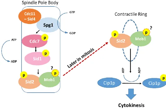
4. The Septation Initiation Network
In the fission yeast S. pombe, mitotic process is spatially different from budding yeast. Nevertheless, the molecules involved in the regulation of cell division are deeply conserved. Studies using this model organism have demonstrated that most of the proteins involved in MEN in budding yeast have counterparts in fission yeast. Here, the signaling cascade that coordinates cell division in which Mob1 protein is involved is the Septation Initiation Network (SIN). SIN has a role in actin-myosin ring contraction and in cytokinesis, being involved in the correct formation of the septum in time and space (for review see (Hergovich & Hemmings, 2012)). The mechanics of the SIN cascade are very similar to MEN. As in the case of MEN, the core components of the SIN network are also a GTPase Spg1, the effector kinases Cdc7 and Sid1 that belong to the Ste20 kinase family, the NDR family kinase and its activator Mob1. The final outcome of the SIN is also the phosphorylation, by Sid2-Mob1 complex, of the Cdc14-like phosphatase Cip1p (for review see (Johnson, McCollum, & Gould, 2012)) (Fig.3).
Figure 4 - The Septation Initiation Network – schematic representation of the S. pombe SIN. The core components of the SIN show a spindle pole body localization throughout mitosis. The activation of the GTPase Spg1 consequently activates the Cdc7 kinase which in turn phosphorylates the Sid1 kinase. At the end of this cascade, Sid2 is activated by phosphorylation and this activation depends on its binding partner Mob1. This complex relocalizes to the region where the septum and the actin-miosin contractile ring is formed, being involved in cytokinesis completion. One of the effects of the active Sid2-Mob1 complex is the maintenance of the Cdc14-like phosphatase in the cytoplasm, which is critical for cytokinesis progression.
18
Similarly to what occurs in MEN, the scaffold bipartite protein complex Cdc11-Sid4 is requested to concentrate SIN molecules at the spindle pole bodies. There, the GTPase Spg1 is activated which in turn leads to the activation of the Sid1 kinase that consequently activates by phosphorylation Sid2 protein in the Sid2-Mob1 complex. The activity of the complex Sid2-Mob1 reaches its maximum level just before septation, and it localizes at the septum at the end of mitosis. Still, the details of this relocalization of Sid2-Mob1 are unknown. Active Sid2-Mob1 is critical to maintain the Cdc14-like phosphatase Cip1p in the cytoplasm which allows cytokinesis completion (Chen et al., 2008). Further investigation regarding Sid2 substrates at the contractile ring will certainty contribute to a better understanding of cytokinesis regulation by this molecule.
5. The Hippo pathway
In multicellular organisms, orthologs of the core proteins of both MEN and SIN are also present and most of them are implicated in the major signaling cascade controlling the balance between cell proliferation and cell death, the Hippo pathway. This complex signaling pathway was firstly described in D. melanogaster and has since been described also in mammalians (for review see (Mo, Park, & Guan, 2014)). Taking into account the fundamental role of the Hippo pathway it is not surprising that the correct function of this cascade is critical both for embryonic development and adult tissues homeostasis.
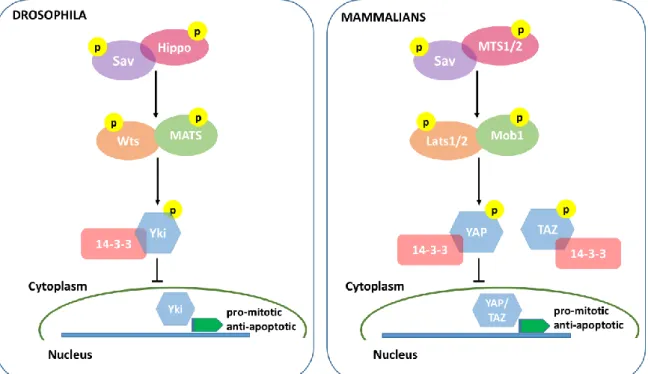
In D. melanogaster, the core components of the Hippo pathway were identified in genetic screenings searching for tumor suppressor genes, and in fact, inactivation of any of these core proteins always results in cell over proliferation. The fundamental players of this pathway in the fly are the Ste20-like kinase Hippo (Hpo), its scaffolding protein Salvador (Sav), the NDR family kinase Warts (Wts) and finally, its activator Mats (Mob1 nomenclature in the fly) (Harvey, Pfleger & Hariharan, 2003; Justice, Zilian, Woods, Noll & Bryant, 1995; Lai et al., 2005; Pantalacci, Tapon & Leopold, 2003; Udan, Kango-Singh, Nolo, Tao & Halder, 2003). The final target of the pathway is the transcription factor activator Yorkie (Yki) whose nuclear localization depends on its phosphorylation status. Yki acts as an oncongene, since it promotes the transcription of pro-mitotic and anti-apoptotic genes (Huang, Wu, Barrera, Matthews & Pan, 2005; Oh & Irvine, 2008) (Fig. 4).
During development, the Hippo pathway kinase module is inactive, Yki is not phosphorylated by Wts and can migrate to the nucleus to exert its transcriptional
19
activation function. In this case, cells proliferate until tissues and organs reach their normal size. When proliferation needs to be stopped, the core kinases of the Hippo pathway are sequentially activated, as follows: Hippo is activated upon receiving upstream information that proliferation should be repressed. Active Hippo forms a complex with the scaffold protein Sav and the complex can in turn phosphorylate the Wts kinase that, after forming a complex with its activator Mats, can phosphorylate Yki (Zeng & Hong, 2008). Phosphorylated Yki is sequestered in the cytoplasm mainly via the binding to 14-3-3 proteins (Ren, Zhang & Jiang, 2010).
All the core proteins described in the fly have homologues in mammals, that work in the same fashion as described in D. melanogaster (for a review see (Mo et al., 2014)). In mammals, the core components of the Hippo pathways are the ste-20 kinase MST1/2 (Hippo in the fly), the scaffolding protein Sav (also known as hWW45), the NDR family kinase Lats1/2 (stands for large tumor suppressor) and finally, its activator Mob1. The final effector of the Hippo cascade in mammalians is the protein YAP or TAZ (Yki in the fly) that are also transcription activator factors whose activity in the nucleus induces the expression of anti-apoptotic and proliferation associated genes such YAP family members BIRC5/survivin and BIRC2/cIAP1 and the BCL2 family gene MCL1 (Dong et al., 2007; Zhang, Smolen & Haber, 2008). To facilitate the understanding of the Hippo pathway cascade both in D. melanogaster and in mammals, comparative schemes of the two pathways containing their core molecules are shown in figure 4.
Taking into account the role that Yki/YAP activity has on proliferation induction, it is not surprising that the deregulation of its activity is associated with cancer development. Effectively, while the activity of the Yap protein as a transcription activator is critical for growth and correct development in different organs (Gee, Milgram, Kramer, Conlon & Moody, 2011; Septer et al., 2012; Zhang et al., 2010) and also for stem cell pluripotency (Lian et al., 2010), a great amount of studies have shown that the uncontrolled activity of YAP is associated with tumor formation and cancer progression in several different tissues such as the liver, intestine, lung, skin and brain (Camargo et al., 2007; Orr et al., 2011; Wang et al., 2010; Zhang, Wu, & Xing, 2011). As the cellular effector of Hippo signaling is an oncoprotein it is easy to understand why the deregulation of any of the Hippo pathway core kinases leads to uncontrolled cell proliferation. In fact, while YAP is classified as oncogene, all the other Hippo core proteins that regulate its activity are well established as tumor suppressors.
20
Even though the main events involving the Hippo pathway central kinase module are the ones described above, the importance of the pathway in tissues homeostasis led to an increasing interest in investigating the upstream regulators of the pathway. Which external signals or signaling pathways impinge on the Hippo cascade to promote its activation?
In recent years, several molecules and pathways have been implicated in the Hippo signaling response, most of them related to cell and tissue organization.
Indeed, the Hippo pathway can accommodate information coming from cell pathways responsible for maintenance of the polar structure of epithelia such as components of the apical-basal polarity protein complexes and also components of the planar cell polarity machinery.
Furthermore, the Hippo pathway can also respond to mechanical alterations or tensions, transducing signals coming from cytoskeleton molecules, and the extracellular matrix (for review see (Yu & Guan, 2013)).
Figure 5 - The Hippo pathway in D. melanogaster and Mammals. A schematic representation of the core molecules involved in Hippo signaling both in D. melanogaster and in mammals. The pathway impinges on the transcription factor activator Yki/YAP that in its phosphorylated form is maintained in the cytoplasm not inducing pro-mitotic genes transcription. So, when activated, the Hippo cascade inhibits cell proliferation and increase of tissues/organs size.
21
The details of this regulation will be discussed later in more detail. Understanding the regulation of tissue homeostasis and how individual cell function/structure contributes to it is a major issue both in basic cell biology as well as in the understanding of some human diseases. Indeed, the disruption of the tightly controlled structure of tissues such as epithelia can lead to cellular transformation that, when uncontrolled, is associated, for example, with cancer development.
23