45 Effects of radiation on the nervous system of rats
Radiol Bras. 2008;41(1):45–47 Original Article
Effects of low X-radiation doses on the central nervous
system development: an experimental study in rats*
Efeitos de baixas doses de radiação-X no desenvolvimento do sistema nervoso central: estudo experimental em ratos
Liliane Lins1, Laís Gomes2, Lis Gomes2,Marcele Trindade2, Leonardo Dias2, Ricardo Bragança2, Rodrigo Pimentel2
OBJECTIVE: The present study analyzes the consequences of X-irradiation for the development of the ner-vous system of rat fetuses. MATERIALS AND METHODS: The sample included ten eight-week-old, preg-nant Rattus norvegicus albinus, Wistar. Five female rats constituted the control group and other five had their abdominal region exposed for 30 seconds to a single 0.3 Gy radiation dose from a 70 kV, 10 mA Gnatus odontological apparatus. At the 17th gestational day both groups were submitted to hysterectomy. Selected sections were examined for comparative brain analysis of both groups. RESULTS: The clinical evaluation demonstrated no morphological difference between the control and the experimental groups. Twenty-seven percent of the animals in the experimental group presented mild brain hemorrhage, while 73% of the ani-mals had severe cerebral cortex hemorrhage and nervous tissue damage. None of the aniani-mals in the control group presented cerebral hemorrhage or nervous tissue damage. CONCLUSION: These evidences demon-strate that low X-radiation doses may cause brain hemorrhage and, consequently, nervous tissue damage.
Keywords: Irradiation; Malformations; Hemorrhage; Central nervous system.
OBJETIVO: Este trabalho analisa as conseqüências da irradiação-X no desenvolvimento do sistema nervoso de fetos de ratos. MATERIAIS E MÉTODOS: O trabalho foi constituído de 10 Rattus norvegicus albinos, Wistar, fêmeas, grávidas, com idade de oito semanas. Cinco ratas fêmeas constituíram o grupo controle e outras cinco tiveram suas regiões abdominais expostas por 30 segundos a uma dose de 0,3 Gy proveniente de um aparelho odontológico Gnatus de 70 kV e 10 mA. No 17º dia gestacional, ambos os grupos foram submetidos a histerectomia. As seções selecionadas foram examinadas para análise cerebral comparativa entre os grupos. RESULTADOS: O exame clínico revelou não haver diferenças morfológicas entre os grupos controle e experimental e nenhum dos animais apresentou anormalidades grosseiras. Vinte e sete por cento dos animais do grupo experimental apresentaram hemorragia cerebral moderada e 73% apresentaram he-morragia severa e danos no tecido nervoso. Nenhum animal do grupo controle apresentou hehe-morragia cere-bral ou danificações de tecido nervoso. CONCLUSÃO: Estas evidências demonstram que pequenas doses de radiação-X podem causar hemorragias cerebrais e, conseqüentemente, lesão tecidual nervosa.
Unitermos: Irradiação; Malformações; Hemorragia; Sistema nervoso central. Abstract
Resumo
* Study developed at Escola Bahiana de Medicina e Saúde Pública, Salvador, BA, Brazil.
1. PhD, Titular Professor of Embriology at Escola Bahiana de Medicina e Saúde Pública, Salvador, BA, Brazil.
2. Graduate Students of Medicine at Escola Bahiana de Me-dicina e Saúde Pública, Salvador, BA, Brazil.
Mailing address: Dra. Liliane Lins. Departamento de Morfolo-gia, Escola Bahiana de Medicina e Saúde Pública. Rua Frei Henrique, 8, Nazaré. Salvador, BA, Brazil, 40050 420. E-mail: [email protected].
Received May 15, 2006. Accepted after revision August 16, 2007.
INTRODUCTION
Neuroblasts, probably the most abun-dant type of cell in mammals embryo-fe-tuses, are extremely radiosensitive and rep-resent an intermediate stage between neu-roepithelial cells and neurons. In mice,
neuroblasts appear at the 7th day of gesta-tion, while in the human species they are formed 18 days after fecundation. Both in the human species and in mice, these cells are present from the gestation to the neo-natal period, interconnecting the develop-ing tissues and organs(1). So, the use of ra-diation during the embryonal period where there is the highest concentration of neuroblasts, frequently results in congeni-tal anomalies of the central nervous system and associated structures, such as mi-crophthalmos, anophtalmia, micrencephaly and anencephaly. Studies demonstrate that an X-ray dose of some hundreds Gy may induce a significant number of encephalic
hemorrhages, the quantity of lesion being exponentially proportional to the dose in-crease(2). However, there is still a necessity for analysis of the effect of low X-radiation doses on the development of the nervous system of rat fetuses.
The effect of radiation on the embryo in the pre-implantation phase may be best described as “everything or nothing”, dichotomically represented either by an early embryo death or the normal develop-ment of the embryo. It is considered that the chromosomal damage caused by X-irra-diation is preponderant in determining the embryo death because of the resulting primitive cells degeneration.
Lins L, Gomes L, Gomes L, Trindade M, Dias L, Bragança R, Pimentel R. Efeitos de baixas doses de radiação-X no desenvolvimento do sistema nervoso central: estudo experimental em ratos. Radiol Bras. 2008;41(1):45–47.
Radiol Bras. Jan/Fev 2008;41(1)
46
Lins L et al.
Radiol Bras. 2008;41(1):45–47 The primary effect of the radiation
ex-posure on the organogenesis is the devel-opment of malformations(3). Such abnor-malities are related to the structures in-volved in the organogenesis at the moment of irradiation, the differentiation stage and the dose administered(4). Severe and persis-tent deficits in the adult brain, disorganized cortical architecture, decrease in the corti-cal size and cerebral weight, micrenceph-aly and motor dysfunction have been ob-served in the study developed by Miki et al.(5), who have successively applied X-ra-diation at doses ranging between 1.0 Gy and 2.0 Gy in the period between the 13th and 18th day of gestation.
Takaiet al.(6) have exposed adult rats to an X-radiation 1.5 Gy dose on the hippoc-ampus, resulting in cognitive dysfunction related to cell ectopia. Still, as regards hip-pocampal alterations, Schmitz et al.(7), studying gamma radiation, have reported Purkinje cells decrease, and increase in the cerebral volume as a result of a cumulative dose of 3.0 Gy applied in the period be-tween the 13th and 16th days of gestation. Considering the need for additional histomorphological studies on the effects of X-irradiation on the embryonal develop-ment, the present study is aimed at evalu-ating the effects of low X-radiation doses on the development of the nervous system of rat fetuses.
MATERIALS AND METHODS
The sample studied included 15 (10 fe-male and 5 fe-male) eight-week-old Rattus norvegicus albinus, Wistar, weighting on average 275 g. Both male and female rats were kept under a 12-hour light-dark cycle, with unrestricted access to food and water. The female animal models were fertilized according to an adaptation from the method of Chahoud & Kwasigroch(8). An otoscope was utilized for detecting the vaginal plug to determine the day of conception(9).
Five female animal models at the 8th gestational day, with 37 embryos, were kept under standard survival conditions, constituting the control group. Other five female animal models, also at the 8th ges-tational day, with 39 embryos, had their ab-dominal region exposed to a single 0.3 Gy X-radiation dose for 30 seconds. A Gnatus,
70 kV, 10 mA odontological X-ray equip-ment. All of the rats were kept in separate cages, anesthetized with ether and submit-ted to the irradiation and perfusion proce-dures. On the 17th day of gestation, both groups were submitted to hysterectomy and sacrificed with an ether injection into the abdominal cavity under 0.2 ml/100 g ketamine anesthesia.
The animals’ uteri were fixed in a 10% formaldehyde solution, dehydrated and embedded in paraffin. A microtome was utilized to obtain several 4 µm-tissue sec-tions. Selected sections were hematoxilin-eosin stained and evaluated for compara-tively analyzing the fetuses brains in both groups. The macroscopic analysis was based on the closure of the rostral neu-ropore, while the microscopic analysis con-sisted in a morphological evaluation of the brains, utilizing a ten-fold magnification. The analysis covered the hippocampus and cerebellum, besides neuronal ectopia, loss of the tissue architecture and hemorrhage, rated as mild, moderate and severe accord-ing to the vessels congestion intensity and blood cells flow out.
The Epi-Info 2004 software (CDC. 3.3 version) was utilized for statistical analy-sis, according to the chi-square test for hemorrhage variables and alterations in the nervous tissue architecture, comparing both the experimental and control groups. A p value < 0.05 was considered as statis-tically significant.
RESULTS
Thirty-seven and 39 embryos, respec-tively in the control and experimental groups, were evaluated, with no follow-up loss being observed. Clinical examinations demonstrated absence of macroscopic dif-ferences between groups, considering that 100% of the animal models presented a normal closure of the rostral neuropore, and none (0%) presented signs of anoph-talmia, micrencephaly and anencephaly.
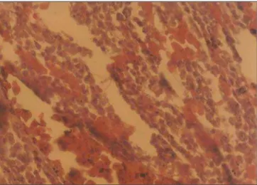
Microscopic analyses demonstrated ce-rebral cortex hemorrhage ranging between moderate and severe. Twenty-seven per cent of the animal models in the experimen-tal group presented moderate cerebral hem-orrhage, and 77% presented severe hemor-rhage (p < 0.05; Figure 1). All the cases of
severe cerebral hemorrhage were associ-ated with loss of the nervous tissue archi-tecture (p = 0.05). None of the animals in the control group presented cerebral hem-orrhage or loss of nervous tissue architec-ture (Figure 2).
DISCUSSION
All of the gestations developed nor-mally up to the analyses period, without any behavioral alteration being observed in the experimental group as compared with the control group. An otoscope was effec-tively utilized for detecting the vaginal plug, allowing the accurate determination of the exact day of conception, and increas-ing the predictability of the gestation, as demonstrated by Voipio & Nevalainen(9). This method did not cause any stress or pain to the animals, besides being rapid and easy to perform. Therefore the otoscope in-sertion did not present any morphological effect on the gestation development, and the rats were not affected by any external interference during the study.
Considering that the nervous system cells are extremely sensitive to radiation, nervous tissue lesions and hemorrhages were the main findings in the present study. However, other effects such as motor and cognitive dysfunctions, Purkinje cells atro-phy, micrencephaly and hippocampal ecto-pia have been observed and reported by other authors(5,6), nevertheless they were not investigated in the present study.
Hemorrhages detected in the brain of rats exposed to radiation constitute evi-dences compatible with the findings of Yang & Tobias(2), who have demonstrated that a low X-radiation dose may cause ce-rebral hemorrhage and, consequently, ner-vous tissue lesion. According to the find-ings of Friedberg et al.(4), none of the ani-mal models presented any abnorani-mality such as anophtalmia, micrencephaly and anencephaly, probably because of the low radiation dose applied.
47 Effects of radiation on the nervous system of rats
Radiol Bras. 2008;41(1):45–47
The description of the effect of X-irra-diation during the pre-implantation period as “everything or nothing”(3) was con-firmed in the present study, considering that the embryos developed normally up to the 17th gestational day, and no follow-up loss was recorded. Vos(3) also reports that the primary effect of X-ray exposure during the organogenesis phase is the development of malformations. Nevertheless, in the present study, the control group was irradiated dur-ing the organogenesis phase and no malfor-mation could be evidenced. However, loss of nervous tissue architecture and hemor-rhage probably would have resulted in later developmental disorders.
According to Schmitz et al.(7), the pre-natal irradiation results not only in neuronal loss, but also in a massive volumetric de-crease in the cerebral regions investigated. Probably because of the low radiation dose applied, this could not be observed. It is known that progenitor cells may be injured by irradiation and cannot divide during radiation exposure, but, maybe, a single
X-radiation dose is not enough to cause dam-ages during the cells development stdam-ages. These authors have utilized different lev-els of irradiation, and this may have af-fected the growth factors.
Finally, based on their findings, the au-thors support the assertion that X-radiation in the form and conditions described in the present study may determine the occur-rence of cerebral hemorrhages and nervous tissues lesions in rat fetuses.
Acknowledgement
The present study was partially sup-ported by Fundação Bahiana para Desen-volvimento das Ciências.
REFERENCES
1. Rugh R. X-ray-induced teratogenesis in the mouse and its possible significance to man. Ra-diology. 1971;99:433–43.
2. Yang T, Tobias C. Effects of heavy ion irradiation on the brain vascular system and embryonic de-velopment. Adv Space Res. 1984;4:239–45. 3. Vos O. Effects and consequences of prenatal
ir-radiation. Boll Soc Ital Biol Sper. 1989;65:481– 500.
4. Friedberg W, Faulkner DN, Neas BR, et al. Dose-incidence relationships for exencephalia, anoph-thalmia and prenatal mortality in mouse embryos irradiated with fission neutrons or 250 kV X-rays. Int J Radiat Biol Relat Stud Phys Chem Med. 1987;52:223–36.
5. Miki T, Fukui Y, Takeuchi Y, et al. A quantitative study of the effects of prenatal X-irradiation on the development of cerebral cortex in rats. Neurosci Res. 1995;23:241–7.
6. Takai N, Sun XZ, Ando K, et al. Ectopic neurons in the hippocampus may be a cause of learning disability after prenatal exposure to X-rays in rats. J Radiat Res. 2004;45:563–9.
7. Schmitz C, Born M, Dolezel P, et al. Prenatal protracted irradiation at very low dose rate in-duces severe neuronal loss in rat hippocampus and cerebellum. Neuroscience. 2005;130:935– 48.
8. Chahoud I, Kwasigroch TE. Controlled breeding of laboratory animals In: Neubert D, Merker HJ, Kwasigroch TE, editors. Methods in prenatal toxicology. Stuttgart: Georg Thieme; 1977. p.78– 91.
9. Voipio HM, Nevalainen T. Improved method for vaginal plug detection in rats. Scand J Lab Anim Sci. 1998;25:5–9.
10. Wang H, Chen D, Gao C, et al. Effects of low level prenatal 60Co gamma-irradiation on postnatal
growth and behavior in mice. Teratology. 1993; 48:451–7.
Figure 1. Photomicrography of the encephalic region of a 17-day experimen-tal rat fetus. Severe, diffuse cerebral hemorrhage, and loss of the nervous tissue architecture can be observed. (Hematoxilin-eosin; ten-fold magnifica-tion).