XI
Radiol Bras. Mar/Abr;44(2):XI–XIII
Marcelo Souto Nacif1, Nadine Kawel2, Christopher T. Sibley3, Anna Zavodni2, João A. C. Lima4, David A. Bluemke5
Study developed at Department of Radiology and Imaging Sciences, National Institutes of Health (NIH) Clinical Center, Bethesda, MD, USA. 1. MD, PhD, Professor at Radiology Department – Universidade Federal Fluminense (UFF), Niterói, RJ, Brazil, Fellow of Radiology and Imaging Sciences, National Institutes of Health (NIH) Clinical Center, Bethesda, MD, USA. 2. MD, Fellow of Radiology and Imaging Sciences, National Institutes of Health (NIH) Clinical Center, Bethesda, MD, USA. 3. MD, Staff Clinician, Department of Radiology and Imaging Sciences, National Institutes of Health (NIH) Clinical Center, Bethesda, MD, USA. 4. MD, Professor at Division of Cardiology, Johns Hopkins University School of Medicine, Baltimore, MD, USA. 5.MD, PhD, Director, Department of Radiology and Imaging Sci-ences, National Institutes of Health (NIH) Clinical Center, Bethesda, MD, USA. Mailing Address: Dr.Marcelo Souto Nacif. Cordell Avenue 4583, 20814 Bethesda, MD, USA. E-mail: [email protected]. Web site: www.msnacif.med.br
0100-3984 © Colégio Brasileiro de Radiologia e Diagnóstico por Imagem
WHICH IS YOUR DIAGNOSIS?
Nacif MS, Kawel N, Sibley CT, Zavodni A, Lima JAC, Bluemke DA. Which is your diagnosis? Radiol Bras. 2011 Mar/Abr;44(2):XI–XIII.
Forty-one year old hypertensive (African American) male patient, with a history of drug abuse for a “short period” at the age of 29 years, was referred to the Radiology Department at National Institutes of Health Clinical Center for evaluation by cardiac computed tomographic angiography (CTA) and cardiac magnetic resonance imaging (CMR).
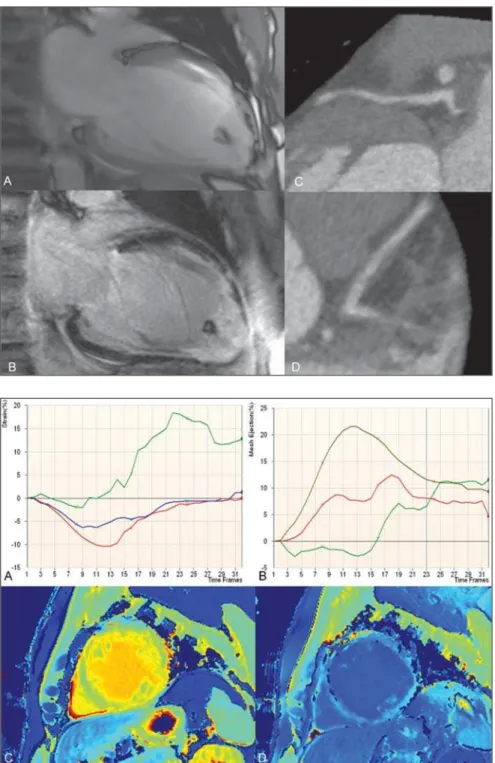
Figure 1. ECG-gated CMR (A,B) and CTA (C,D). Two chamber cine-CMR (A) and late enhancement (B). Proximal left anterior descending coronary ar-tery (C,D).
XII Radiol Bras. 2011 Mar/Abr;44(2):XI–XIII Images description
Figure 1. ECG-gated CMR (A,B) and CTA (C,D). Cine-CMR, two chamber view (A) and late enhancement (B). Proximal left anterior descending coronary artery (C,D). Observe marked left ventricular di-latation associated with wall thinning in the territory of the left anterior descending coronary artery related to previous infarc-tion caused by vasospasm. Enlargement of the apical trabeculated region with calci-fied thrombus. CTA (C,D) demonstrating a soft plaque in the proximal left anterior descending coronary artery with positive remodeling and subtle luminal narrowing (30% stenosis).
Figure 2. Tagging analysis (A,B). Strain rate (A) and mesh ejection fraction (B). Pre-contrast (C) and post-contrast (D) MOLLI T1 color maps. The tagging analy-sis showed poor segmental ejection frac-tion (base = 21.4%, midcavity = 7.7%, and apex = –2.4%), and the peak Ecc strain (%) at the base, midcavity and apex were re-spectively –10.6%, –5.7% and 0.6% of the average LV midwall. Pre-contrast (C) and post-contrast (D) MOLLI T1 color coded maps for visual (qualitative) analysis: in the regions of delayed gadolinium enhance-ment, T1 color maps demonstrate yellow color on pre-contrast and dark blue on post-contrast maps according to a longer T1 time in the pre-contrast phase and a shorter T1 time in the post-contrast phase. As the pre-contrast green myocardium (shorter T1 time) and the post-contrast blue myocar-dium (longer T1 time) are compared, re-gions with larger amounts of fibrous tissue and extracellular space enlargement can be differentiated. Analysis of T1 mapping: extracellular volume fraction for the myo-cardium at the base of the heart without late enhancement corresponded to 32%, while, in the apical region with late enhancement, it corresponded to 57%.
Background: The normal range for mid-wall peak Ecc strain (%) is –17.5 ± 4%(1). The extracellular volume fraction is calculated from the ratio of the myocardial relaxation rates and blood corrected by the hematocrit value:
(∆R1myocardium/∆R1blood)*(1 – Ht).
Healthy volunteers assessed with the same protocol in our lab demonstrated that
the normal extracellular volume fraction (± standard deviation) corresponds to 27 ± 3%.
Diagnosis: Cocaine-related myocardial infarction in association with poor global strain rate and increased extracellular vol-ume fraction calculated on T1 maps.
COMMENTS
Association between cocaine use and myocardial ischemia/infarction was first reported by Coleman et al. in 1989(2). Fre-quently, the diagnosis depends on correct information about the patient’s history of drug abuse. The assisting physician should be aware of the differential and try to con-firm the diagnosis with blood or urine tests. The clinical history will always be difficult to assess, depending on the patient’s coop-eration(3).
In spite of the fact that the most common form of myocardial infarction is related to the rupture of an unstable (vulnerable) ath-erosclerotic plaque, coronary vasospasm, either in association or not with the devel-opment of a secondary thrombus is also a potential cause for myocardial necrosis(4). Cocaine acts as a powerful sympatho-mimetic agent, producing central and pe-ripheral adrenergic stimulation which in-duces coronary vasoconstriction that is usually related to the peak blood concen-tration of cocaine. However, a late effect may occur as a result of endothelial activa-tion and the synergic prothrombotic activ-ity, causing hypercoagulability and acute thrombus formation in the coronary ves-sel(5,6).
The early development of coronary ath-erosclerosis in chronic cocaine abusers has been reported in the literature(7,8). In the present case, a soft plaque with mild steno-sis was detected in the proximal left ante-rior descending coronary artery.
Minimal data are available to guide the evidence-based treatment of cocaine-re-lated myocardial infarction. Usually, the treatment is clinical, with benzodiazepines, aspirin, nitroglycerin, verapamil, besides stimulus for behavioral change . However, some authors such as Sharma et al.(7) be-lieve that a percutaneous coronary inter-vention at the moment of the vasospasm is the treatment of choice for such patients.
In the present case, at the time of the acute precordial pain at the age of 29, the patient underwent percutaneous coronary inter-vention (angioplasty), and was hospitalized in a coronary care unit for three weeks.
Among Americans over 12 year of age, 27.7 million, corresponding to 12% of the population, have used cocaine at some point in their lifetime; and more than 5.9 million, or 2.5% of the entire US population, have used cocaine in the last year(9,10). Cocaine use is a major public health problem world-wide and has increased in popularity in the last decades(5,8). The potential impact on the public health policies is worrisome(11).
At long term, the best strategy to be adopted would be to enter patients and us-ers into a substance abuse program to edu-cate them about the irreversible harmful effects of cocaine on the cardiac muscle 5). We strongly support a massive public health policy to reduce the number of co-caine users and avoid the related compli-cations.
Non invasive cardiac imaging
Computed tomography coronary an-giography can non-invasively and accurately detect and quantify coronary plaque (12). In the present case, it was straightforward to detect the soft plaque in the proximal left anterior descending coronary artery on CTA images.
Many precise software tools are avail-able for the assessment of ventricular func-tion using tagging(1) and diffuse or focal fibrosis with modified Lock-Locker inver-sion recovery (MOLLI)(13–15) imaging.
Cardiac magnetic resonance tagging is a powerful non-invasive diagnostic tool for quantifying regional systolic and diastolic myocardial function. Ongoing technologi-cal developments, particularly in imaging techniques, equipment and analytical tools utilized to implement CMR tagging, will improve the analyses of regional myocar-dial function. This will be useful for com-parison and precise follow up of patients. In the present case, we could accurately assess the regional function.
fi-XIII
Radiol Bras. Mar/Abr;44(2):XI–XIII
brosis, and T1 mapping is a powerful tool for quantifying the extent and aggressive-ness of such abnormality. The T1 mapping technique has already been validated by means of myocardial biopsies in the clini-cal context of patients with nonischemic fibrosis who were referred for cardiac transplant(16). Myocardial extracellular vol-ume measurements quantify diffuse fibro-sis not readily detectable by conventional late gadolinium enhancement(13,14). In the present case, we could differentiate the fi-brosis detected by late enhancement on the apex of the left ventricle (high extracellu-lar volume – ECV, 57%) from the fibrosis present on the bottom of the left ventricle (high ECV, 32%) in comparison with healthy hearts (ECV = 27%).
Important considerations
Non-invasive studies, such as CTA and CMR are fundamental in the evaluation of subclinical atherosclerotic disease and quantification of myocardial fibrosis.
Randomized, prospective studies would be extremely important to better understand these powerful techniques.
REFERENCES
1. Fernandes VR, Cheng S, Cheng YJ, et al. Racial and ethnic differences in subclinical myocardial function: the Multi-Ethnic Study of Atheroscle-rosis. Heart. 2011;97:405–10.
2. Coleman DL, Ross TF, Naughton JL. Myocardial ischemia and infarction related to recreational co-caine use. West J Med. 1982;136:444–6. 3. Rao SC, Main ML. Acute severe functional
mi-tral regurgitation secondary to cocaine-mediated anterior myocardial infarction. J Am Soc Echo-cardiogr. 2008;21:297.e3–4.
4. Kloner RA. Natural and unnatural triggers of myocardial infarction. Prog Cardiovasc Dis. 2006;48:285–300.
5. Rezkalla SH, Kloner RA. Cocaine-induced acute myocardial infarction. Clin Med Res. 2007;5: 172–6.
6. Raddino R, Pedrinazzi C, Zanini G, et al. Acute myocardial infarction in a young woman with antiphospholipid syndrome and occasional co-caine abuse. Int J Cardiol. 2005;105:236–8. 7. Sharma AK, Hamwi SM, Garg N, et al.
Percuta-neous interventions in patients with cocaine-asso-ciated myocardial infarction: a case series and re-view. Catheter Cardiovasc Interv. 2002;56:346–52. 8. Sonne C, Stempfle HU, Klauss V, et al. Intravas-cular ultrasound-guided percutaneous coronary intervention in a human immunodeficiency virus-positive patient with cocaine-associated acute myocardial infarction: case report and review. Heart Lung Circ. 2005;14:197–200.
9. Bansal D, Eigenbrodt M, Gupta E, et al.
Tradi-tional risk factors and acute myocardial infarction in patients hospitalized with cocaine-associated chest pain. Clin Cardiol. 2007;30:290–4. 10. Aslibekyan S, Levitan EB, Mittleman MA.
Preva-lent cocaine use and myocardial infarction. Am J Cardiol. 2008;102:966–9.
11. Coombs M. Cocaine-induced myocardial infarc-tion. Nurs Crit Care. 2007;12:176–80. 12. Rinehart S, Vazquez G, Qian Z, et al.
Quantita-tive measurements of coronary arterial stenosis, plaque geometry, and composition are highly re-producible with a standardized coronary arterial computed tomographic approach in high-quality CT datasets. J Cardiovasc Comput Tomogr. 2011;5:35–43.
13. Piechnik SK, Ferreira VM, Dall’Armellina E, et al. Shortened Modified Look-Locker Inversion re-covery (ShMOLLI) for clinical myocardial T1-mapping at 1.5 and 3 T within a 9 heartbeat breath-hold. J Cardiovasc Magn Reson. 2010;12:69. 14. Schelbert EB, Testa SM, Meier CG, et al.
Myocar-dial extravascular extracellular volume fraction measurement by gadolinium cardiovascular mag-netic resonance in humans: slow infusion versus bolus. J Cardiovasc Magn Reson. 2011;13:16. 15. Messroghli DR, Radjenovic A, Kozerke S, et al.
Modified Look-Locker inversion recovery (MOLLI) for high-resolution T1 mapping of the heart. Magn Reson Med. 2004;52:141–6. 16. Nacif MS, Noureldin RAA, Sibley CT, et al.