ABSTRACT
orthodontic bonding materials
Érika Machado CALDEIRA1, Amanda OSÓRIO1, Edna Lúcia Couto OBEROSLER2, Delmo Santiago VAITSMAN3,
Daniela Sales ALVIANO4, Matilde da Cunha Gonçalves NOJIMA5
1- DDS, MSD, School of Dentistry, Department of Orthodontics, Federal University of Rio de Janeiro, Rio de Janeiro, RJ, Brazil.
2- Biologist and Technical Chemistry, Institute of Chemistry, Department of Analytical Chemistry, Federal University of Rio de Janeiro, Rio de Janeiro, RJ, Brazil. 3- MSD, PhD, Professor, Institute of Chemistry, Department of Analytical Chemistry, Federal University of Rio de Janeiro, Rio de Janeiro, RJ, Brazil. 4- PhD, Professor, Institute of Microbiology Prof. Paulo de Góes, Federal University of Rio de Janeiro, Rio de Janeiro, RJ, Brazil.
5- MSD, PhD, Associate Professor, School of Dentistry, Department of Orthodontics, Federal University of Rio de Janeiro, Rio de Janeiro, RJ, Brazil.
Corresponding address: Matilde da Cunha Gonçalves Nojima - Programa de Pós-Graduação em Odontologia (Ortodontia) - Faculdade de Odontologia - Universidade Federal do Rio de Janeiro - Rua Professor Rodolpho Paulo Rocco, no 325 - Ilha do Fundão - Rio de Janeiro - RJ - Brasil - 21941-617 - Phone: +55 21 2590-2727 - Fax: +55 21 25909771 - e-mail: [email protected]
O
capacity of 3 bonding materials. Material and Methods: Thirty nine specimens with standardized surface smoothness and dimensions were prepared. The antimicrobial capacity of the materials against S. mutans, L. casei and C. albicans was evaluated by determining the percentage of growth inhibition of these microorganisms in an inoculated medium, obtained by optical density readouts on a spectrophotometer. The potential to interfere in microbial growth on the surface of the studied materials was observed by means of !"# $% days was analyzed by means of ion chromatography. Results: The PLUS group presented the highest percentage of microbial inhibition and the most contamination-free surface.
&'() #* TM
Plus Color Change was the one that presented the best general behavior considering the evaluated aspects.
Key words: Orthodontics. Microbiology. Fluorides. Dental bonding.
INTRODUCTION
) + orthodontic treatment, the retentive surfaces of the appliance make them difficult to clean + accumulation. Imbalances such as gingivitis, gingival hyperplasia and caries may occur during orthodontic treatment6,17,28, especially when
patients do not cooperate with regard to performing the recommended cleaning protocols. In this context, there is an increase in the acidogenic bacterial populations, such as Streptococcus mutans and Lactobacillus sp in the plaque and saliva25,28. The set formed by retentive niches,
+ rich in fermentable carbohydrates also favors the increase in the Candida albicans population in the oral cavity, particularly if there is caries activity19.
There are reports in the literature that microorganisms of the oral microbiome adhere + # It is also known that components present in the matrix of adhesive materials may accelerate bacterial growth, increasing the levels of pathogens extremely harmful to tissues3. Therefore, the ideal bonding material must be capable of withstanding salivary biochemistry, constant changes in pH, different temperatures, and especially the resident oral microbiota. Thus, the importance must be pointed out of maintaining essential properties, such as:
activity3.
Fluoridated bonding materials have been inserted outstandingly in Orthodontics because of / patients’ cooperation. Nevertheless, it is important to point out that these materials are recommended as supplementary to and not as substitutes for conventional prophylactic alternatives6, since
their anticariogenic capacity is still being widely discussed2,6,23. In view of the context presented,
the aim of this study was to evaluate the microbial inhibition capacity of three orthodontic bonding materials, and their fluoride release in vitro. Thus, the hypothesis considered in the present study was that bonding materials play a biological # prevent microbial contamination can be noted in the prevention and maintenance of the integrity of the oral tissues.
MATERIAL AND METHODS
Preparation of specimens
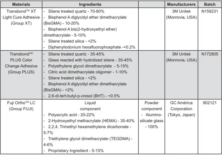
To conduct the study, three materials extensively applied in bonding orthodontic accessories were selected: TransbondTM XT (XT Group), a traditional
light cure adhesive (3M Unitek, Monrovia, CA, USA), TransbondTM Plus Color Change (PLUS Group), a
moisture tolerant light cure adhesive (3M Unitek, Monrovia, CA, USA) and Fuji OrthoTM LC (FUJI
Group), a resin reinforced glass ionomer cement (GC America Corporation, Tokyo, Japan) (Figure 1). For fabricating the specimens with standardized / < perforations measuring 5 mm in diameter and 2 mm in thickness was used. The materials were manipulated in accordance with the manufacturers’ instructions and then inserted into the matrix in a single increment, applying the pressure required for producing specimens with smooth surfaces (Figure 2). Light polymerization was performed for 40 seconds on each surface, using a halogen light with 550 mW/cm2 (Foto Optilight LD Max, Gnatus
– 50/60 Hz, Ribeirão Preto, SP, Brazil), with the active tip of the equipment touching directly on the glass slides26.
Microbiologic test
Microbiologic analyses were performed in two stages. Firstly, the antimicrobial activity of the materials was evaluated by means of spectrophotometry (quantitative aspect). For this, 18 specimens were prepared, divided in duplicate
Materials Ingredients Manufacturers Batch
TransbondTM XT
Light Cure Adhesive (Group XT)
- Silane treated quartz - 70-80%
- Bisphenol A diglycidyl ether dimethacrylate (BisGMA) - 10-20%
- Bisphenol A bis(2-hydroxyethyl ether) dimethacrylate - 5-10%
3M Unitek (Monrovia, USA)
N159231
TransbondTM
PLUS Color Change Adhesive
(Group PLUS)
- Silane treated quartz - 35-45%
- Glass reacted with hydrolized silane - 35-45% - Polyethylene glycol dimethacrylate - 5-15% - Citric acid dimethacrylate oligomer - 1-10%
- Bisphenol A diglycidyl ether dimethacrylate !"#
$& ' */#;
3M Unitek (Monrovia, USA)
N172805
Fuji OrthoTM LC
(Group FUJI)
Liquid component - Polyacrylic acid - 20-22%
- 2-Hydroxyethyl methacrylate (HEMA) - 35-40% - 2,2,4, Trimethyl hexamethylene dicarbonate - 5-7%
- Triethylene glycol dimethacrylate (TEGDMA) - 4-6%
- Proprietary Ingredient - 5-15%
Powder component - Alumino-silicate glass
- 100%
GC América Corporation (Tokyo, Japan)
902121
for each tested microorganism.
With regard to the qualitative aspect of analyses, after fabricating one control specimen for each material (baseline), the initial surface (contamination-free) record was made by scanning electron microscopy (SEM). After evaluation on the spectrophotometer, one specimen per microorganism was randomly selected from each group, to be submitted to a SEM analysis to verify microbial growth on its surfaces after contamination, and therefore, the inherent antimicrobial potential of each material. For each specimen evaluated by the SEM, various images were obtained in order to contemplate a large scanning area.
Quantitative analysis of antimicrobial activity
The following strains were used in the experiment: S. mutans (ATCC 25175), L. casei
(ATCC 4646) and C. albicans (ATCC 10231), kept in BHI broth (Brain Heart Infusion, Himedia, São Paulo, SP, Brazil). Initially, the BHI broth contaminated with the microorganisms of choice (positive control) was placed into 24 well plates, interspersed with wells containing sterile medium (negative control) and inoculated medium in conjunction with the specimens that would have their antimicrobial capacity tested. The quantity of cells in the experiment was standardized in order to attain the desired concentration of 5x10³ cells per well (500 μl). The plates were
incubated in an oven at 37°C, and the plate with S. mutans, was placed in an anaerobic jar. After 24 h, the optical density (OD) readout of each well was taken, starting by calibrating the spectrophotometer to 550 nm (Beckman Coulter DU 530 Spectrophotometer, Fullerton, CA, USA) by taking the sterile medium readout. The inoculated medium readout consisted of the positive control of growth for each microorganism1,13.
To obtain the percentage of microbial growth inhibition of the different groups studied, the
following equation was used, with: (M) mean of optical densities of each material tested; (C) positive control of growth: % growth inhibition =100-(M/C)x100.
Qualitative analysis of antimicrobial activity
In addition to quantifying the antimicrobial activity by spectrophotometry, the specimens were analyzed by SEM, in order to visualize the microbial proliferation on the surface of each studied material. To obtain the initial record of the material surfaces (baseline), one specimen from each group was prepared on aluminum stubs, and gold sputtered for examination by scanning electron microscopy (JEOL-JSM; 5800LV, Tokyo, Japan). A voltage of 30 kV and low vacuum mode (45 Pa) was used. Immediately after the quantitative analysis of the antimicrobial activity,
one specimen per microorganism from each group was randomly selected and +<stored in 2.5% glutaraldehyde and 0.1 M cacodylate buffer. After 2 h, the specimens were removed, washed with PBS (phosphate buffered saline) solution and dried. Y</ + records under conditions similar to the initial ones.
Various records of each specimen were obtained with the goal of extensive scanning of the surface. Z[[[<+ #
Fluoride release analysis
& /\ were prepared for each group (XT, PLUS, FUJI), totaling 18 specimens. After each specimen was </ surfaces were sealed with sticky wax, and it was ]+/ 19.62 mm2 remained exposed.For each specimen,
15 tubes of the Eppendorf® type, previously
washed for ionic decontamination, were filled with 1.5 mL of ultrapure water. The Eppendorf®
tubes corresponded to the 15 predetermined time / $ hour to the 14th/+ $/$/
2 d, 3 d, 4 d, 5 d, 6 d, 7 d, 8 d, 9 d, 10 d, 11 d, 12 d, 13 d and 14 d. During this cycle, the tubes were kept in a microbiologic oven at 37°C, separately in receptacles which were named XT, PLUS and FUJI.
The daily time when the specimens were moved into their new tubes was standardized. The specimens were manipulated with tweezers and they had been delicately dried with absorbent paper before they were reinserted into the sequence of the cycle. As the changes occurred, the solutions in which the specimens had been immersed were sent for laboratory analysis. It should be pointed out that the ultrapure water was the renewed vehicle, since the same specimen was kept from the beginning to the end of the experiment. The purpose of this
/ by exhaustion of this same material throughout the cycle. During the experiment, no ionic supplement whatsoever was inserted. The ultrapure water was renewed in an attempt to eliminate any possible saturation of the medium.
This study generated a total of 270 accumulated fluoride release readouts, performed by ion Chromatography (DX 80 Ion Analyzer, Dionex, Sunnyvale, CA, USA), in order to perform the in each solution. Before the readouts, the ion Chromatograph was calibrated with 5 specific patterns, these being: 1- ultrapure water; 2- pattern 1 (F- concentration=0.02 mg/L); 3-
pattern 2 (F- concentration=0.2 mg/L); 4- pattern
3 (F- concentration=1.0 mg/L); 5- pattern 4
(F- concentration=2.0 mg/L). The data obtained
were transferred and processed by an integrator, automatic module Dionex 4400 that promotes complete automation of the system. The time that + ion was approximately 12 minutes for each 1.5 mL sample of ultrapure water.
Statistical analysis
The percentage of growth inhibition data for each specimen of the XT, PLUS and FUJI groups obtained in the microbiologic analysis, as well as / analysis of variance ANOVA and Tukey Test in the SPSS 17.0 software (Statistical Package for Social Sciences, SPSS Inc., Chicago, IL, USA).
The difference between the means was considered + ^[#[Z #
The results obtained for the qualitative analysis of antimicrobial capacity were descriptively
recorded.
RESULTS
Quantitative analysis of antimicrobial activity
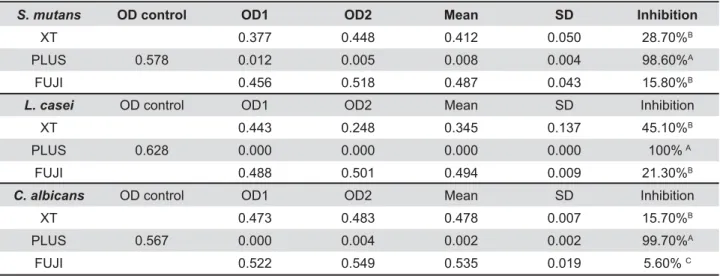
Table 1 shows the results obtained in the microbiologic test, specifying the activity of the materials in relation to the microorganisms analyzed, with respect to the microbial growth and inhibition.
To evaluate the microbiologic action of materials against S. mutans and L. casei, it was shown that _`'+ growth (p<0.05). With regards to action against
C. albicans, the materials showed significant differences among them (p<0.05), with emphasis on the PLUS, since it was the material that + the fungus (p<0.001).
Qualitative analysis of antimicrobial activity
The SEM photomicrographs shown in Figure 3 (1A, 2A, 3A) reveal the images of the negative control specimen surfaces from the XT, PLUS and FUJI groups, respectively. Images 1B and 3B denote S. mutans growth on XT and FUJI specimens, in this order. Visually, the greatest microbial growth occurred in the FUJI group (3B), whereas the PLUS group was found to have a contamination-free surface (2B), which also occurred in image 2C, in which no L. casei was observed on the surface of the specimen from the PLUS group. Images 1C (XT) and 3C (FUJI) also point out microbial growth, which was more expressive in the FUJI group.Lastly, it is possible to visualize great C. albicans proliferation on the
S. mutans OD control OD1 OD2 Mean SD Inhibition
XT 0.377 0.448 0.412 0.050 28.70%B
PLUS 0.578 0.012 0.005 0.008 0.004 98.60%A
FUJI 0.456 0.518 0.487 0.043 15.80%B
L. casei OD control OD1 OD2 Mean SD Inhibition
XT 0.443 0.248 0.345 0.137 45.10%B
PLUS 0.628 0.000 0.000 0.000 0.000 100% A
FUJI 0.488 0.501 0.494 0.009 21.30%B
C. albicans OD control OD1 OD2 Mean SD Inhibition
XT 0.473 0.483 0.478 0.007 15.70%B
PLUS 0.567 0.000 0.004 0.002 0.002 99.70%A
FUJI 0.522 0.549 0.535 0.019 5.60% C
<< Q>? ;#$' Q"VVW
FUJI surface (3D), followed by XT (1D) and PLUS |}"/ + #
Fluoride release analysis
$% of the experiment are shown in Table 2, and are expressed comparatively in Figure 4. One observes that the FUJI was the most outstanding material for fluoride release, especially in the first 24 hours, followed by the PLUS and XT, respectively. Although the latter characteristically does not / +/ of the technique. On the tenth day of the cycle, the values obtained for the FUJI and PLUS were similar (p>0.05).
DISCUSSION
There has been much discussion about the maintenance of the oral health in patients submitted to orthodontic treatment, such as the formation of initial caries lesions, which even today, is a potential risk. In a study recently conducted with 230 individuals, 71.7% developed from 1 to 12 white spot lesions at the end of treatment, and 69.8% presented adequate oral hygiene conditions at the beginning of orthodontic therapy17. These
data are indicative of the importance of instituting a good preventive program in these patients. Should there be clinical signs of disease progression, efforts must be redoubled in an attempt to
F- 1 h 1 d 2 d 3 d 4 d 5 d 6 d 7 d 8 d 9 d 10 d 11 d 12d 13 d 14 d
XT 0.007C 0.004C 0.001C 0.000C 0.002C 0.001C 0.001C 0.001C 0.002C 0.002C 0.002B 0.002C 0.001C 0.001C 0.002C
(0.005) (0.002) (0.001) (0.000) (0.001) (0.001) (0.000) (0.002) (0.001) (0.002) (0.002) (0.001) (0.001) (0.001) (0.001)
PLUS 0.087B 1.007B 0.628B 0.465B 0.440B 0.428B 0.424B 0.446B 0.448B 0.618B 0.609A 0.583B 0.502B 0.479B 0.454B
(0.029) (0.420) (0.750) (0.876) (0.032) (0.024) (0.058) (0.187) (0.163) (0.105) (0.106) (0.097) (0.070) (0.089) (0.077)
FUJI 0.267A 2.564A 1.276A 1.129A 1.031A 1.106A 0.839A 0.829A 0.938A 0.908A 0.691A 0.780A 0.632A 0.634A 0.845A
(0.087) (0.821) (0.293) (0.309) (0.193) (0.260) (0.152) (0.221) (0.190) (0.125) (0.148) (0.094) (0.128) (0.090) (0.121)
Y! <Z '<< << ? <Q> <;< same time analyzed, being A>B>C).
Table 2-! Y ?#< < 'Q \&#Q ]; ?
Figure 3- Scanning electron microscopy photomicrographs showing the surfaces of tested materials: (1) XT Group, (2) PLUS Group and (3) FUJI Group (columns); (A) Baseline, negative control, (B) S.mutans, (C) L.casei and (D) C.albicans
revert the situation of demineralization. Thus, it their composition, with the purpose of helping to prevent caries development. Nevertheless, still widely discussed, in spite of it clearly having remineralization potential24.
The results obtained demonstrated the greater + TM Plus Color Change as
an antimicrobial material, since it showed the best percentages of growth inhibition for all the species evaluated. The Fuji OrthoTM LC presented the most
unfavorable result with regards to antimicrobial / / release, which raises questions about the direct relationship of fluoride with microbial growth #) for the antimicrobial potential of the material, &'()/ comparison with the other groups with regards to antimicrobial activity, which was not demonstrated + #
Considering physicochemical characteristics10,
Lee, et al.16(2009) in their study, concluded that
there is greater microbial adherence to the surfaces of the bonding materials than to the orthodontic appliance accessories, and associated this fact with the free surface energy of these materials. Greater adhesion of S. mutans to the Fuji OrthoTM LC was
also found than on the composites, corroborating ] ] + study. It is also known that the surface roughness of materials is directly proportional to the increase in microbial adhesion5,20. Therefore, the fact that
glass ionomer cement is a material with low resistance and greater roughness, when compared / + the FUJI in the present study. With regard to chemical composition, the Fuji OrthoTM LC contains
the co-monomer TEGDMA, described in the literature as being capable of stimulating microbial proliferation13,15,18.
Authors have described the main characteristics
of ionomer materials as being anticariogenic and having antimicrobial properties. The former, is directly related to the preventive and remineralizing / 14.
/ / aluminum of the Fuji OrthoTM LC was shown to
+ and adhesion on its surface. This denotes the prevalence of the other previously described +#
Factors such as the initial pH of the material, hydrophobicity and chemical substances released are also related to antimicrobial activity10,14.
Chin, et al.10 (2006) studied the hydrophobicity
of some orthodontic bonding materials, among them the TransbondTM XT and Fuji OrthoTM LC, both
demonstrating similar hydrophobicity values, and representing the most expressive values in the #* ]/ + was found in these materials. Similar results were found by Ahn, et al.1 (2009), who observed
that both the TransbondTM XT and Fuji OrthoTM LC
exercised no antimicrobial activity, with the FUJI having shown the greatest microbial proliferation, + #
Although the cytotoxicity of the material may be related to microbial growth inhibition, it cannot + found in this research, because all the materials tested released substances such as bisphenol A, Bis-EMA, EGDMA, TEGDMA, HEMA, among others, capable of promoting adverse biologic reactions15,18. Therefore, it would not be conclusive to attribute the bests results found for the PLUS exclusively to cytotoxicity.
/ studies with the goal of quantifying it use the ion-selective electrode in their methodology2,7, since
there is a scarcity of the use of ion chromatography in the literature to proceed with such an analysis. This technique is frequently used when one desires to obtain the other components in the sample, because of its efficient ion detector system. In the present study, the chosen methodology for measuring fluoride ions was shown to be adequate and to have excellent precision because it determined anions and cations at trace levels4.
During the 15 time intervals of analysis for ]+ / increase in release in the 24 h7, there were some
/ + the passage of the time considered (Figure 4). ] < by the fact that the components of the external layer of the material became exhausted, and were dissolved in the water. With regard to the PLUS
/ matrix is a little limited by the resin components + # / matrix of the FUJI is more sensitive to hydration, there is less limitation of ion displacement8. This
+ / the more recurrent peaks, in spite of it having been equivalent to the PLUS on the 10th day of the cycle.
) release from the surface and the later release from the subsurface of the material matrix. The initial + / the release afterwards, said to be late, is allowed through the micropores and from the matrix of the material itself7,11.
+] TM XT, and
the better performance of the Fuji OrthoTM LC
when compared with the TransbondTM Plus Color
Change. 0 modified glass ionomer cements (RMGIC) is attributed to the acid-base reaction between the
and polyacid liquid, which results in the release of 2,7. The porosity of the material also has ] / FUJI in comparison with the composites is therefore +29. Ahn, et al.2 (2011) observed that !)* and recharge capacity, and these ions penetrate precisely into the spaces previously occupied by #) emphasize that the process of matrix erosion is release27.
In spite of the more expressive fluoride + / on the enamel surface to this factor, which is of clinical importance in protecting the tooth against demineralization2,7,21. In a previous study by
Gorton and Featherstone12 (2003), it was proved
that irrespective of the antimicrobial potential, the & TM LC, used for
the bracket bonding, was capable of promoting a cariostatic effect in vivo, when compared with the TransbondTM XT.
In view of the above discussion, the presence of fluoride available to patients submitted to orthodontic treatment is of extreme importance in preventing the appearance of caries lesions9,
highlighting the bonding material capable of exercising this function22. In a similar manner, the
interesting factor with regards to the antimicrobial activity of the material also refers to the protection against its surface degradation. The material capable of reducing microbial growth, and
particularly microorganism adhesion on its surface, is able to prevent a cycle of deterioration, since bacterial growth on the material is capable of + accumulation3.
CONCLUSION
This study supports the hypothesis that bonding materials can contribute positively to orthodontic treatment outcomes, with an essential role in the maintenance of health and balance in the oral cavity. The importance is emphasized of the material being able to withstand microbial attacks in order to maintain its integrity and that of the adjacent tissues. In this respect, the TransbondTM
Plus Color Change was outstanding among the others, and the Fuji OrthoTM LC presented less
satisfactory results. However, with regard to the /&'() more expressive results, emphasizing its relevant role in anticariogenic activity. Therefore, one must consider that there is no ideal material, and the ++ clinical experience, respecting the individuality of each case.
REFERENCES
1- Ahn S, Lee S, Kook J, Lim B. Experimental antimicrobial + # Dent Mater. 2009;25:206-13.
|0(/`(/`}/`# recharging protocols on fluoride ion release from various orthodontic adhesives. J Dent. 2011;39:196-201.
3- Beyth N, Bahir R, Matalon S, Domb AJ, Weiss EI. Streptococcus
mutans +0 #
Dent Mater. 2008;24:732-6.
4- Biemer TA, Asral N, Sippy A. Ion chromatographic procedures #(* A. 1997;771:355-9.
5- Bollen CM, Lambrechts P, Quirynen M. Comparison of surface roughness of oral hard materials to the threshold surface roughness for bacterial plaque retention: a review of the literature. Dent Mater. 1997;13:258-69.
\0/#*0 0 releasing materials. Adv Dent Res. 1995;9:377-83.
7- Cacciafesta V, Sfondrini MF, Tagliani P, Klersy C. In-vitro release rates from 9 orthodontic bonding adhesives. Am J Orthod Dentofacial Orthop. 2007;132:656-62.
8- Calabrich CFC, Barbosa MC, Simionato MRL, Ferreira RFA. Evaluation of antimicrobial activity of orthodontic adhesive associated with chlorhexidine-thymol varnish in bracket bonding. Dental Press J Orthod. 2010;15:62-8.
9- Caldeira EM, Fidalgo TKS, Passalini P, Marquezan M, Maia LC, Y !*# brackets. Braz Dent J. 2012;23:581-85.
$$0 + !/ * / ] `/ _ / _ */ Mongiorgi R. Fluoride release and absorption at different pH from glass-ionomer cements. Dent Mater. 2006;22:441-9.
12- Gorton J, Featherstone J. In vivo inhibition of demineralization around orthodontic brackets. Am J Orthod Dentofacial Orthop. 2003;123:10-4.
13- Hansel C, Leyhausen G, Mai UE, Geurtsen W. Effects of various resin composite (co)monomers and extracts on two caries-associated micro-organisms in vitro. J Dent Res. 1998;77:60-7. 14- Hayacibara MF, Rosa OP, Koo H, Torres AS, Costa B, Cury JA.
S.
mutans +#(}#|[[||\0$#
15- Huang TH, Tsai CY, Chen SL, Kao CT. An evaluation of the cytotoxic effects of orthodontic bonding adhesives upon a primary + / oral cancer-cell line. J Biomed Mater Res Part B Appl Biomater. 2002;63:814-21.
16- Lee SP, Lee SJ, Lim BS, Ahn SJ. Surface characteristics of orthodontic materials and their effects on adhesion of mutans streptococci. Angle Orthod. 2009;79:353-60.
17- Maaitah EFA, Adeyemi AA, Higham SM, Pender N, Harrison JE. Factors affecting demineralization during orthodontic treatment: a post-hoc analysis of RCT recruits. Am J Orthod Dentofacial Orthop. 2011;139:181-91.
$0! /* /'/!/#* < effects of orthodontic composites. Angle Orthod. 2010;80:759-64. 19- Mardegan RC, Klein MI, Golvea MB, Rodrigues JAO, Gonçalves / (&# Candida albicans strains from caries-free and caries-active healthy children. Braz J Microbiol. 2006;37:26-32.
|[0!`/(/!*/#) roughness on streptococcal adhesion forces to composite resins. Dent Mater. 2011;27:770-8.
|$0!}/Y(// _#}+ in relation to brackets bonded with glass ionomer cement or a resin adhesive. Angle Orthod. 1999;69:65-70.
22- Passalini P, Fidalgo TKS, Caldeira EM, Gleiser R, Nojima MCG, !`*#! 0 orthodontic resins submitted to different pH cycling regimes. Braz Oral Res. 2010;24:197-203.
23- Passalini P, Fidalgo TKS, Caldeira EM, Gleiser R, Nojima MCG, Maia LC. Preventive effect of fluoridated orthodontic resins subjected to high cariogenic challenges. Braz Dent J. 2010;21:211-5.
24- Sanders BJ, Gregory RL, Moore K, Avery DR. Antibacterial and + 0 with chlorhexidine. J Oral Rehabil. 2002;29:553-8.
25- Sanpei S, Endo T, Shimooka S. Caries risk factors in children under treatment with sectional brackets. Angle Orthod. 2010;80:509-14.
26- Silva KG, Pedrini D, Delbem ACB, Cannon M. Effect of pH variations in a cycling model on the properties of restorative materials. Oper Dent. 2007;32:328-35.
27- Silva KG, Pedrini D, Delbem AC, Cannon M. Microhardness media. Braz Dent J. 2007;18:309-13.
28- Tufekci E, Dixon JS, Gunsolley JC, Lindauer SJ. Prevalence +< appliances. Angle Orthod. 2011;81:206-10.
![Table 2-! Y ?#< < 'Q \&#Q ]; ? Figure 3- Scanning electron microscopy photomicrographs showing the surfaces of tested materials: (1) XT Group, (2) PLUS Group and (3) FUJI Group (columns); (A) Baseline, negative control, (B) S.mutans, (C) L.ca](https://thumb-eu.123doks.com/thumbv2/123dok_br/14982777.511214/5.892.227.664.115.547/scanning-electron-microscopy-photomicrographs-surfaces-materials-baseline-negative.webp)
