230
Araújo Filho JAB et al. Inflammatory myofibroblastic tumor of the bladder in a child
Radiol Bras. 2012 Jul/Ago;45(4):230–232
Inflammatory myofibroblastic tumor of the bladder in a child:
a case report
*
Tumor miofibroblástico inflamatório da bexiga em criança: relato de caso
José de Arimatéia Batista Araújo Filho1, João Augusto dos Santos Martines2, Brenda Margatho
Ramos Martines2, Marcella Santos Cavalcanti3, Giovanni Guido Cerri4, Cláudio Campi de Castro5
Inflammatory myofibroblastic tumors rarely affect the urinary tract or children, and frequently mimic malignancy on imaging studies. According to the recent literature, only 35 cases of such bladder tumors in children have been reported. The authors present the case of a child with a bladder myofibroblastic tumor with favorable progression following complete surgical resection.
Keywords: Bladder tumor; Inflammatory myofibroblastic tumor; Computed tomography.
Tumores miofibroblásticos inflamatórios raramente acometem vias urinárias ou crianças, comumente mimetizando neoplasias malignas nos exames de imagem. Foram descritos apenas 35 casos desses tumores na bexiga de crian-ças, segundo a literatura recente. Os autores apresentam o caso de uma criança com um tumor miofibroblástico ve-sical que evoluiu favoravelmente após ressecção cirúrgica completa.
Unitermos: Tumor de bexiga; Tumor miofibroblástico inflamatório; Tomografia computadorizada.
Abstract
Resumo
* Study developed at Hospital Universitário – Universidade de São Paulo (HU-USP) and Hospital das Clínicas da Faculdade de Medicina da Universidade de São Paulo (HC-FMUSP), São Paulo, SP, Brazil.
1. MD, Resident of Radiology and Imaging Diagnosis, Instituto do Coração (InCor) – Hospital das Clínicas da Faculdade de Medicina da Universidade de São Paulo (HC-FMUSP), São Paulo, SP, Brazil.
2. MDs, Physicians Assistants at Imaging Unit of Hospital Universitário – Universidade de São Paulo (HU-USP), São Paulo, SP, Brazil.
3. MD, Resident in Pathology, Hospital das Clínicas da Facul-dade de Medicina da UniversiFacul-dade de São Paulo (HC-FMUSP), São Paulo, SP, Brazil.
4. Full Professor, Department of Radiology, Faculdade de Medicina da Universidade de São Paulo (FMUSP), São Paulo, SP, Brazil.
5. Private Docent, Professor, Department of Radiology, Facul-dade de Medicina da UniversiFacul-dade de São Paulo (FMUSP), Di-rector, Unit of Imaging, Hospital Universitário – Universidade de São Paulo (HU-USP), São Paulo, SP, Brazil.
Mailing Address: Dr. José de Arimatéia Batista Araújo Filho. Alameda Santos, 2534, ap. 51, Cerqueira César. São Paulo, SP, Brazil, 01418-200. E-mail: [email protected]
Received February 24, 2012. Accepted after revision May 24, 2012.
Araújo Filho JAB, Martines JAS, Martines BMR, Cavalcanti MS, Cerri GG, Castro CC. Inflammatory myofibroblastic tumor of the bladder in a child: a case report. Radiol Bras. 2012 Jul/Ago;45(4):230–232.
0100-3984 © Colégio Brasileiro de Radiologia e Diagnóstico por Imagem
CASE REPORT
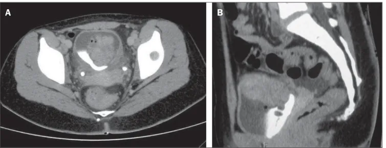
expansile, heterogeneous, lobulated lesion measuring 6.0 × 4.1 cm on the upper wall, with blurring of the adjacent fat (Figure 2). Freezing biopsy and tumor resection were performed. The surgical specimen measured 10.0 × 7.5 × 4.0 cm, was brown-ish colored and had a pedicle measuring 2.0 × 1.5 cm. Anatomopathological analysis confirmed IMT with areas of ulceration of the mucosa extending up to the lamina pro-pria and adjacent mucosa, presenting urethelial hyperplasia without atypias.
The postoperative period was unevent-ful. Two months later, pelvic magnetic resonance imaging demonstrated postop-erative fibrotic changes, with no sign of relapse. Follow-up with abdominal ultra-sonography did not demonstrate any change.
DISCUSSION
IMTs rarely affect the urinary tract and frequently are associated with trauma, in-fections or genitourinary tract instrumen-tation(1). Such tumors are most commonly found in adult individuals and rarely in children. A recent meta-analysis has iden-tified only 35 cases describing IMT of blad-der in this age range(4).
suggest the involvement of the chromo-some 2p23(2) and co-infection with
Myco-bacterium tuberculosis(3) in the IMT’s pathogenesis.
The authors report the case of a child ad-mitted to the Hospital Universitário and Hospital das Clínicas da Universidade de São Paulo with diagnosis of IMT of blad-der, focusing on radiological findings and the differential diagnosis of the disease.
CASE REPORT
A female, 13-year-old patient present-ing abdominal pain for one month, weight loss (6 kg) and macroscopic hematuria. The patient was in good general conditions, with paleness (++/4+), and painful abdo-men at palpation. Laboratory tests demon-strated normocytic/normochromic anemia (hemoglobin = 7.8 mg/dl), hematuria and leukocyturia, with negative uroculture. Unaltered leukogram and renal and hepatic function tests.
Ultrasonography revealed the presence of a heterogeneous mass measuring 6.0 × 3.0 cm on the vesical wall, with internal vascularization at Doppler (Figure 1). Computed tomography (CT) demonstrated diffuse vesical wall thickening and an
INTRODUCTION
231
Araújo Filho JAB et al. Inflammatory myofibroblastic tumor of the bladder in a child
Radiol Bras. 2012 Jul/Ago;45(4):230–232
IMTs of bladder may be locally aggres-sive, mimicking malignant neoplasms at cytoscopy and imaging studies, the latter being its main differential diagnosis. Al-though any vesical site can be affected, there is a subtle predilection for the upper vesical wall(5). Essential criteria for the anatomopathological diagnosis include proliferation of fusiform myoepithelial cells and lymphocytic infiltrate(2). Immuno-histochemical analysis may reveal positiv-ity for cytokeratin, vimentin and anaplas-tic lymphoma kinase (ALK)(2).
At imaging evaluation, both cystic and solid variants have already been reported,
although generally a single exophytic or polypoid mass is observed, almost always sparing the vesical trigone(6). At CT and ultrasonography, many lesions are ill-de-fined and invade circumjacent tissues, with internal vascularization identified at color Doppler(6). Large-sized lesions may present an extravesical component with difficult differentiation from malignant tumors. Generally, the lesion comprises necrotic tissue in the central region and fusiform cells in an edematous stroma with periph-eral vessels and inflammatory cells, which explains the ring-shaped contrast-enhance-ment at CT. At MRI, T2-weighted images
show hyperintense central area, with pe-ripheral low signal intensity, while bladder carcinomas generally present hyperintense signal. After contrast agent injection, the signal intensity increases significantly in the periphery while in the central region little increase is observed(6).
The treatment consists of surgical resec-tion, corticotherapy, radiotherapy or con-servative treatment. Relapse after resection is generally rare.
Given the considerable overlap of im-aging findings among IMT, rhabdomyosa-rcoma and leiomyosarhabdomyosa-rcoma, the anatomo-pathological differentiation is critical in the Figure 1.A: Pelvic ultrasonography demonstrating the presence of heterogeneous, lobulated mass on the vesical wall. B: Doppler mapping of a vesical mass, demonstrating arterial flow pattern within the lesion.
B
A
Figure 2.A: Axial computed tomography of pelvis after intravenous contrast injection (excretory phase) showing expansile, heterogeneous, lobulated lesion on the upper vesical wall, with heterogeneous enhancement and blurring of the adjacent fat. B: Sagittal computed tomography of pelvis after intravenous contrast injection (excretory phase) demonstrating diffuse bladder wall thickening and the mentioned expansile lesion.
232
Araújo Filho JAB et al. Inflammatory myofibroblastic tumor of the bladder in a child
Radiol Bras. 2012 Jul/Ago;45(4):230–232
presence of the above described findings to reduce the number of unnecessary radical surgeries.
REFERENCES
1. Lecuona AT, Van Wyk AC, Smit SG, et al. Inflam-matory myofibroblastic tumor of the bladder in a 3-year-old boy. Urology. 2012;79:215–8. 2. Yagnik V, Chadha A, Chaudhari S, et al.
Inflam-matory myofibroblastic tumor of the urinary blad-der. Urol Ann. 2010;2:78–9.
3. Androulaki A, Papathomas TG, Liapis G, et al. In-flammatory pseudotumor associated with Myco-bacterium tuberculosis infection. Int J Infect Dis. 2008;12:607–10.
4. Houben CH, Chan A, Lee KH, et al. Inflammatory myofibroblastic tumor of the bladder in children: what can be expected? Pediatr Surg Int. 2007;23: 815–9.
5. Kim H, Oh SN, Rha SE, et al. Inflammatory myofibroblastic tumor of the bladder: report of two cases. J Korean Soc Radiol. 2010;63:261–5. 6. Wong-You-Cheong JJ, Woodward PJ, Manning