IX
Radiol Bras. Jul/Ago;44(4):IX–X
Pedro José de Santana Júnior1, Jorgeana Milhomem Bandeira2, Ana Caroline Vieira Aurione3, Renato Duarte Carneiro4, Rubens Carneiro dos Santos Júnior5, Kim-Ir-Sen Santos Teixeira6
Study developed at Hospital das Clínicas da Universidade Federal de Goiás (UFG), Goiânia, GO, Brazil. 1. MD, Radiologist, Trainee at the Unit of Magnetic Resonance Imaging of Santa Casa de Misericórdia de São Paulo, São Paulo, SP, Brazil. 2. MD, Radiologist, Trainee at Unit of Magnetic Resonance Imaging of CRER – Centro de Reabilitação e Readaptação Henrique Santillo, Goiânia, GO, Brazil. 3. Gradu-ate Student of Medicine, Universidade Federal de Goiás (UFG), Goiânia, GO, Brazil. 4. Titular Member of Colégio Brasileiro de Radiologia e Diagnóstico por Imagem (CBR), MD, Radiologist at Multimagem Diagnósticos, Goiânia, GO, Brazil. 5. Master in Radiology, Professor, Department of Radiology, Hospital das Clínicas da Universidade Federal de Goiás (UFG), Head for the Unit of Magnetic Resonance Im-aging at Instituto de Neurologia de Goiânia, Goiânia, GO, Brazil. 6. PhD of Radiology, Associate Professor, Department of Radiology at Hospital das Clínicas da Universidade Federal de Goiás (UFG), Goiânia, GO, Brazil. Mailing Address: Dr. Pedro José de Santana Júnior. Rua T-36, nº 3485, ap. 104, Edifício Solar dos Tocantins, Setor Bueno. Goiânia, GO, Brazil, 74223-050. E-mail: [email protected]
0100-3984 © Colégio Brasileiro de Radiologia e Diagnóstico por Imagem
WHICH IS YOUR DIAGNOSIS?
Santana Júnior PJ, Bandeira JM, Aurione ACV, Carneiro RD, Santos Júnior RC, Teixeira KISS. Which is your diagnosis? Radiol Bras. 2011 Jul/Ago;44(4):IX–X.
A female, 67-year-old patients presented at the Hospital das Clínicas ambulatory – Universidade Federal de Goiás (HC-UFG) complaining of pain and paresthesia in the right lower limb, with progressive inten-sity over the last five months. The patient remained limited to the bed, in decubitus position with the af-fected limb in semi-flexed position. She denied the occurrence of trauma. At clinical examination, a pal-pable mass of about 15 cm in diameter, with undefined margins was found in the right flank. The patient reported previous history of uterine cervix carcinoma diagnosed in 2007. Abdominal radiography and com-puted tomography (CT) were performed at the Department of Radiology of HC-UFG.
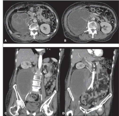
Figure 1. Abdominal radiography. Figure 2. Abdominal computed tomography, contrast-enhanced axial sections (A,B) and coronal
sec-tions (C,D).
A B
X Radiol Bras. 2011 Jul/Ago;44(4):IX–X
Images description
Figure 1. Abdominal radiography dem-onstrates fading of the psoas muscle out-line associated with opacity in the right hemiabdomen. Note the expansile effect characterized by displacement of bowel loops to the left and undefined ascending colon.
Figure 2. Abdominal computed tomog-raphy. Contrast-enhanced axial (A,B) and coronal (C,D) sections demonstrate an expansile, multiseptated, heterogeneous cystic lesion with peripheral iodinated con-trast uptake, located in the right iliopsoas compartment. Also, densification of adja-cent fat planes (on A,B,C,D) and displace-ment of the ipsilateral kidney (on C,D) are observed.
Diagnosis: Metastasis from squamous cell carcinoma of uterine cervix to the right iliopsoas compartment.
COMMENTS
The iliopsoas compartment may be in-volved by different pathological processes including infection, tumor and hemor-rhage. The patients may present several nonspecific signs and symptoms, causing delay in the diagnosis. The imaging assess-ment of such compartassess-ment, particularly by means of CT has been a relevant landmark in the diagnosis of such lesions. However, it is important to note that radiological find-ings may be common to several etiologies, which most of times.
The involvement of the iliopsoas com-partment is usually secondary to a direct ex-tension of adjacent tumors of retroperito-neal, abdominal, pelvic, neuronal, osseous and lymphonodal origin(1,2). Primary tu-mors of the iliopsoas muscle (liposarcoma, fibrosarcoma, leiomyosarcoma and hemangiopericytoma) are rarely found. The retroperitoneal fascial planes do not offer a barrier to tumor dissemination, with di-rect and random invasion in contrast to the inflammatory/infectious involvement(3). At CT and magnetic resonance imaging, le-sions with either homogeneous or
hetero-geneous appearance are observed as a func-tion of the presence of necrosis, hemor-rhage and alterations in the cellular struc-ture(3). Presence of hypoattenuating area, irregular margins, adenopathy, bone de-struction and discontinuity of fascial planes represent the most significant findings in the distinction among tumors, abscesses and hematomas(2). In the series described by Muttarak & Peh(1), among the 14 re-ported cases of tumor, there was secondary involvement in ten, with metastasis from uterine cervix carcinoma being the most common origin, similarly to the present report.
The infectious involvement of the iliop-soas compartment may be primary or sec-ondary. Primary abscesses are rare and gen-erally idiopathic. Staphylococcus aureus
and gram-negative microorganisms are the organisms most frequently involved. Immunocompromised patients, especially those undergoing corticotherapy, chemo-therapy, as well as HIV-positive patients are particularly predisposed to infection(1,3). The secondary involvement of the iliopsoas compartment is much more frequent, gen-erally resulting from dissemination of in-fectious processes originated in kidneys (perinephric abscesses), bones (osteomy-elitis and tuberculosis), and in intestinal loops (appendicitis, diverticulitis, Crohn’s disease and perforated colon carcinoma(1). The most common tomographic findings are enlargement of the iliopsoas muscle with hypoattenuating Center, as well as parietal contrast enhancement. Such signs are nonspecific and non-distinguishable from metastases and lymphoma. Secondary findings that may corroborate such diagno-sis include densification in adjacent fat planes, bone destruction and intermingled gas bubbles.
Iliopsoas compartment hematomas may be either spontaneous or secondary to hem-orrhagic diathesis, anticoagulant therapy, trauma, tumor, recent surgery or biopsy, or even resulting from extension of bleeding in adjacent organs and vessels(1,2). As causal factor and patient’s age were
corre-lated, most noticeable association with coagulopathy and trauma was observed in the youngest age range (fourth decade of life), and with aortic aneurysm rupture, anticoagulant therapy for arteriosclerotic disease and thromboembolism in the most advanced age range (seventh decade of life)(3). Acute hemorrhage is seen as a spon-taneously hypoattenuating lesion. Fluid-fluid level may be present because of he-matocrit effect. It is important to highlight that chronic hematomas are hardly differ-entiated from abscesses and tumors. Percu-taneous aspiration is useful in such differ-entiation.
Based on the present case, it is possible to observe the critical role of CT in the de-termination of the involvement of the ili-opsoas compartment by the lesion. At one end, one observes the difficulty in deter-mining which radiological findings are suggestive of disease in such location. In a high number of cases, such findings are insufficient to determine the disease etiol-ogy. On the other hand, the diagnostic ac-curacy significantly increases as radiologi-cal findings are associated with cliniradiologi-cal data. In the present case, the patient was submitted to laparotomy with biopsy of the retroperitoneal mass radiologically identi-fied, with a result compatible with a poorly differentiated infiltrating squamous cell carcinoma and extensive areas of necrosis. Hence the significance of the present re-port, where the previous history of invasive squamous cell carcinoma of uterine cervix, in association with imaging findings may favor the diagnosis of metastatic lesion in the iliopsoas muscle.
REFERENCES
1. Muttarak M, Peh WCG. CT of unusual iliopsoas compartment lesions. Radiographics. 2000;20: S53–66.
2. Leão ARS, Amaral RPG, Abud TG, et al. Patolo-gias do compartimento iliopsoas: avaliação radio-lógica. Radiol Bras. 2007;40:267–72.