www.jcol.org.br
Journal of
Coloproctology
* Corresponding author.
E-mail: [email protected] (J.A. Reis Neto).
2237-9363/$ - see front matter. © 2014 Sociedade Brasileira de Coloproctologia. Published by Elsevier Editora Ltda. All rights reserved. http://dx.doi.org/10.1016/j.jcol.2013.11.001
Original article
Laparoscopic treatment of acquired megacolon
Oliveira LH
a, Banci SO
a, Kagohara O
a, Simões Neto J
a,b, Reis Junior JA
a,b, Reis Neto JA
a,*
a Sociedade Brasileira de Coloproctologia, Rio de Janeiro, RJ, Brazil
b Pontifícia Universidade Católica de Campinas (PUC-Campinas), Campinas, SP, Brazil
a r t i c l e i n f o
Article history:
Received 21 September 2013 Accepted 25 November 2013
Keywords:
Obstipation Constipation Acquired megacolon Laparoscopy Duhamel surgery
Mechanic colon-recto-anal anastomosis
a b s t r a c t
In spite of the large experience acquired in the last 50 years with the surgical treatment of the Chagasic megacolon, the use of colorectal video laparoscopic surgery brought some controversy in several aspects of the treatment that already had been considered as re-solved. One of the basic aspects to the establishment of the colorectal video laparoscopic surgery is to maintain the same procedure of the conventional surgery, since the results obtained in this operation were considered as curative. Constipation is only a symptom of a multisymptomatic disease, and the surgical treatment of acquired megacolon must be considered as dei nitive in the cure of this symptom; recurrence of the constipation or dilatation after a short period of time must be considered deleterious to the patient. Based in 41 years of experience with the Duhamel procedure in the treatment of 912 patients with acquired megacolon, the authors propose to apply the same technique in the surgical laparoscopic approach of acquired megacolon, including the same colon-recto-anal anas-tomosis. The results obtained in 56 patients operated on by laparoscopic approach showed the same curative results, but with lower morbidity.
© 2014 Sociedade Brasileira de Coloproctologia. Published by Elsevier Editora Ltda. All rights reserved.
Palavras-chave:
Obstipação Constipação Megacolo adquirido Laparoscopia Cirurgia de Duhamel Anastomose cólon-reto-anal mecânica
r e s u m o
Tratamento laparoscópico do megacolo adquirido
que possibilite ao paciente livrar-se deinitivamente de um sintoma, visto que é possível que em curto espaço de tempo ele venha a necessitar tratar outra manifestação sintoma-tológica (cardíaca ou esofágica) da enfermidade causal. Baseados na experiência adquirida nos últimos 50 anos (912 pacientes) com a técnica de Duhamel, em que o ponto importante é a realização de uma ampla anastomose da parede anterior do cólon abaixado à parede posterior (mucosa) do reto, ao mesmo tempo em que se anastomosa a parede posterior do cólon abaixado ao canal anal, são analisados os resultados obtidos com esta mesma técnica realizada por laparoscopia. Esta mesma incisão no canal anal serve para a retirada do seg-mento cólico ressecado, sem necessidade de laparotomia auxiliar. Os resultados observa-dos em 56 pacientes quanto à cura da obstipação são similares aos registraobserva-dos na cirurgia convencional, porém com um menor índice de morbidade, seja intra ou pós-operatória. .
© 2014 Sociedade Brasileira de Coloproctologia. Publicado por Elsevier Editora Ltda. Todos os direitos reservados.
Introduction
Despite the vast experience gained over the last 50 years with the surgical treatment of acquired megacolon, the introduction of laparoscopic surgery brought some con-troversy in some points that already had been considered resolved.1-22
One of them refers to the signiicance of lack of synchroni-zation of sigmoid-rectal contraction in the genesis of acquired megacolon, incoordination which is responsible for the symp-tom of constipation and by the pathological alteration of dila-tation and elongation of viscera.8,10,20,23-30 This point is crucial to the understanding of the surgical treatment, because the residence of the rectum in the intestinal path results in recur-rence of megacolon, whether in the short, medium or long term.9-22,31
Another important point is about the need to lower a “macroscopically normal” colon in the sake of the colon-anal anastomosis procedure. The colon is called “macroscopically normal” because is common knowledge, and experimental evidence reveals, that microscopic mioenteric lesions occur universally in a Chagasic colon.23,24,26-28,32,33
Early studies of Bernardes35 and Reis Neto and Cunha15,36 in 1963 introduced in Brazil and Latin America the proposi-tion of using the Duhamel technique for surgical treatment of acquired megacolon. The results obtained proved that the surgery solves the problem of the symptom of consti-pation, although without resolution of Chagas disease. With the advent of laparoscopic surgery, this technique began to be used in a more frequent basis,15,18,21,22,30,31 mainly for the ease of retro-rectal detachment from pneumoperitoneum and the possibility of colonic resection by a perineal route, without opening the abdominal cavity for removal of the colon.15,18,21,31
However, to facilitate the operation and in order to pre-vent the lowering of the colon, some authors have chosen to conduct a posterior colorectal anastomosis with circular mechanical suture, with the colon in the posterior wall of the rectum. In some patients, this is done just above the dentate line; in others, 4-5 cm above this line; and, in some cases, at the level of peritoneal relection or even immedi-ately above it.37-42
This work aims to present the results obtained with Du-hamel surgery performed laparoscopically for the treatment of constipation arising from acquired megacolon, using abso-lutely the same technique and tactics used in the convention-al surgery. Based on the experience acquired over the last 50 years with 912 patients operated on with the Duhamel tech-nique, in which the important point is the accomplishment of a wide anastomosis of the anterior wall of the lowered colon to the posterior wall (mucosa) of the rectum and, at the same time, with the anastomosis of the posterior wall of the low-ered colon to the anal canal is performed. We must stress the absolute necessity of maintaining this anastomosis to obtain the cure of constipation.
Patients and methods
The results obtained in 56 patients with acquired megacolon operated laparoscopically between 1993 and 2013 were ana-lyzed. Of the patients, 31 (55.3%) were female and 25 (44.6%) were male. The average age was 56 years (23-72 years). Of these patients, 21 (37.5%) were operated as a technical dem-onstration in specialization courses of colorectal laparoscopic surgery, both in Brazil and in Latin America, or in live demon-strations in Conferences of the specialty.
No patient in this series had concomitant megesophagus. Four patients (7.1%) exhibited cardiac abnormalities compat-ible with Chagas disease.
The bowel preparation was in accordance to the protocol used in conventional surgery:
• 48 hours before surgery, high caloric liquid diet, which will continue until the day before surgery;
• 24 hours prior to surgery, anterograde bowel preparation with mannitol 10%;
• 3 enemas of phospho-soda (Fleet enema*) applied respec-tively 24, 20 and 16 hours before surgery;
• Two rectoscopies, one the night before surgery and the other immediately before surgery to evaluate the condi-tions of rectal ampulla cleaning and to promote vacuum-ing of possible gas or secretions;
• hair shaving and urinary catheterization in the operating room.
Operative technique (laparoscopic surgery)
After being anesthetized, the patient is placed in the lithot-omy-Trendelenburg position, with concomitant exposure of abdomen and perineum.
The pneumoperitoneum is held in the left upper quad-rant with Veress needle or Hasson trocar, depending on the conditions of the abdomen. The average pressure of CO2 dur-ing surgery should be maintained between 11 and 13 mm Hg. The endoscope, generally at 30°, is inserted in the left up-per quadrant, into a trocar of 10-12 mm.
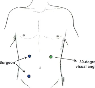
Two other trocars 10-12 mm are placed into the right hy-pochondrium and right iliac fossa, both to be used by the surgeon. In general, these trocars are sufi cient to complete the surgery. However, a fourth trocar of 10-12 mm may be placed into the left iliac fossa for use by an assistant as needed (Fig. 1).
Abdominal phase
• In female patients the histeropexia, with elevation of the uterine body to the abdominal wall, is important for an ad-equate exposure of the pelvis and of the posterior fornix. This is done by the introduction of a straight needle trans-i xtrans-ing the abdomtrans-inal wall and that trans-is setrans-ized by the surgeon under direct vision. The uterine leal et is pierced by the needle, and that raises the round ligaments, pinning them to the abdominal wall;
• an incision of the parietal leal et on the inner face of the mesosigmoid with identii cation of the left ureter and of inferior mesenteric artery and its branches. This maneu-ver is facilitated by a forced Trendelenburg and a small right lateral decubitus position;
• left parietal colon detachment, mobilizing the entire left hemicolon to the splenic l exure; when necessary, mobili-zation of the splenic l exure;
• opening of the pelvic peritoneum to the patient’s right and detachment of retro-rectal space with scissors to the level of ano-recto-coccygeal ligament;
• identii cation of the colon to be lowered and of the point where the intestinal section will be performed. Ligation of the marginal arcade of Drummond from this point; usually is sufi cient the ligation of the superior rectal artery and the last branch of sigmoid artery;
• an analysis of the degree of mobility of the colon: to test the length of the loop to be lowered, evaluating if the loop goes, without stress, to the anal canal;
• introduction of a rectal probe in order to aspirate possible intestinal residues, especially gas, thus reducing the diam-eter of the colon and rectum;
• opening of the perirectal fat at the level of the peritoneal rel ection, to isolate the rectal ampulla and facilitate its section. Ligation of the ascending rectal arteries with met-al clips. This dissection should completely isolate the rec-tal ampulla, including the peritoneum on its anterior wall, leaving exposed only the muscle layer.
• section of the rectal ampulla at the level of the peritoneal rel ection with use of a linear endostappler; two or more shots are often needed, in view of the diameter of the rec-tal ampulla;
• isolated sutures covering the sectioned rectal ampulla with the peritoneum of the posterior fornix (2-4 sutures are sufi cient).
Perineal phase
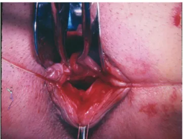
• exposure of the anal canal and pectinate line with Collins' (or similar) valve. If the retrorectal detachment was com-plete, the mucosa is stretched by CO2 and this facilitates the dissection (Fig. 2).
• posterior semicircular incision, 1 cm above the dentate line; and dissection in the plane between the mucosa and inter-nal sphincter to the Milligan and Morgan ring (elevators’ ring). At this point the retrorectal space is reached, with possibility of an abrupt elimination of all the CO2 gas from the cavity. This is sufi cient reason to proceed with caution and block this dissection with a small pledget (Fig. 3);
Fig. 1 – Position of the trocars in Duhamel surgery by
laparoscopic approach. Fig. 2 – Exposure of the anal canal and dentate line.
Surgeon 30-degree
• introduction of a long forceps through the channel to-ward the abdomen. The surgeon identii es the clamp and applies the device on the same loop that was sectioned at the level of the peritoneal rel ection (this is the seg-ment of colon to be lowered and that will be resected by a perineal route);
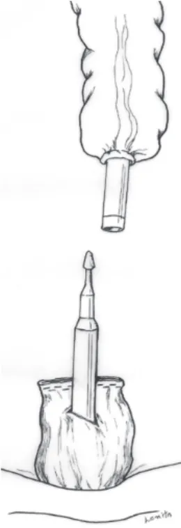
• carefully and slowly, this segment of the colon is pulled up to the perineum, through the retrorectal space be-tween the rectal mucosa and the internal sphincter. The entire loop to be ressected is exposed, and the demarcat-ed area is resectdemarcat-ed (Fig. 4 A and B). The fact of the colonic contents have been aspirated, with the colon collapsed, facilitates the passage of the organ through the space de-scribed;
• after the remotion of the previously marked colonic seg-ment, the posterior hemi-circumference of the lowered colon is anastomosed to the mucosa of the anal canal at the place where the surgeon began dissecting the muco-sa. This anastomosis is performed with interrupted
su-tures (polivycril 0000) between the seromuscular of the lowered colon and the mucosa and anal sphincter (Fig. 5);
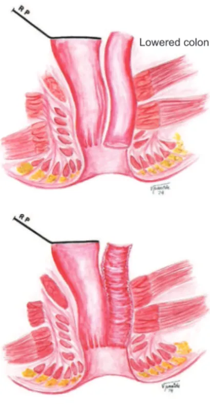
• the anterior hemicircunference of the lowered colon is anastomosed to the posterior rectal wall by means of a linear stapler of 7.5 cm (Fig. 6);
• closure of sites of introduction of the three abdominal trocars.
Results
To evaluate the results of surgery, especially to compare with the results of conventional surgery, some characteris-tic postoperative features of the intervention were observed, namely:
• Fluid resuscitation
• Feasibility of the lowered colon
• Colon-rectum-anal anastomosis patency
Fig. 3 – Exposure of the internal anal sphincter. The rectal mucosa is lifted by two clamps. At this point, CO2 gas
exhaustion from the abdominal cavity occurs.
Fig. 4 – A, The distended loop, to be desiccated, exposed in the perineum. The loop came down the rectorectal space and traversed the area between the rectal mucosa and the internal sphincter. No matter the size of the loop, as long as it is syllabled, it will pass through this space without great diffi culty. B, The fragment removed by the perineal route and infl ated with air.
Fig. 5 – Anastomosis of the lowered colon and anal canal (posterior wall of the lowered colon and posterior hemicircunference of anal canal) via interrupted sutures. The anterior rectal wall (mucosa) and the anterior wall of the lowered colon are demonstrated by anatomical forceps.
Fig. 6 – Latero-lateral anastomosis between the anterior wall of the lowered colon and the posterior wall of the rectum with a linear stapler device.
• Dehiscence of rectal ampulla closure
• General complications
In no patient of this series a postoperative blood replace-ment was required, either in the intra- or postoperatory pe-riod.
No case of necrosis of the lowered colon or of infection of pre-sacral space was observed. In 5 (5.8%) patients, a release of the splenic lexure was needed.
The operating time ranged from a minimum of 95 min-utes to a maximum of 240 minmin-utes, with an average of 142 minutes.
The closure of the rectal ampulla was performed with a green-loaded mechanical stapler. The number of applied loads varies according to the diameter of the rectum, from a minimum of 2 to a maximum of 4 loads. However, no dehis-cence of the rectum, even in patients treated with the maxi-mum number of loads, was observed. The gas elimination time ranged from one to three days, and the faecal elimina-tion time ranged from one to six days.
The food was reintroduced after 1-3 days, with an average of 1.7 days.
The hospital stay ranged from a minimum of two days to a maximum of 11 days, with an average of 3 days.
All colon-rectum-anal anastomoses were considered per-vious, being crossed with ease both by digital examination as by endoscopy.
Complications
1) Intraoperative: there was no alternance to laparotomy sur-gery;
2) Immediate postoperative period: two female patients (3.5%) had postoperative urinary infection;
3) Mediate postoperative period: two patients (3.5%) had re-sidual plicoma and one (1.7%) had mucosal prolapse. One patient (1.7%) developed temporary partial incontinence (latus and liquid stool) for four months, being asymptom-atic after this period of time.
Late progression
Of the patients studied, 21 (37.6%) were not followed by the surgical staff (these patients were operated during courses or meetings). The follow-up ranged from a maximum of 10 years to a minimum of four months, and 35 (62.5%) patients had a median follow-up of 10 years. The contrast radiographic study (barium enema) was performed in 35 (62.5%) patients; the colon-rectum-anal anastomosis patency and a lowered colon of normal caliber were demonstrated. The contrast ra-diographic study (bowel movements) was performed in two patients (3.5%), demonstrating that the contrast enters into the rectal ampulla, however, with total elimination after 48 hours. Rigid sigmoidoscopy was performed in 35 (62.5%) pa-tients three months after surgery on average, demonstrating colon-rectum-anal anastomosis patency.
Discussion
It would be dificult to understand the dilation of a hollow muscular viscera, in the absence of an organic obstacle down-stream, without the existence of a dynamic pathology, that is,
without the understanding that the inadequacy of the pro-pulsive movements of the colons may cause a delay of the faecal movement, leading to stasis and a consequent dila-tion.23,26-28,32-34,44
The complex coordination of these colonic movements has been extensively studied and only an exact notion of the physiological side may lead to the understanding of the pathological side, i.e., to understand whether the megacolon present is of the acquired type is necessary to understand the pathophysiology of the disease.45,24,25
Since Adler45 established the concept of segmental func-tional unit, it is believed that two adjacent segments represent a functional unit when a proximal contraction is followed by another similar contraction distally, thereby enabling the pro-pulsion of intestinal contents.
If the contraction of the proximal segment is not integrat-ed or coordinatintegrat-ed with the distal activity, and if an uncoordi-nated and disorganized contraction occurs, the faecal propul-sion is not processed.
These propulsive movements observed in the various co-lon segments consist of waves of contraction in an aboral di-rection, generating a long functional segment in opposition to the true peristalsis; these contractions are preceded by a zone of increased pressure, usually caused by gas.
However, this wave of contraction, which takes place at a speed of 25 mm/sec, stops when it encounters a colon region already contracted. The contractions observed in normal co-lons are isotonic (no change in intraluminal pressure), which allows a free movement of the intestinal contents. This con-tractile activity is coordinated by the intramural plexuses; in-jury or absence of such complexes leads to a failure in the transmission of the contractile wave.45
Studies of the sigmoid-rectal motility showed that the nor-mal activity of the two segments is independent and that this distinct intersegmental activity constitutes the motor coordi-nation.24,25 The increased activity of the rectum is a functional barrier to overdistension of this organ.39
The surgical treatment of acquired megacolon evolved with the progress of this physiological knowledge and with the etiopathogenic and pathophysiological understanding of the disease.1-21,46
Histological and electromanometric studies represented an extremely important contribution to the establishment of the effective foundations of the modern surgical treat-ment.24,25,29
The histologic study demonstrated denervation of the myenteric plexus in all colon segments, due to an inlamma-tory process that ends up by causing destruction of ganglion cells.23,26-28,32-34,44 As a consequence of this destruction, changes in motility emerge, especially in the sigmoid-recto-anal seg-ment, compromising the synergy of coordination of move-ments of muscle contraction, making dificult the progression and expulsion of faecal matter.24 The sigmoid-rectal interseg-mental activity changes, and the wave of relaxation following contraction ceases to exist.24
Hyper-motility, hyperexcitability and a consequent motor incoordina-tion occur.
This lack of pace of colonic contractions causes muscle hypertrophy, due to over-effort compensating the lack of co-ordination. Faecal matter stagnation and dilation of the loop occur.10,12,26-28,44,46
Originally, the surgeries were limited to the resection of the dilated segments, establishing a link between the treatment of acquired megacolon and Hirschsprung disease.1-4 It was soon recognized that the segment responsible for the onset of dila-tion was the rectosigmoid and that, therefore, this would be the segment to be removed to cure the bowel movement symp-toms.2,3,44
Only since 19532 retossigmoidectomy became the operation of choice for the surgical treatment of acquired megacolon. Initially, however, the colorectal anastomosis was practiced exclusively by the abdominal route, resulting in high rates of complications, mainly anastomotic dehiscence.3,47
The changes introduced by a “delayed” colorectal anasto-mosis diminished considerably these complications.3,4 How-ever, the pelvic dissection of the rectum remained an obsta-cle to surgical management, particularly in less experienced hands.1,15,36,47 This motivated the search for a surgical alterna-tive that would avoid or diminish the risks inherent to this dis-section.
In 1963 Bernardes35 and Reis Neto and Cunha,15,36 in con-current studies, introduced in Brazil and Latin America the proposition of using the Duhamel surgery for the surgical treat-ment of acquired megacolon. The initial results were gradually turning to one certainty – of an adequate surgical proposi-tion.5-20,31,35,36,46,48-59
The use of the stapler, eliminating the need for a colostomy perineal, brought the inal elements to the almost universal ac-ceptance of this technique, as the best proposed surgical treat-ment of acquired megacolon in our days.16,17,20,21,46,58,59
The great advantage of this surgery is that while it carries out a colon-rectum-anal anastomosis, eliminating the segment responsible for the lack of motor coordination, also eliminates the need for a large pelvic dissection.9-17,21,31,36,48-53,57-59
Undoubtedly this colon-rectum-anal anastomosis is the im-portant detail of the surgery, since it excludes the sigmoid-rec-tal segment with respect to the faecal transit.12,15-21,31 By allow-ing the faecal transit directly into the inferior rectus segment, the colon-rectum-anal anastomosis maintains continence, while excluding the segment of the superior rectus responsible by the dyskinesia and subsequent faecal retention.60-62
The use of laparoscopy in the treatment of acquired mega-colon, with mechanical colon-rectum-anal anastomosis and removal of colonic segment through the anal canal, was intro-duced in 1994,17,31 bringing great beneit to the patient, especial-ly in terms of period of hospitalization and post-operative pain. However, despite of being a widespread technique, several studies have shown technical and tactical variations, and there is no uniformity of thought with respect to certain aspects.
The most important controversial issues to be discussed are:
1) anastomosis of the colon to the posterior wall of the rec-tum or anal canal?
2) removal of resected colon by abdominal incision or by the anal canal?
3) what segment of the colon should be preserved for anas-tomosis?
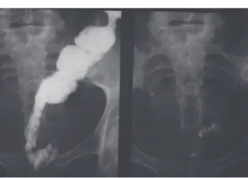
The most relevant fact is a higher anastomosis of the colon to the upper rectum segment, which could lead to dyskinesia, faecal retention and relapse of the symptoms of constipation. Radiologic studies of the isolated rectum after Duhamel operation, with colon-rectum-anal anastomosis, proved that there is a passage of stool through the rectum, but without faecal stasis (Figs. 7 and 8).12-14,18-21,52,56
The comparative study between the techniques of Du-hamel and of DuDu-hamel-Haddad.7 showed that both the direct anastomosis of the lowered colon to the anus as the delayed technique lead to the resolution of the symptoms of obstipa-tion, thanks to the exclusion of the sigmoid-rectal segment,
Fig. 7 – Intestinal transit held in patient operated for acquired megacolon by Duhamel technique. It can be seen that the contrast enters the lower rectum, being totally eliminated without retention.
by carrying out the anastomosis to the level of inferior rec-tum.
The comprehensive analysis of the mechanical end-to-lateral colorectal anastomosis constructed on the posterior wall of the pelvic and/or abdominal segment of the rectum, recently proposed mainly for the laparoscopic operation of Duhamel, led to the following conjectures (Fig. 9):
1) the anastomosis is performed between two thick walls (following the muscular hypertrophy of colon and rec-tum walls), which may hinder the overall penetration of the staples in the mechanical anastomosis;1,9,23,25,37,41,53,57 2) this anastomotic stenosis has a high incidence. In a
pre-liminary study, upon the analysis of the results obtained in 20 patients operated with this type of anastomosis by a conventional route, an incidence of 30% of stenotic complications (6 patients) was found in the short and medium terms.37-39 This was the irst published trial re-garding this type of anastomosis. The high rate of com-plications observed in this experiment led to a modii-cation of the technique: after the circular anastomosis, its ampliation with a mechanical longitudinal suture is performed.39
3) the anastomosis is performed far above the pectineal line; in some cases, depending on the surgeon's experi-ence, to 6 or more centimetres above the anal margin – thus, almost in the upper rectus. If the rectum is resect-ed above the peritoneal relection, this anastomosis may be situated in the upper rectus, with greater possibility of recurrence of constipation and megacolon, according to the pathophysiology of this disease.2,3,10,12,24,44,48 Ra-diological and electromanometric studies to assess the long-term behaviour of the rectal ampulla and the pos-sible incidence of recurrence of dyskinesia are scarce.
4) the cost-beneit analysis of this type of anastomosis in relation to longitudinal colon-rectum-anal anastomosis, both mechanical procedures, must also be taken into ac-count, even considering the results as similar: the circu-lar anastomosis is more expensive than the longitudinal anastomosis.
To inalize this really complex and controversial issue about the best type of anastomosis (circular or longitudinal) and how high it should be performed (posterior wall of rectum, or anal canal), some considerations should be taken into account with respect to the longitudinal colon-rectum-anal anastomosis:
1) the anastomosis is performed between the rectal mu-cosa and the lateral wall of the lowered colon; therefore, between two walls of lesser thickness, which makes more precise and eficient the perforation by metal sta-ples;12-14,52,53,55-57
2) the longitudinal anastomosis measures 7 centimetres long, with a minimum indice of stenosis and zero dehis-cence.12-14,52,53,55-57 In 198916,58 the irst results obtained in 87 patients with acquired megacolon and operated between 1981 and 1989 by conventional approach with colon-rec-tum-anal anastomosis with longitudinal mechanical sta-pler (type PLC50) were presented. This group of patients was assessed solely on the conditions of the mechanical colon-rectum-anal anastomosis, in the short and medium terms. The evaluation analysis was performed using digi-tal touch, proctoscopy and radiological examination. The digital touch and the proctoscopy were performed at the time of hospital discharge and in postoperatory ambula-tory visits at 30, 60 and 90 days. The anastomosis was con-sidered patent in all 87 patients examined. In no patient anastomotic leakage was found. Radiological studies (bar-ium enema, or intestinal transit) were performed in only 13.8% of patients. Five patients were examined by barium enema and seven by intestinal transit. All were examined after more than ive years of surgery. All patients evaluated by radiological examination showed a patent anastomosis (Fig. 10 A and B);
3) the anastomosis goes straight to the anal canal, preserving the inferior rectus. The long-term follow-up reveals nor-mal continence (Fig. 10 A and B).12-14,52,53,55-57
4) the internal sphincter should be preserved in the dis-section of the anal canal; the surgical plan should be lo-cated between the rectal mucosa and internal sphincter (Fig. 3). The initial studies on the use of the surgery of Duhamel15,35,36 for the treatment of acquired megacolon exhibited distinct characteristics, exactly in this dissec-tion: according to Bernardes6 the resection of the internal sphincter, keeping the original proposition of Duhamel,63 was essential for the cure of dyskinesia, while for Reis Neto15,36 the preservation of that structure, in addition to not interfere with the recurrence of the disease, is essen-tial for a proper continence (modiication of Grob64 for the Duhamel technique). In the long term, it was found that the resection of the internal sphincter is inconvenient and unnecessary.5,10,12,48,65
5) the cost-beneit analysis is favourable to the anastomosis with the longitudinal stapler;
6) in experienced hands, the colon-rectum-anal anastomosis can be performed manually, with excellent results.5,6
As the resection of the colon, the great advantage of the colon-rectum-anal anastomosis lies in the possibility of re-moval of the colonic segment to be resected by perineal route, without the need of an abdominal incision. The colon passes with ease through the retro-rectal space, being exteriorized through the perineal incision in the anal canal.
This maneuver, used to remove the colonic segment to be resected, has some advantages: it allows the identii ca-tion of the exact point of colon secca-tion, identifying the vi-ability of the colon; it allows that the surgeon, by an abdomi-nal approach, control the tension on the margiabdomi-nal arcade of Drummond, preventing manoeuvres that lead to improper stretching thereof; and avoids an additional abdominal inci-sion (Fig. 4).
Because the colon is distended, the introduction of a rectal probe during surgery, with suction of the gaseous contents, allows the proper emptying of the colon and eases its re-moval by perineal approach. In order to bring the colon to the perineum, with transposition of the tunnel between the rectal mucosa and the internal sphincter without causing vascular
injuries, the segment must be tractioned through the anterior wall and not through meso.
The third controversial aspect refers to the segment to be lowered: the authors disagree about the routine mobilization of the splenic l exure, about which segment must be lowered, and about the length of the colon to be resected.
When mobilizing the colon and choosing the segment to be lowered, some considerations should be taken into ac-count:
• the plexus injury is universal and similar throughout the length of the colon;23,25,32-34,38,41,65
• although the diameter of the colon may appear normal, a plexus lesion is present;23,25,32-34,38,41,65
• the cause of constipation and colonic dilatation is ex-actly this plexus injury, which compromises the synergy of movement coordination of muscle contraction, estab-lishing the lack of coordination between sigmoid and rec-tum;23,25,32-34,38,41,65
• the removal of the rectum off the faecal path elimi-nates the symptom of constipation and dilata-tion.1,2,5,7-15,24-27,29,35,36,40-43,47,49-57,60-62,64,65 Clinical observations have shown that a simple colostomy, when performed for the treatment of necrotic volvulus of sigmoid, causes the colon to regain a normal diameter;
• In open surgery, the lowering of a segment of dilated co-lon leads to the cure of constipation without recurrence of symptoms, even with long-term follow-up;5,12,26,27,51,53,55-57
• the study of the marginal arcade must determine in whose patient the ligation of the inferior mesenteric artery and the mobilization of the splenic l exure are necessary.3 There are elongated colons (dolicocolons) in which the lowering of the sigmoid, with the removal only of the rec-tus abdominis and of a short segment of sigmoid, is suf-i csuf-ient to cure the constsuf-ipatsuf-ion.5,12,26,27,51,53,55-57
Although extensive, all these commentaries on the results of the surgical treatment of acquired megacolon with the technique described by Duhamel, in particular the restoration of intestinal transit with the colon-rectum-anal anastomosis, are important to analyze the type of technique to be used in videolaparoscopy.21
One of the basic rules for the introduction of videolaparos-copy in the treatment of colorectal diseases has been to keep the original technique used in surgeries by the conventional ap-proach, provided that the results observed with the same tech-nique lead to the cure of symptoms or of the causative disease. In particular, with regard to the surgical treatment of ac-quired megacolon, the surgeon should keep in mind (in the proposal of the surgical treatment) that unlike what happens with the surgery for the treatment of other diseases, benign or malignant, in this case the objective is not the causal treat-ment of the disease, but essentially the cure of the manifesta-tion of one of its symptoms.
Therefore, it is really important to consider a treatment that will not result in good results just for a short time, but that will allow the patient to get rid dei nitely of a symptom, since it is possible that in the near future he (or she) may present other relevant symptoms of Chagas disease, when a second surgical procedure would be in order.
Fig. 10 – Schematic representation of the colon-rectum-anal anastomosis. A, The lowered colon goes down the retrorectal space to the channel and then through the dissected tunnel between the rectal mucosa and internal sphincter. B, Upon completion of the latero-lateral anastomosis between the anterior wall of the lowered colon and rectal mucosa and of the end-to-end anastomosis between the anal canal and the posterior wall of the lowered colon, a patent anastomosis is obtained, communicating the colon with inferior rectum and anal canal.
Lowered colon
Likewise, the option should be in favour of a conduct that may come to permanently cure the constipation, albeit with fewer complications.
In proposing the current approach for laparoscopic treat-ment of acquired megacolon, there was an extremely solid and scientiic basis of over 40 years of using the same conventional approach.21
Although the number of cases treated by laparoscopy is less than 10% of the experience acquired with the conventional surgery, similar results were obtained with respect to the cure, but with fewer complications. One must consider, however, that in recent years the number of patients with Chagas dis-ease and megacolon shows a tendency to fall.
In this series of patients operated by laparoscopy, no cases of dehiscence of closure of the rectal ampulla, as well as of low-ered colon necrosis, were observed. There was no alternance of laparoscopic surgery for the so-called conventional surgery.
In all patients the colon-rectum-anal anastomosis was con-sidered patent. A temporary partial incontinence occurred in one (1.78%) patient, with restoration of normalcy in the short term (four months).
R E F E R E N C E S
1. Cardoso AA. A retossigmoidectomia no tratamento do megacólon chagásico. Rev Goiana Med 5:103-127,1959. 2. Cutait, DE. Tratamento do megassigma pela
retossigmoidectomia. Tese Livre-Docência, USP, 1953. 3. Cutait, DE. Megacolo. Nova técnica de retossigmoidectomia
abdominoperineal sem colostomia. Anais I Cong ALAP vol 2: 831-834, 1960.
4. Cutait DE, Figliolini FJ. Megacolo adquirido: nova técnica de anastomose colorretal na retossigmoidectomia abdômino-perineal. Rev Paul Med, vol. 60:447-458,1962.
5. Lins Neto MAF. Operação de Duhamel modiicada com anastomose colorretal imediata para o tratamento do megacólon chagásico: técnica e resultados. Tese Mestrado, USP, São Paulo, 1997.
6. Lins Neto MAF, Cansanção CLC, Farias LRC. Anastomose colorretal imediata na operação de Duhamel. Rev Bras Coloproctologia, vol 8:14-16, 1988.
7. Medeiros RR, Reis Neto JA, Leonardi LS, Pires AM, Accorroni ME. Estudo comparativo entre as técnicas de Duhamel e Duhamel-Haddad na cirurgia do megacólon adquirido. Rev Paul Med, set-out vol. 96(3/4):61-65, 1980.
8. Moreira H. Contribuição ao estudo da isiopatologia no
tratamento cirúrgico do megacólon chagásico. In Patologia
Colorretal, Manzione A. ed Kronos, São Paulo:243-250, 1974. 9. Moreira H. Tratamento cirúrgico do megacolo chagásico
pela técnica de Duhamel-Haddad. Aspectos técnicos. In.
Atualização Cirúrgica. Pinotti HW, ed Robe, São Paulo, vol VII:161-182,1982.
10. Moreira H. Bases isiopatológicas para o tratamento cirúrgico do megacólon chagásico. Rev Gioana Med, 32:73-78, 1986. 11. Moreira H, Rezende JM. Megacolo Chagásico. Clínica,
diagnóstico e tratamento. In Coloproctologia. Conceitos.
Moreira H, ed. Escaleno, Goiânia: 15-60, 1993.
12. Reis Neto, JA. Contribuição ao tratamento cirúrgico do megacolo adquirido. Emprego do abaixamento retro-retal e trans-anal do colo. (técnica de Duhamel). Tese Doutorado, Unicamp 1968.
13. Reis Neto, JA. Resultados tardios da operação de Duhamel no tratamento do megacolo adquirido. Rev Ass Med Bras 18:57-61, 1972.
14. Reis Neto JA. Duhamel procedure in the treatment of acquired megacolon. Int Surg vol. 60:399-402, 1975. 15. Reis Neto JA, Cunha AR. Empleo de la tecnica de Duhamel
para el tratamiento del megacolon chagasico. Anais II Cong ALAP, vol. 3: 1063-1065, 1963.
16. Reis Neto JA, Quilici FA, Cordeiro, F. Anastomose Mecânica com RL-90 e PLC-50 na Operação de Duhamel. XV Cong Nac Col Int Cir, São Paulo, 1989.
17. Reis Neto JA, Quilici FA. Suturas mecânicas em cirurgia
video-laparoscópica colorretal. In Margarido NF, Saad JRR,
Cecconelllo I, Martins JL, Paula RA, Soares LA. Vídeo-Cirurgia. ed Robe; São Paulo, Brasil; 393-411, 1994.
18. Reis Neto JA, Reis Jr. JA. Acquired Megacolon. In International
Meeting of Coloproctology, Colorectal Eporediensis Centre, ed. ATTI, Itália, cap. Constipation 143-147, 2000.
19. Reis Neto JA, Reis Jr. JA. Acquired Megacolon. In New trends
in coloproctology, J A Reis Neto, ed. Revinter, cap 9-1:329-335, 2000.
20. Reis Neto JA, Reis Jr. já. Tratamento cirúrgico do Megacolo
adquirido. In Terapêutica Cirúrgica. A Petroianu, ed
Guanabara-Koogan, vol 1: 465-484, 2001.
21. Reis Neto JA, Pedroso MA, Lupinacci RA, Reis, Junior JA, Ciquini S, Lupinacci RM et al.. Megacolo Adquirido. Perspectivas isiopatológicas para o tratamento
laparoscópico. Rev Bras Coloproctologia jan/março 2004, vol 24:49-62.
22. Souza JS, Martins FA, Carmel APW. Laparoscopic colectomy
for megacolon. In New Trends in Coloproctology, J A Reis Neto,
ed Revinter, cap 9-2:336-338, 2000.
23. Andrade ZA. Anatomia patológica da doença de Chagas. Rev Goiana Med 4:103-119, 1958.
24. Habr-Gama, A. Motilidade do cólon sigmóide e do reto (contribuição à isiopatologia do megacolo chagásico ). Tese Doutorado, USP, 1966.
25. Habr-Gama A, Cecconello I, Souza Jr AHS. Eletromanometria
do cólon. In Cecconello I, Zilberstein B, Habr-Gama A, Felix
N, Pollara WM, Pinotti HW, Betarello A. Atividade motora do aparelho digestivo, São Paulo, cap. VII, 1986.
26. Köberle, F. Patogênese dos megas. Rev Goiana Med, 2:101,1956.
27. Köberle F. Patologia do megacolo adquirido. Anais I Cong ALAP. Vol I:269-277, 1960.
28. Okumura M, Correa Neto A. Etiopatogenia do megacolo chagásico. Rev Hosp Clin vol. XVIII(5):351-360, 1963. 29. Moreira, H. Estudo eletromanométrico da atividade
motora do coto retal e do cólon descendente em pacientes chagásicos submetidos às operações de Hartmann e de Duhamel. Tese Doutorado, Fac Med UF Goiás, 1970. 30. Souza JS, Martins FA, Carmel APW. Tratamento cirúrgico
do Megacolon Chagásico por videolaparoscopia. In RAMOS,
J.R., REGADAS, F.S.P. SOUZA, J.S. Cirurgia Colorretal por Videolaparoscopia . Ed. Revinter, Rio de Janeiro, 1997. 31. Reis Neto JA, Cordeiro F, Quilici FA, Reis Jr JA. Cirurgia
de Duhamel para Tratamento do Megacólon por Via Laparoscópica. VII Curso Anual Cl. Cir., Col. Inter. Cir., 1994. 32. Alcantara FG, Oliveira JAM. Fase crônica da moléstia de
Chagas em ratos Wistar; pesquisa quantitativa dos neurônios no plexo de Meissner. Rev Inst Med Trop São Paulo, 6:204-206, 1964.
33. Alcantara FG, Oliveira JAM. Destruição neuronal no plexo de Auerbach em ratos chagásicos crônicos. Rev Inst Med Trop São Paulo, 6:207-210, 1964.
34. Andrade SG, Andrade ZA. Doença de Chagas e alterações neuronais no plexo de Auerbach (estudo experimental em camundongos). Rev Inst Med Trop São Paulo, 8:219-224,1966. 35. Bernardes, AO. Tratamento cirúrgico do megacólon pela
36. Reis Neto JA. Emprêgo da técnica de Duhamel para o tratamento cirúrgico do Megacolo. Tema Livre. XIII Congresso Brasileiro de Proctologia, São Paulo, 1963.
37. Ciquini S, Quilici FA, Reis Neto JA. Nova abordagem no tratamento do megacolo chagásico. Estudos preliminares. Tema Livre Rev Bras Coloproctologia, setembro, vol 13(1):40, 1993. Suplemento 1.
38. Ciquini S, Quilici FA, Reis Neto JA. Nova abordagem cirúrgica para o megacólon chagásico. Estudos preliminares. Tema Livre. V Cong Bras Cir Dig, novembro, 1993.
39. Ciquini S, Quilici FA, Reis Neto JA. Duhamel com sutura mecânica no tratamento do megacólon chagásico. Tema Livre. Rev Bras Coloproctologia, outubro vol. 14:28, 1994. Suplemento 1.
40. Habr-Gama A, Bocchini SF, Kiss DR, Souza Jr AMS.
Retossigmoidectomia abdominal com anastomose mecânica colorretal na parede anterior do reto para cirurgia do megacólon. Tema Livre. Rev Bras Coloproctologia, vol. 10:38, 1990. Suplemento 1.
41. Habr-Gama A, Kiss DR, Bochichini SF, Teixeira MG, Pinotti HW. Megacolon chagásico – tratamento pela retossigmoidectomia abdominal com anastomose mecânica colorretal termino-lateral: resultados preliminares. Rev Hosp Clin Fac Med São Paulo, vol 49: 199-203,1994.
42. Nahas SC. Tratamento cirúrgico do megacólon chagásico pela retossigmoidectomia abdominal com anastomose mecânica colorretal término-lateral posterior imediata. Tese Professor Livre Docente, USP, São Paulo, 2000.
43. Costa RB, Lima ECF. Plexos submucoso e mioentérico do cólon humano na moléstia de Chagas. Rev Inst Med Trop São Paulo, 6(5):211-218, 1964.
44. Rezende JM. Etiopatogenia do megacolo adquirido. Anais I Cong ALAP vol 1:259-268, 1960.
45. Adler HF, Atkinson AJ, Ivy AC. Motility of the human colon: the normal pattern, dyskinesia and the effect of drugs. JAMA,121:6646-6654,1943.
46. Valle Jr HN. Tratamento do megacólon chagásico. In
Coloproctologia Terapêutica, Cruz GMG. Ed Revinter, vol. III, cap.162:2127-2138, 2000.
47. Raia A, Haddad J, Simonsen O, Correa Neto A. Complicações da retossigmoidetomia abdômino-perineal no tratamento do megacólon adquirido. Rev Paul Med vol. 59(1):8-11, 1961. 48. Haddad, J. Tratamento do megacolo adquirido pelo
abaixamento retro-retal do colo com colostomia perineal. Operação de Duhamel modiicada. Tese Doutorado, USP, 1968. 49. Haddad J, Raia A, Correa Neto A. Abaixamento retro-retal do
cólon com colostomia perineal no tratamento do megacólon adquirido. Rev Assoc Med Bras vol. 2 (3):83-88,1965.
50. Reis Neto JA. Emprêgo da técnica de Duhamel no tratamento cirúrgico do Megacolo Adquirido. Tema Livre. XIV Congresso Brasileiro de Proctologia, Porto Alegre, 1964.
51. Reis Neto JA. Emprêgo da técnica de Duhamel no
tratamento cirúrgico do Megacolo Adquirido. Análise de 43 casos. Tema Livre. XV Congresso Brasileiro de Proctologia, Rio de Janeiro, 1965.
52. Reis Neto JA. Estudo crítico do reto isolado na operação de Duhamel. Tema Livre. XVI Congresso Brasileiro de Proctologia, São Paulo, 1966.
53. Reis Neto JA. Emprêgo da técnica de Duhamel no tratamento do Megacolon Adquirido. Anales de la Asociación Latino Americana de Proctologia, Chile, 285-294, 1966.
54. Reis Neto JA. Tratamento Cirúrgico do Megacolo.Tema Livre. X Congresso Brasileiro de Cirurgia, Rio de Janeiro, 1967. 55. Reis Neto JA. Tratamento cirúrgico do Megacolo Adquirido.
Emprego da técnica de Duhamel. XI Congresso Brasileiro de Cirurgia, São Paulo, 1969.
56. Reis Neto JA. Estudo radiológico da ampola retal pós-operação de Duhamel. XI Congresso Brasileiro de Cirurgia, São Paulo, 1969.
57. Reis Neto JA. Tratamento cirúrgico do Megacolo Adquirido. Emprego da técnica de Duhamel. Análise de 110 pacientes operados. Tema Livre. XXI Congresso Brasileiro de Gastroenterologia, Recife, 1969.
58. Reis Neto JA. Tratamento cirúrgico do megacólon adquirido. Uso da sututa mecânica. Fórum continuado de VT, sessão V, XXXVIII Cong Bras Coloproctologia, Rio de Janeiro, 1989. 59. Reis Neto JA. Cuidados com próteses, suturas e
anastomoses mecânicas. In Cirurgia Geral. Pré e
pós-operatório. Jorge IF, ed. Atheneu, cap. 12:88-94, 1995. 60. Chiflet A. Anatomia funcional del recto. Anales III Cong
ALAP, vol I:318-339, 1963.
61. Gorsch RV. Proctology anatomy. Second Edit., The Williams & Wilkins Co. Baltimore, 1955.
62. Praite F, Giraud D, Dupret S. Práctica anatomoquirúrgica
ilustrada. Fascículo II:Región abdominal media y recto.
Salvat Ed S.A. Barcelona, 1937.
63. Duhamel B. Une nouvelle opération pour le megacolon congenital: l’abaissement retro-rectal et trans-anal du colon et son application possible au traitement de quelques autres malformations. La Presse Med 64(95):2240-2250, 1956. 64. Grob M. Intestinal obstruction in the newborn infant. Arch
Dis Child, 35:4042, 1960.
65. Habr-Gama A, Costa Curta L, Raia A. Anatomia e isiologia do esfíncter interno do ânus. Rev Soc Bras Proctol. Vol 3:21-30,1970.
66. Degni M. Estudo anátomo-cirúrgico das artérias do cólon sigmóideo e segmento retossigmóideo. Tese Prof. Catedátrico, Fac Med Porto Alegre, 1947.