Results and complications of CT-guided transthoracic
fine-needle aspiration biopsy of pulmonary lesions*
Biópsia aspirativa transtorácica por agulha fina guiada por TC de lesões pulmonares: resultados e complicações
Cristiano Dias de Lima, Rodolfo Acatauassu Nunes, Eduardo Haruo Saito, Cláudio Higa, Zanier José Fernando Cardona, Denise Barbosa dos Santos
Abstract
Objective: To analyze the cytological findings of CT-guided percutaneous fine-needle aspiration biopsies of the lung, to demonstrate the diagnostic feasibility of the method in the investigation of pulmonary lesions, and to determine the complications of the procedure, evaluating its safety. Methods: A retrospective analysis of 89 patients with various types of pulmonary lesions who underwent 97 procedures over a period of five years. The patients were divided into groups regarding the indication for the procedure: suspicion of primary lung cancer (stages IIIB or IV); suspicion of lung cancer (stages I, II, or IIIA) and clinical contraindications for surgery; suspicion of pulmonary metastasis from other organs; and pulmonary lesions with benign radiological aspect. All of the procedures were performed with 25-gauge needles and were guided by spiral CT. The final diagnosis was confirmed by surgical biopsy and clinical/oncological follow-up. For the analysis of complications, the total number of procedures was considered. Results: The main indication for the procedure was suspicion of advanced-stage primary lung cancer. The accuracy of the method for malignant lesions was 91.5%. The lesion was confirmed as cancer in 73% of the patients. The major complication was pneumothorax (27.8%), which required chest tube drainage in 12.4% of the procedures. Conclusions: The principal indication for CT-guided fine-needle biopsy was suspicion of primary lung cancer in patients who were not surgical candidates. The procedure has high diagnostic feasibility for malignant pulmonary diseases. The most prevalent complication was pneumothorax. However, in most cases, chest tube drainage was unnecessary. No deaths were related to the procedure.
Keywords: Biopsy, fine-needle; Tomography, spiral computed; Lung neoplasms; Pneumothorax.
Resumo
Objetivo: Analisar os resultados citológicos de biópsias aspirativas percutâneas por agulha fina guiada por TC de pulmão, demonstrar a viabilidade diagnóstica do método na investigação de lesões pulmonares e determinar as complicações do procedimento, avaliando sua segurança. Métodos: Análise retrospectiva com 89 pacientes com tipos diversos de lesões pulmonares que foram submetidos a 97 procedimentos em um período de cinco anos. Os pacientes foram divididos em grupos de acordo com a indicação para o procedimento: suspeita de neoplasia pulmonar primária (estádios IIIB e IV); suspeita de neoplasia pulmonar (estádios I, II e IIIA) e contraindicações clínicas para cirurgia; suspeita de metástase pulmonar oriunda de outros órgãos; e lesões pulmonares com aspecto radiológico benigno. O método foi padronizado com agulha fina de 25 gauge. Todos os procedimentos foram guiados por TC helicoidal. O diagnóstico final foi confirmado por biópsias cirúrgicas e acompanhamento clínico/ oncológico. Para a análise das complicações, foi considerado o número total de procedimentos. Resultados: A principal indicação do procedimento foi a suspeita de neoplasia pulmonar primária avançada. O método apresentou acurácia de 91,5% para lesões malignas. A lesão foi confirmada como neoplásica em 73% dos pacientes. A principal complicação foi o pneumotórax (27,8%), com necessidade de drenagem tubular em 12,4% do total de procedimentos. Conclusões: A principal indicação para biópsia por agulha fina guiada por TC foi a suspeita de doença neoplásica pulmonar primária sem possibilidade de tratamento cirúrgico. O procedimento tem alta viabilidade diagnóstica para doenças pulmonares de origem neoplásica. A mais prevalente complicação foi o pneumotórax, sem necessidade de drenagem tubular na maioria dos casos. Não ocorreram óbitos relacionados ao procedimento.
Descritores: Biópsia por agulha fina; Tomografia computadorizada espiral; Neoplasias pulmonares; Pneumotórax.
* Study carried out in the Department of Thoracic Surgery, Universidade do Estado do Rio de Janeiro – UERJ, Rio de Janeiro State University – Pedro Ernesto University Hospital, Rio de Janeiro, Brazil.
Correspondence to: Eduardo Haruo Saito. Disciplina de Cirurgia Torácica, Hospital Universitário Pedro Ernesto, Avenida 28 de setembro, 77, 4 andar, Vila Isabel, CEP 20551-030, Rio de Janeiro, RJ, Brasil.
Tel: 55 21 2868-8093. E-mail: cristianodiaslima@gmail.com Financial support: None.
of the procedure and the mortality associated with it, especially in patients at high risk.
Methods
We performed a retrospective analysis and selected 89 patients with pulmonary lesions who had undergone CT-guided fine-needle aspiration biopsy only. Of the 89 patients, 8 had undergone 2 fine-needle aspiration biopsies (total of 97 biopsies). Of the 89 patients, 56 (62.9%) were male, and 33 (37.1%) were female. The mean age was 64 years (range: 28-87 years). The present study was approved by the Research Ethics Committee of the Rio de Janeiro State University Pedro Ernesto University Hospital.
Coagulation disorders, anticoagulant use, and contralateral pneumonectomy were considered contraindications for fine-needle aspiration biopsy. Bullous emphysema, severe pulmonary emphysema, nodules smaller than 1 cm in diameter, and primary pulmonary hypertension were considered to constitute relative contraindications when the pulmonary lesion was not near the pleura.
The needle used in the procedures was a standard 25-gauge needle of 8-9 cm in length introduced with the aid of a needle used for spinal anesthesia. In 2 patients, however, the procedures were performed with needles that were of a larger caliber (20 gauge), because the patients were obese, and were longer because of the depth of the lesions. No external (larger caliber) guides, such as those used in cutting needle biopsies, were used.
All of the procedures were guided by spiral CT. Neither fluoroscopy nor ultrasound was used. A cytopathologist was on-site during the procedure in order to analyze the material.
All of the biopsies included in the present study complied with the requirement that CT images be recorded and stored as digital files.
The technique employed varied little, and the procedures were always performed by the same team. The needle was always inserted in such a way as to pass through as little atelectasis-free lung parenchyma as possible. The exceptions were cases in which variations in decubitus were required (in patients with limited movement) and those in which there was discomfort related to pain—due to tumor invasion of bone structures or to bone metastasis.
Introduction
In a large number of patients with various pulmonary lesions, including nodules and lung masses, further investigation might be required if bronchoscopy fails to confirm the diagnosis.
In patients for whom surgical procedures are clinically contraindicated, as well as in those being screened for advanced-stage lung cancer (IIIB and IV) or other types of cancer with lung metastases—most of whom require a diagnosis in order to receive palliative treatment— CT-guided fine-needle aspiration biopsy of pulmonary lesions has become a diagnostic alternative. The procedure was first employed by Leyden in 1883, followed by Menetrier in 1886; the complication rates were high but decreased with time, due to a wider availability of biopsy material and imaging techniques.(1) In Brazil,
transthoracic fine-needle aspiration biopsy was widely disseminated in the 1970s, being guided by routine X-rays or fluoroscopy and showing a sensitivity of 79.2% for lung cancer, as reported in the studies presented by Porto.(2)
The discovery of new chemotherapeutic agents, the development of new techniques, and the modernization of radiotherapy equipment have resulted in a dramatic improvement in quality of life and markedly greater survival among patients with stage IIIB or IV lung cancer, as well as among those with inoperable lung metastases from other organs.
When bronchoscopy fails to confirm the diagnosis in patients suspected of having primary lung cancer, or in those suspected of having lung cancer at clinical and radiological stages I, II, or IIIA who are candidates for surgical treatment but present with clinical risk factors that constitute contraindications for surgery, CT-guided biopsy becomes the only diagnostic option. Such risk factors are related to smoking, COPD being the most common.
As previously mentioned, the groups of patients were subdivided according to the cytological diagnosis:
• malignancy group: diagnosis of cancer
based on fine-needle aspiration biopsy findings; strong suspicion of cancer based on fine-needle aspiration biopsy findings and subsequent oncological treatment or surgical confirmation (or both); and cancer confirmed by surgical biopsy and cytological findings suggestive of benignity (false-negatives)
• benignity group: diagnosis of specific
benign disease based on fine-needle aspiration biopsy findings; diagnosis of nonspecific benign disease based on fine-needle aspiration biopsy findings confirmed by surgical biopsy or clinical follow-up; confirmed benign disease with fine-needle aspiration biopsy findings suggestive of cancer (false-positives)
The patients in the unconfirmed diagnosis group presented with cytological findings that were suggestive of benignity or malignancy. However, those patients were lost to clinical follow-up.
The diagnostic accuracy of CT-guided fine-needle aspiration biopsy was calculated (with 95% CI) against the gold standard (surgical biopsy with clinical/oncological follow-up). The kappa statistic was used to measure the concordance between the diagnostic techniques.
Complications were analyzed on the basis of the number of procedures rather than the total number of patients. Pneumothorax was expected to be the major complication, and the cases of pneumothorax were divided into those that required chest tube drainage and those that did not. Cases of pneumothorax requiring chest tube drainage were divided into those that required immediate drainage (performed immediately after the biopsy) and those that did not.
Results
Of the 89 patients, 51 (57%) were allocated to the suspicion of primary lung cancer group, 21 (24%) were allocated to the suspicion of stage I, II, or IIIA lung cancer/surgery contraindicated group, 4 (4%) were allocated to the suspicion of pulmonary metastasis from other organs group, In 39% of the procedures, the patients were
in the supine position, whereas they were in the prone position in 48% and the lateral decubitus position in 13%. In all cases, every effort was made to avoid normal lung tissue along the needle path to the lesion to be biopsied.
Anatomical landmarks on the CT slice were used in order to determine the best point of entry into the skin: the sternum in patients in the supine position; and the spinous process of the thoracic vertebrae in those in the prone position. The distance between the skin and the lesion was also determined by CT. In patients in the lateral decubitus position, external markings were routinely used because the lack of anatomical landmarks. The midline of the sagittal plane, as indicated by CT, was often ignored because patients had difficulty in maintaining the same position during the procedure.
For fine-needle aspiration biopsy, after local anesthesia, the needle was initially positioned in such a way that it did not penetrate the thoracic cavity, and its trajectory was confirmed by another CT slice. If the trajectory was approved, needle insertion proceeded until the pulmonary lesion was reached. Subsequently, another series of CT scans was obtained in order to determine whether the tip of the needle was correctly positioned in the desired area of the lesion. No sedation was used during the biopsies.
The patients were divided into groups regarding the indication for the procedure: suspicion of primary lung cancer (stages IIIB or IV); suspicion of lung cancer (stages I, II, or IIIA) and presenting with clinical risk factors that contraindicated surgery; suspicion of pulmonary metastasis from other organs; and pulmonary lesions with benign radiological aspect.
aspiration biopsy, the surgical procedure being therefore required in order to confirm the diagnosis. Of those 4 patients, 1 was diagnosed with pulmonary nodules associated with Hodgkin’s disease, 1 was diagnosed with lung metastasis from adenoid cystic carcinoma, and 2 were diagnosed with primary lung cancer.
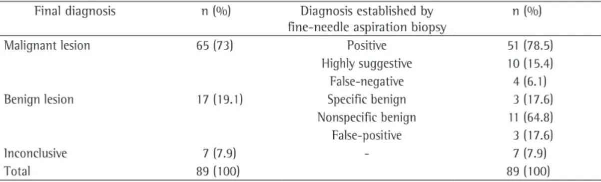
Of the 17 patients with a confirmed diagnosis of benign lesion, 3 (17.6%) presented cytological findings that were suggestive of cancer. Those 3 patients underwent surgical biopsy, 1 being diagnosed with pulmonary fibrosis and 2 being diagnosed with granuloma due to tuberculosis (Figure 1).
Of the 17 cases confirmed as benign, only 3 (17.6%) were specifically diagnosed by fine-needle aspiration biopsy (Figure 2): as hamartoma (in 1); as pulmonary cryptococcosis (in 1 HIV-positive patient); and as pulmonary tuberculosis (positive culture for Mycobacterium tuberculosis) with associated silicosis (in 1 patient). In 11 (64.7%) of those 17 cases, the cytological findings were not specific, the diagnosis being confirmed by clinical follow-up/ imaging tests in 4 and by surgical biopsy in 7. For the patients undergoing clinical follow-up, appointments and CT scans of the chest were regularly made available for a minimum period of 3 years, during which time there were no changes in the clinical profiles or X-ray findings. Among the patients undergoing surgical biopsy, the final diagnoses were as follows: pulmonary tuberculosis (in 3); pulmonary fibrosis (in 2); pulmonary cyst (in 1); and tissue fibrosis from previous surgery, with suspicion of recurrent cancer (in 1).
In 7 (7.9%) of the 89 patients, the diagnosis was not confirmed, because the patients were and 13 (15%) were allocated to the apparently
benign pulmonary lesions group.
There were 16 patients with stage IV cancer, among which the principal sites of metastasis were the brain (in 8), adrenal glands (in 5), and bone (in 3). One of those patients required CT-guided cytological confirmation of the diagnosis of the adrenal mass, because it was a solitary pulmonary nodule without mediastinal lymph node enlargement (T1N0M1). The diagnosis of non-small cell lung cancer was confirmed by the two procedures.
The principal clinical risk factors that contraindicated the surgical procedure were advanced COPD, severe heart disease, and wasting.
Of the 89 patients submitted to CT-guided fine-needle aspiration biopsy, 65 (73%) received a final diagnosis of lung cancer. In 51 (78.5%) of those, the diagnosis was confirmed by the procedure. In 10 (15.4%) of the 65, the cytological findings were highly suggestive of cancer, and 6 of those patients were started on clinical/oncological treatment after the cases had been discussed by the clinical and oncological teams; the treatment was warranted by an advanced clinical/oncological profile and by the impossibility of performing surgical biopsy. Those 6 patients were regularly followed by the clinical and oncological teams and were monitored through imaging studies until their deaths. In the remaining 4 patients, the diagnosis was confirmed surgically, because they presented with a clinical profile that posed lower surgical risk (Table 1).
In 4 (6.1%) of the 65 patients with a confirmed diagnosis of cancer, the diagnosis was not confirmed by CT-guided fine-needle
Table 1 - Final diagnosis compared with the diagnosis established by fine-needle aspiration biopsy in 89 patients.
Final diagnosis n (%) Diagnosis established by fine-needle aspiration biopsy
n (%)
Malignant lesion 65 (73) Positive 51 (78.5)
Highly suggestive 10 (15.4)
False-negative 4 (6.1)
Benign lesion 17 (19.1) Specific benign 3 (17.6)
Nonspecific benign 11 (64.8) False-positive 3 (17.6)
Inconclusive 7 (7.9) - 7 (7.9)
follow-up; and 3 (3.1%) were in patients who required immediate drainage due to rapid clinical decompensation related to a profile of severe COPD. Chest tube drainage was required in 12 (12.4%) of the procedures. One of the patients submitted to chest tube drainage presented with empyema that resolved easily, without associated morbidity. Two patients presented with mild, self-limiting hemoptysis that did not require subsequent bronchoscopy. In 8 patients, a control CT scan taken after fine-needle aspiration biopsy revealed alveolar hemorrhage along the needle path. This was merely a radiological finding and had no clinical impact. No other complications, such as hemothorax, gas embolism, or tumor cell dissemination, were observed. In 62 (63.9%) of the total of procedures, there were no complications of any type. No deaths were related to the procedure.
Discussion
All of the fine-needle aspiration biopsies evaluated were in compliance with the requirement that the needle pass through as little lost to clinical follow-up or died due to clinical
causes after the biopsy.
Table 2 specifies the diagnoses established through cytological examination or culture of the CT-guided fine-needle aspiration biopsy samples. Cases of unconfirmed diagnosis are not shown.
The kappa statistic revealed a significant concordance (p < 0.001) between the diagnosis established by CT-guided fine-needle aspiration biopsy and that established by the gold standard (kappa = 0.75; 95% CI: 0.57-0.93). As shown in Table 3, this concordance can be considered relatively “good” (0.61 < kappa < 0.80).
For the analysis of complications, we considered all 97 procedures performed (in a total of 89 patients). The most common complication was pneumothorax, which occurred in 27 (27.8%) of the 97 procedures. Of those 27, 15 (15.5%) were in patients who were followed clinically and through imaging studies and did not require chest tube drainage; 9 (9.3%) were in patients who required chest tube drainage due to the onset of pneumothorax-related symptoms after the procedure or to large initial pneumothorax, without the need for clinical
Figure 1 - CT images of patients with cytological findings suggestive of cancer of epithelial origin and with final diagnoses, established after surgical biopsy, of pulmonary fibrosis (in 1a) and granuloma with caseous necrosis (in 1b and 1c).
to surgery and intraoperative frozen section analysis in order to avoid diagnostic and treatment delay, as well as to minimize costs and morbidity. In such cases, fine-needle aspiration biopsy does not avoid the need for surgery even in cases of negative cytology, which is due to the radiological features suggestive of neoplastic lesion, commonly associated with active or passive exposure to smoking.
Cytological or histological confirmation of malignant neoplasm is definitive. However, numerous difficulties arise when attempting to establish the definitive diagnosis of a benign lesion. A finding of inflammatory cells is not sufficient to confirm a diagnosis of benignity or to rule out malignancy. To rule out neoplasia, the diagnosis of benign disease must be specific.
(7,8) Therefore, in cases in which the cytological
findings were suggestive of benignity but nonspecific, the investigation proceeded with surgical biopsy or clinical and radiological follow-up. A specific diagnosis of benign disease greatly contributes to the investigation, avoiding surgery or long-term follow-up in patients with high surgical risk. With the exception of specific lesions, such as hamartomas and infections, a specific diagnosis established exclusively by cytological analysis of fine-needle aspiration biopsy samples has a low yield, ranging from 12% to 68%.(7,9,10) In the present study, this type
of diagnosis was made in 17.6% of the cases of confirmed benignity, 1 patient having been definitively diagnosed with hamartoma and 2 with localized infection, results that were consistent with those found in the literature.
(7) However, histopathological analysis of
cutting needle biopsy samples (obtained with larger needles) shows rates that range from 52% to 91%.(10,11) Studies have suggested that,
specifically for benign lesions, the number of final diagnoses increases by as much as 63% when such diagnoses are based on concomitant cytology and histopathology.(9,10,12)
The accuracy of CT-guided needle biopsy in establishing a diagnosis of malignant lesions ranges from 70% to 100%.(7,9,10,13-18) The rate of
false-positive results is typically below 4%.(7) In
the present study, the accuracy of the procedure in establishing a diagnosis of malignant lesion was 91.5%, and the rate of false-positive results was 17.6% (3 cases), all cytological findings being suggestive of, but not confirming the atelectasis-free lung parenchyma as possible,
thus reducing the risk of pneumothorax.(3-6)
The effect that the results of CT-guided fine-needle aspiration biopsy of pulmonary lesions have on the clinical management of patients varies from case to case. The major useful aspects of the procedure include its accuracy in diagnosing malignant lesions, its ability to establish cancer subtypes efficiently, its ability to establish specific benign diagnoses, and, consequently, its ability to reduce the need for surgery (thoracotomy or video-assisted thoracic surgery) for the analysis of cases.(7) Patients with
pulmonary lesions that are highly likely to be malignant and surgically resectable, without clinical or oncological contraindications for the procedure, should be directly submitted
Table 2 - Final diagnosis established by fine-needle aspiration biopsy.
Diagnosis Patients,
n
Malignant 65
Epidermoid carcinoma 25
Malignant neoplasm of epithelial origin 15
Adenocarcinoma 10
Poorly differentiated malignant neoplasm 4
Non-small cell carcinoma 2
Small cell carcinoma 1
Large cell carcinoma 1
Plasmacytoma 1
Metastasis from pancreatic carcinoma 1 Metastasis from prostate adenocarcinoma 1
False-negative 4
Benign 17
Inflammatory/nonspecific changes 11
Hamartoma 1
Cryptococcosis 1
Tuberculosis 1
False-positive 3
Table 3 - Measurements of the ability of fine-needle aspiration biopsy to diagnose neoplasia.
Measurement % 95% CI
Sensitivity 93.8 88.0 99.7
Specificity 82.4 64.2 100.0
Accuracy 91.5 85.4 97.5
Rate of false-positives 17.6 0.0 35.8 Rate of false-negatives 6.2 0.3 12.0 Positive likelihood ratio 5.32
18 gauge in diameter.(7,14) In the present study, as
occurred in other studies involving fine needles, hemoptysis was self-limiting, and its incidence was low. Hemothorax, gas embolism, and tumor cell dissemination are rare complications and were not observed in the present study.
We can conclude that, in the present study, CT-guided fine-needle aspiration biopsy of pulmonary lesions had high diagnostic feasibility for malignant lesions, low morbidity, and no long-term complications. These findings, together with the fact that there were no deaths related to the procedure, makes it an important instrument in the diagnostic investigation of lung cancer. In addition, the procedure is an alternative to surgical biopsy when the latter cannot be performed or will not benefit patients.
Acknowledgments
We are grateful to Fátima Aparecida Magalhães Costa and to all of the thoracic surgery and pathological anatomy residents who participated in the procedures performed and in the analysis of material. We are also grateful to Romuel Braz Rodriguez Romualdo, who was responsible for formatting the manuscript and illustrations.
References
1. Sargent EN, Turner AF, Gordonson J, Schwinn CP, Pashky O. Percutaneous pulmonary needle biopsy. Report of 350 patients. Am J Roentgenol Radium Ther Nucl Med. 1974;122(4):758-68.
2. Prolla JC. Métodos diagnósticos em pneumologia. J Pneumol. 1986;12(3):204-5.
3. Macaulay SE, vanSonnenberg E, Casola G, Harrell JH 2nd. Misdiagnosis or missed diagnosis? Thoracic actinomycosis and carcinoma on sequential CT-guided lung biopsies. AJR Am J Roentgenol. 1990;155(6):1183-4.
4. Khouri NF, Stitik FP, Erozan YS, Gupta PK, Kim WS, Scott WW Jr, et al. Transthoracic needle aspiration biopsy of benign and malignant lung lesions. AJR Am J Roentgenol. 1985;144(2):281-8.
5. vanSonnenberg E, Casola G, Ho M, Neff CC, Varney RR, Wittich GR, et al. Difficult thoracic lesions: CT-guided biopsy experience in 150 cases. Radiology. 1988;167(2):457-61.
6. Boiselle PM, Shepard JA, Mark EJ, Szyfelbein WM, Fan CM, Slanetz PJ, et al. Routine addition of an automated biopsy device to fine-needle aspiration of the lung: a prospective assessment. AJR Am J Roentgenol. 1997;169(3):661-6.
7. Haramati LB, Austin JH. Complications after CT-guided needle biopsy through aerated versus nonaerated lung. Radiology. 1991;181(3):778.
diagnosis of, neoplasia. Those 3 patients underwent surgical biopsy: 2 were definitively diagnosed with pulmonary tuberculosis; and 1 was definitively diagnosed with pulmonary fibrosis.
Pneumothorax is the most common complication of CT-guided lung biopsies regardless of the size/type of needle employed (fine needles or larger, cutting, needles). The incidence of pneumothorax is 17.9-44.0%, 0.4-14.3% of the patients requiring chest tube drainage.(7) In the present study, pneumothorax
occurred in 27.8% of the cases, and chest tube drainage was required in 12.4%.
Various risk factors influence the incidence of pneumothorax after fine-needle aspiration biopsy. The principal patient-related risk factors are severe pulmonary emphysema and COPD, which contribute to an increase in the incidence of pneumothorax and to the need for chest tube drainage after pneumothorax has been detected.
(3-5,19) Two groups of authors have established a
direct relationship between the severity of COPD (as measured by FEV1 during spirometry) and an increased risk of developing pneumothorax, together with a need for chest tube drainage.
(5,19) The relevant lesion-related risk factors for
pneumothorax are the distance between the lesion and the chest wall and the small size of the lesion, which increase the degree of difficulty of the procedure.(3-5,10,15)
In the present study, severe COPD (FEV1 < 1.0 L) and the distance between the lesion and the chest wall were not considered absolute contraindications for the procedure. Three cases required immediate chest tube drainage due to rapid clinical decompensation in patients with severe COPD. The materials for drainage, as well as materials and medications for cardiopulmonary resuscitation, should be available at the site where the biopsy is performed in order to hasten the procedures. Chest tubes of smaller caliber are efficient for treating pneumothorax after lung biopsy.(20) They are easier and quicker to
14. Cox JE, Chiles C, McManus CM, Aquino SL, Choplin RH. Transthoracic needle aspiration biopsy: variables that affect risk of pneumothorax. Radiology. 1999;212(1):165-8.
15. Wallace MJ, Krishnamurthy S, Broemeling LD, Gupta S, Ahrar K, Morello FA Jr, et al. CT-guided percutaneous fine-needle aspiration biopsy of small (< or =1-cm) pulmonary lesions. Radiology. 2002;225(3):823-8. 16. Lourenço R, Camacho R, Barata MJ, Canário D, Gaspar
A, Cyrne C. Biópsia percutânea transtorácica guiada por TC na avaliação de lesões pulmonares de natureza indeterminada. Rev Port Pneumol. 2006;12(9):503-24. 17. Stanley JH, Fish GD, Andriole JG, Gobien RP, Betsill WL,
Laden SA, et al. Lung lesions: cytologic diagnosis by fine-needle biopsy. Radiology. 1987;162(2):389-91. 18. Thornbury JR, Burke DP, Naylor B. Transthoracic
needle aspiration biopsy: accuracy of cytologic typing of malignant neoplasms. AJR Am J Roentgenol. 1981;136(4):719-24.
19. Fish GD, Stanley JH, Miller KS, Schabel SI, Sutherland SE. Postbiopsy pneumothorax: estimating the risk by chest radiography and pulmonary function tests. AJR Am J Roentgenol. 1988;150(1):71-4.
20. Casola G, vanSonnenberg E, Keightley A, Ho M, Withers C, Lee AS. Pneumothorax: radiologic treatment with small catheters. Radiology. 1988;166(1 Pt 1):89-91. 8. Yamagami T, Iida S, Kato T, Tanaka O, Nishimura T.
Combining fine-needle aspiration and core biopsy under CT fluoroscopy guidance: a better way to treat patients with lung nodules? AJR Am J Roentgenol. 2003;180(3):811-5.
9. Murphy JM, Gleeson FV, Flower CD. Percutaneous needle biopsy of the lung and its impact on patient management. World J Surg. 2001;25(3):373-9; discussion 379-80.
10. Kazerooni EA, Lim FT, Mikhail A, Martinez FJ. Risk of pneumothorax in CT-guided transthoracic needle aspiration biopsy of the lung. Radiology. 1996;198(2):371-5.
11. vanSonnenberg E, Goodacre BW, Wittich GR, Logrono R, Kennedy PT, Zwischenberger JB. Image-guided 25-gauge needle biopsy for thoracic lesions: diagnostic feasibility and safety. Radiology. 2003;227(2):414-8. 12. Lucidarme O, Howarth N, Finet JF, Grenier PA.
Intrapulmonary lesions: percutaneous automated biopsy with a detachable, 18-gauge, coaxial cutting needle. Radiology. 1998;207(3):759-65.
13. Laurent F, Michel P, Latrabe V, Tunon de Lara M, Marthan R. Pneumothoraces and chest tube placement after CT-guided transthoracic lung biopsy using a coaxial technique: incidence and risk factors. AJR Am J Roentgenol. 1999;172(4):1049-53.
About the authors
Cristiano Dias de Lima
Physician. Department of Thoracic Surgery, Central Army Hospital, Rio de Janeiro, Brazil.
Rodolfo Acatauassu Nunes
Tenured Adjunct Professor. Department of Thoracic Surgery, Faculdade de Ciências Médicas da Universidade do Estado do Rio de Janeiro – FCM/UERJ, Rio de Janeiro State University School of Medical Sciences – Rio de Janeiro, Brazil.
Eduardo Haruo Saito
Adjunct Professor. Department of Thoracic Surgery, Faculdade de Ciências Médicas da Universidade do Estado do Rio de Janeiro – FCM/UERJ, Rio de Janeiro State University School of Medical Sciences – Rio de Janeiro, Brazil.
Cláudio Higa
Assistant Professor. Department of Thoracic Surgery, Faculdade de Ciências Médicas da Universidade do Estado do Rio de Janeiro – FCM/UERJ, Rio de Janeiro State University School of Medical Sciences – Rio de Janeiro, Brazil.
Zanier José Fernando Cardona
Adjunct Professor. Department of Radiology, Faculdade de Ciências Médicas da Universidade do Estado do Rio de Janeiro – FCM/ UERJ, Rio de Janeiro State University School of Medical Sciences – Rio de Janeiro, Brazil.
Denise Barbosa dos Santos