J Bras Pneumol. 2009;35(6):602-605
Synchronous pulmonary and hepatic nodules in
a patient with previous bronchogenic carcinoma:
the relevance of histopathological confirmation*
Nódulo sincrônico pulmonar e hepático em paciente com antecedente decarcinoma broncogênico: a importância da confirmação histopatológica
José de Jesus Peixoto Camargo, Tiago Noguchi Machuca, Spencer Marcantonio Camargo, Sadi Marcelo Schio, Rodrigo Moreira Bello
Abstract
The synchronous presentation of pulmonary and hepatic nodules in a patient with previously resected bronchogenic carcinoma raises suspicion of recurrence and mandates restaging. We present the case of a 71-year-old male with a history of lobectomy with pericardial resection and mediastinal lymphadenectomy (T3N0M0). At five years after the operation, he presented with a new pulmonary lesion. Restaging detected a synchronous nodule in the liver. Despite the strong suspicion of tumor recurrence, further investigation with a percutaneous liver biopsy revealed hepatocellular carcinoma. In order to investigate the etiology of the pulmonary lesion (hypotheses of recurrent bronchial cancer and of metastatic hepatocellular carcinoma), an open lung biopsy was performed, which revealed chronic inflammatory tissue with foci of anthracosis and dystrophic calcification. The patient was submitted to a non-anatomic resection of the liver lesion. The postoperative course was uneventful, and the patient was discharged on postoperative day 10. This report highlights the relevance of the histopathological diagnosis in patients with a history of bronchogenic carcinoma and suspicion of tumor recurrence. Differential diagnoses and the treatment administered are discussed.
Keywords: Carcinoma, bronchogenic; Neoplasm metastasis; Carcinoma, hepatocellular.
Resumo
A apresentação de lesão sincrônica pulmonar e hepática em um paciente com antecedente de carcinoma bronco-gênico operado gera a suspeita de recidiva tumoral e indica a necessidade de re-estadiamento. Apresentamos o caso de um paciente de 71 anos submetido à lobectomia pulmonar com ressecção de pericárdio e linfadenectomia mediastinal (T3N0M0). Cinco anos após a cirurgia, detectou-se a presença de uma nova lesão pulmonar. No re-estadiamento, foi diagnosticada uma lesão sincrônica no fígado. Apesar da forte suspeita de recidiva tumoral, prosseguiu-se a investigação e uma punção hepática revelou carcinoma hepatocelular. Para esclarecer a etiologia da lesão pulmonar (hipóteses de recidiva de carcinoma brônquico ou de metástase de carcinoma hepatocelular), foi realizada uma biópsia a céu aberto, compatível com reação inflamatória crônica com focos de antracose e de calcificação distrófica. O paciente foi então submetido à ressecção hepática não-regrada com intuito curativo. Teve boa evolução, com alta no 10º dia de pós-operatório. O presente relato destaca a importância do diagnóstico histopatológico em pacientes com antecedente de carcinoma broncogênico e suspeita de recidiva. Hipóteses diag-nósticas e condutas terapêuticas são discutidas.
Descritores: Carcinoma broncogênico; Metástase neoplásica; Carcinoma hepatocelular.
* Study carried out at the Porto Alegre Santa Casa Sisters of Mercy Hospital, Porto Alegre, Brazil.
Correspondence to: Tiago Noguchi Machuca. Av. Independência, 354, apto. 304, CEP 90035-070, Porto Alegre, RS, Brasil. Tel 55 51 9805-0202. Fax 55 51 3214-8316. E-mail: [email protected]
Financial support: None.
Synchronous pulmonary and hepatic nodules in a patient with previous bronchogenic carcinoma: the relevance of histopathological confirmation
J Bras Pneumol. 2009;35(6):602-605 603
Discussion
In clinical practice, physicians often regard new lesions in patients with a history of cancer as tumor recurrence. Consequently, the physicians do not offer curative options to such patients, referring them instead for palliative treatment. In a study involving patients with a history of extrapulmonary cancer in which a pulmonary nodule was incidentally found or found during a follow-up examination, surgical intervention confirmed the presence of metastases in only 41.3% of the cases.(2) In 33%, the histological
findings were consistent with primary lung cancer. In that study, most of the nodules were
Introduction
Synchronous pulmonary and hepatic nodules are commonly found in patients with advanced-stage cancer, principally when the primary tumor is located in the colorectal region.(1) In any other
scenario, a finding of synchronous pulmonary and hepatic nodules is not common in clinical practice and might pose a diagnostic dilemma.
Case report
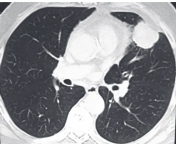
We present the case of a 71-year-old male patient submitted to left upper lobe lobectomy with pericardial resection due to localized inva-sion and mediastinal lymphadenectomy five years ago (Figure 1). The pathological stage was IIB (T3N0M0). A contralateral pulmonary lesion (0.8 cm) was found in the upper segment of the right lower lobe during a follow-up imaging study (Figure 2). This lesion had not been seen in previous imaging studies. The patient was asymptomatic, had no comorbidi-ties and presented no history of liver disease. He was a former smoker (30 pack-years) who had not smoked for 20 years and described himself as a nonalcoholic. Physical examination at admission revealed no particularities, except for a scar from a left posterolateral thoracotomy. Laboratory test results were normal, serology for viral hepatitis was negative, and alpha-fe-toprotein was 3 ng/mL. Restaging detected a synchronous nodule (2.5 cm) in segment 6 of the liver (Figure 3). Tumor recurrence was the principal working diagnosis, and we opted to initially investigate the liver. Percutaneous liver biopsy was performed and revealed hepatocel-lular carcinoma. Since it was necessary to clarify the diagnosis of the pulmonary lesion (principal hypotheses of recurrent bronchial cancer and of metastatic hepatocellular carcinoma), open lung biopsy was performed. The histological diag-nosis was chronic inflammatory reaction with anthracosis and foci of dystrophic calcification. The patient was submitted to non-anatomic resection of the liver lesion (sixth segment). The postoperative course was uneventful, and the patient was discharged on postoperative day 10. Histological analysis of the sample revealed hepatocellular carcinoma, 2.3 × 2.3 × 1.8 cm, classified as Edmondson-Steiner grade II, with tumor-free surgical margins.
Figure 1 - CT scan of the thorax performed in 2002, revealing a nodular lesion with dense soft parts and lobulated, slightly spiculated borders, located in the lingula and measuring approximately 4.0 cm.
604 Camargo JJP, Machuca TN, Camargo SM, Schio SM, Bello RM
J Bras Pneumol. 2009;35(6):602-605
dissemination to other organs) and controlled hepatic disease.(7) The authors reported one-,
three- and five-year survival rates of 80%, 61% and 36%, respectively. The analysis of prog-nostic factors revealed the negative impact of alpha-fetoprotein levels > 500 ng/mL and of recurrence-free intervals < 12 months. In addi-tion, it was shown that the number of lesions and the bilateral presentation of such lesions had no prognostic significance, provided that there was the possibility of resection of all metastases. Other case series have shown that pulmonary resection is indicated for well-selected patients with metastatic hepatocellular carcinoma, even for patients submitted to liver transplant.(8,9)
In the present case, the predicted five-year survival of the patient, classified as stage I according to the International Union Against Cancer staging of hepatocellular carcinoma, was over 60%.(10) In addition, the patient did not have
signs of poor prognosis, such as a lesion >5 cm, vascular invasion or satellite lesions. There was a great possibility of cure, since the initial working diagnosis was bronchial cancer recurrence with metastasis. Therefore, we highlight the relevance of histological confirmation and of following a diagnostic protocol in patients with synchronous nodules (pulmonary and hepatic) and a history of bronchial cancer. The principal differential diagnoses and the correct management of the condition should also be discussed.
References
1. Miller G, Biernacki P, Kemeny NE, Gonen M, Downey R, Jarnagin WR, et al. Outcomes after resection of synchronous or metachronous hepatic and pulmonary colorectal metastases. J Am Coll Surg. 2007;205(2):231-8.
2. Sortini A, Carcoforo P, Ascanelli S, Sortini D, Pozza E. Significance of a single pulmonary nodule in patients with previous history of malignancy. Eur J Cardiothorac Surg. 2001;20(6):1101-5.
3. Mery CM, Pappas AN, Bueno R, Mentzer SJ, Lukanich JM, Sugarbaker DJ, et al. Relationship between a history of antecedent cancer and the probability of malignancy for a solitary pulmonary nodule. Chest. 2004;125(6):2175-81.
4. Di Carlo I, Grasso G, Patane’ D, Russello D, Latteri F. Liver metastases from lung cancer: is surgical resection justified? Ann Thorac Surg. 2003;76(1):291-3. 5. Nagashima A, Abe Y, Yamada S, Nakagawa M, Yoshimatsu
T. Long-term survival after surgical resection of liver metastasis from lung cancer. Jpn J Thorac Cardiovasc Surg. 2004;52(6):311-3.
6. Ercolani G, Ravaioli M, Grazi GL, Cescon M, Varotti G, Gaudio MD, et al. The role of liver resections
treated by video-assisted thoracoscopy. In the case presented here, the location of the nodule, and its size in particular (smaller than 1 cm), were not appropriate for this procedure.
In patients with a history of bronchial cancer, the finding of a new pulmonary lesion raises suspicion of another primary tumor or of tumor recurrence. When investigating this specific population, one group of authors observed that 80% of the patients had another primary tumor and 2% had tumor recurrence.(3) That
shows a great probability of cancer, which merits investigation.
In the present case, the two principal working diagnoses were pulmonary lesion with liver metastasis or, after the liver biopsy results, hepa-tocellular carcinoma with pulmonary metastasis. With regard to the first hypothesis, although there have been few reports of liver resection for lung cancer metastasis, the results have been encouraging, with high rates of five-year recurrence-free survival.(4,5) In most of the cases
reported, a metachronous liver lesion was found; however, in one case with one-year survival, synchronous lesions were found, and the two lesions were treated surgically.(6) It should be
noted that, in all cases, a thorough investigation was conducted to determine whether the liver was the only site of secondary deposition.
Synchronous pulmonary and hepatic nodules in a patient with previous bronchogenic carcinoma: the relevance of histopathological confirmation
J Bras Pneumol. 2009;35(6):602-605 605
9. Bates MJ, Farkas E, Taylor D, McFadden PM. Pulmonary resection of metastatic hepatocellular carcinoma after liver transplantation. Ann Thorac Surg. 2008;85(2):412-5.
10. Cho CS, Gonen M, Shia J, Kattan MW, Klimstra DS, Jarnagin WR, et al. A novel prognostic nomogram is more accurate than conventional staging systems for predicting survival after resection of hepatocellular carcinoma. J Am Coll Surg. 2008;206(2):281-91. for metastases from lung carcinoma. HPB (Oxford).
2006;8(2):114-5.
7. Nakagawa T, Kamiyama T, Nakanishi K, Yokoo H, Kamachi H, Matsushita M, et al. Pulmonary resection for metastases from hepatocellular carcinoma: factors influencing prognosis. J Thorac Cardiovasc Surg. 2006;131(6):1248-54.
8. Tomimaru Y, Sasaki Y, Yamada T, Eguchi H, Takami K, Ohigashi H, et al. The significance of surgical resection for pulmonary metastasis from hepatocellular carcinoma. Am J Surg. 2006;192(1):46-51.
About the authors
José de Jesus Peixoto Camargo
Head of the Department of Thoracic Surgery. Porto Alegre Santa Casa Sisters of Mercy Hospital, Porto Alegre, Brazil.
Tiago Noguchi Machuca
Resident in Thoracic Surgery. Porto Alegre Santa Casa Sisters of Mercy Hospital, Porto Alegre, Brazil.
Spencer Marcantonio Camargo
Surgeon of the Department of Thoracic Surgery. Porto Alegre Santa Casa Sisters of Mercy Hospital, Porto Alegre, Brazil.
Sadi Marcelo Schio
Clinical Coordinator of the Lung Transplant Team. Porto Alegre Santa Casa Sisters of Mercy Hospital, Porto Alegre, Brazil.
Rodrigo Moreira Bello