São Paulo M edical Journal - Revista Paulista de M edicina
Squamous cell carcinoma (SCC) of the gallbladder is a rare and aggressive affection and is responsible for up to 12.7% of the malignant neoplasms of this organ.1,2,3
It characteristically presents invasive growth, a low tendency towards lymph node metastases and a high incidence of local infiltration and hepatic metastases, presenting a worse prognosis than adenocarcinoma of the gallbladder.1,2
In the period between 1968 and 1998, three patients suffer-ing from squamous cell carcinoma of the gallbladder were oper-ated on in our Department. T hey died between 1 and 6 months after the surgery (Table 1).
SCC of the gallbladder is predominantly incident among females, in a proportion of 3:1 over males, and between the fourth and sixth decades of life,1,2,3 as was found in our cases.
Its rapid growth, early metastatic dissemination and diffuse local and regional infiltration characterize the biological behavior of the lesion. Such tumors tend to grow laterally along the fossa of the gallbladder, forming large infiltrative masses and invading the liver and adjacent organs (stomach, duodenum and trans-verse colon) by direct expansion.1,2 T his pattern was verified in the cases here described. Despite this local and regional infiltra-tion, it usually does not present metastases in lymph nodes, and
seeding in the peritoneum is rare.3 Nevertheless, hepatic
metastases, as seen in case 2, are more frequently found in SCC than in adenocarcinoma of the gallbladder.1,2,3
Most studies accept that the squamous cells originate from pre-existing metaplastic squamous epithelium; some others be-lieve that SCC of the gallbladder originates from squamous dif-ferentiation of the adenocarcinoma cells, via expression of mixed phenotypes within a single tumor.1,2,3 Characteristically, the du-plication time for SCC is half that of adenocarcinoma, such that the growth of SCC cells may overtake and substitute that of adenocarcinoma.1,2
Disease is suspected when the lesion reaches a large size and is locally advanced, which was also observed in the cases we studied.
T he surgical options available depend mainly on the degree of local and regional involvement and consist of cholecystec-tomy with resection of a wedge of adjacent liver tissue or direct liver resection allied with regional lymphadenectomy and skel-etization of the hepatic hilum.2,4
Resection of the organs involved as part of the radical op-eration is justified in cases of localized lesion, without metastases
or peritoneal dissemination. Hepato-pancreatic duodenectomy was introduced as a radical treatment option for SCC of the gallbladder because of the type of dissemination seen in squa-mous cell carcinomas.4 However, its long-term benefits have not yet been satisfactorily documented.
Adjuvant postoperative radiotherapy and chemotherapy may be used, although their results are inconsistent and only palliative.1,2
T he extent of the tumor at the time of diagnosis is the most important parameter in determining survival.2,3 T he majority of the patients die around six months after diagnosis when radical surgery is not performed,1,4 as occurred with the patients de-scribed. T hese data reinforce the idea that early diagnosis is the most important parameter for improving the survival indices among patients with SCC of the gallbladder.
Ja ques W a isberg, M D. Department o f Dig estive Surg ery, Ho spital do Servido r Público Estadual, São Paulo , Braz il.
Sa nsom Henrique Bromberg, M D. Department o f Dig estive Surg ery, Ho spital do Servido r Público Estadual, São Paulo , Braz il.
M a ria Isete Fa res Fra nco, M D. Department o f Patho lo g y, Ho spital do Servido r Público Estadual, São Paulo , Braz il.
N agamassa Yamagushi, M D. Department of Digestive Surgery, Hospital do Servidor Público Estadual, São Paulo , Brazil.
Pa ulo Ama ra l dos Sa ntos. Student o f Medicine, Faculdade de Medicina do ABC, Santo André, Braz il.
M á rio Augusto Pa dulo Ca stro. Student o f Medicine, Faculdade de Medicina do ABC, Santo André, Braz il.
○ ○ ○ ○ ○ ○ ○ ○ ○ ○ ○ ○ ○ ○ ○ ○ ○ ○ ○ ○ ○ ○ ○ ○ ○ ○ ○
R EFER EN C ES
1. Hanada M, Shimizu H, Takami M. Squamous cell carcinoma of the gallbladder associated with squamous metaplasia and adenocarcinoma in situ of the mucosal columnar epithelium. Acta Pathol Jpn 1986;36:1879-86.
2. Karasawa T, Itoh K, Komukai M et al. Squamous cell carcinoma of gallbladder- Report of
two cases and review of literature. Acta Pathol Jpn 1981;31:299-308.
3. Khaira HS, Awad RW, T hompson AK. Squamous cell carcinoma of the gallbladder
present-ing with a biliary-colic fistula. Eur J Surg Oncol 1995;21:581-2.
4. Miyazaki K, Tsutsumi N, Kitahara K, et al. Hepatopancreatoduodenectomy for squamous and
adenosquamous carcinoma of the gallbladder. Hepato-Gastroenterol 1995;42:47-50.
Squamous cell carcinoma of the gallbladder
Departments of Digestive Surgery and Pathology, Hospital do Servidor Público Estadual, São Paulo, Brazil
R
e
se
a
r
c
h
L
e
tt
e
r
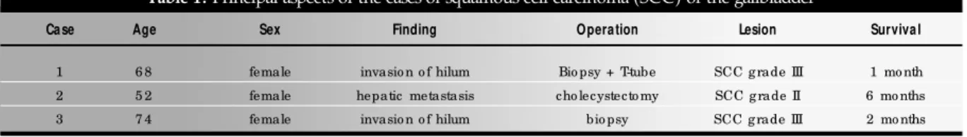
Table 1. Principal aspects of the cases of squamous cell carcinoma (SCC) of the gallbladder
Case Age Sex Finding Operation Lesion Survival
1 6 8 female invasio n o f hilum Bio psy + T-tube SCC g rade III 1 mo nth
2 5 2 female hepatic metastasis cho lecystecto my SCC g rade II 6 mo nths
3 7 4 female invasio n o f hilum bio psy SCC g rade III 2 mo nths
Sao Paulo M ed J/ Rev Paul M ed 2 0 0 1 ; 1 1 9 (1 ):4 3
Address for correspondence: Jaques W aisberg