33
p-ISSN:2231-6140,eA Study of Cervical Pap Sm
Dr Kruti Bhut1*, Dr Payal Shah2, 1
Second year Resident, 2 Assistant Pr of Pathology, P.D.U. Govt Medical C
Abstract:
Background: Cancer of Cancer of cervix is readily preve adequate and repetitive cytologic This is a retrospective study ai examined at a teaching tertiary clinical data and Pap smear cyto proforma. All the smears were re 1436 Pap smears were examined 40 years (fourth decade). There w of 1187 smears were reported as and 390(22.7%) were inflammato were reported to have epithelial cases with ASC-US, 27 cases of carcinoma. Conclusion: Premal easily by Pap smears. The epithel
Keywords: Cervical cytology, P
Introduction:
Cervical cancer is the seco in women worldwide after breast countries, the leading cause of de cancers in the early stage of deve situ, are highly treatable because located in a layer of cells in or aro not spread to other parts of the bo metastasize to other parts of the b difficult to treat and cervical canc more complex2.
The Papanicolaou (Pap) screening test for cervical cancer was used for smears made out of and pre-cancer lesions. But pre collected from vagina, endocerv
*
Corresponding Author:
Dr Kruti Bhut,
E-mail: [email protected]
e-ISSN:2395-7859 Origi
Smears in a Tertiary Care Hospital.
, Dr Milan Purohit3, Dr Gauravi Dhruva4
t Professor, 3 Associate Professor, 4 Professor & Hea al College and Hospital, Rajkot, Gujarat, India.
of the cervix is the leading cancer among fema eventable, and can be diagnosed at the pre-invasiv
gical screening with Papanicolaou (Pap) smears. aimed to evaluate all previously conducted cer ry care hospital during one year period. Metho ytology reports were obtained and data noted in reported as per the 2001 Bethesda system. Resul ed. Maximum number of patients was in the age g e were 133(9.2%) unsatisfactory or inadequate sam as Negative for Intraepithelial Lesion or Maligna atory. Out of a total of 1436 Pap smears, only 116 ial cell abnormality. The 116 abnormal cases com of LSIL, 36 cases of HSIL, 7 cases of invasive sq alignant and malignant lesions of cervix can b helial cell abnormality rate in our study was 8.0%.
Pap smear, Screening, Squamous Intraepithelial L
econd most common cancer ast cancer, and in developing death by cancer1. Cervical
velopment, or carcinomas in se the cancer cells are around the cervix and have body. Once the cancer cells e body the disease is more ancer treatment becomes
p) smear was introduced in 1941 and became cer and premalignant lesions3. Originally, the term of posterior fornix material for purpose of detect presently, the term is used for smear made fr
ervical canal, ectocervix or vaginal vault. The examination of cervical PAP smears in vaginal discharge showed that cervica intra epithelial neoplasia of various .com
iginal Article ead, Department
males of India. sive stage with rs. Objectives: cervical smears thods: Detailed in a structured sults: A total of e group of 31 – samples. A total gnancy (NILM) 16(8.0%) cases omprised of 36 squamous cell n be diagnosed %.
Lesion (SIL)
34
p-ISSN:2231-6140,e-ISSN:2395-7859 Original Article invasive cervical carcinoma are much more common in India as compared to the Western countries. The possible reason for this fact is the absence of cervical screening program, low social-economic status, and lack of awareness of cervical cancer prevention by PAP smears4. The simplicity, effectiveness and versatility of Pap test have made it an integral part of routine clinical examination and large chunk of workload in gynecological and pathological practice is due to this test5.Aims and Objectives
The aim of our study is to evaluate the
1. To explore various lesions of uterine cervix (inflammatory and neoplastic)
2. To find out target age group in which screening effort scan be concentrated for early detection as well as reduction of incidence of cervical cancer, in our setup.
Material and Methods
In this Retrospective Study, we have conducted Pap test in 1436 female at Cytology Laboratory, Dept. of Pathology, P.D.U. Government Hospital, Rajkot, Gujarat with prior consent between August 2015 to July 2. Sampling Technique is systemic random sampling.
All women coming to gynaecology OPD between the ages of 21 to 60 years consenting for Pap smear were included in the study. Those who presented with excessive white discharge per vagina, bleeding per vagina, irregular menstruation, pelvic pain and dyspareunia were considered as symptomatic.
Before taking Pap smear, it was ensured that no local douche, antiseptic cream and no local internal examination was done on day of test. The patient was placed in dorsal lithotomy position and a Cusco’s bivalve speculum was introduced through vagina and cervix was visualized. The longer projection of the Ayre’s spatula was placed in the cervix near squamo-columnar junction and rotated through 360 degree6.
35
p-ISSN:2231-6140,e-ISSN:2395-7859 Original Article Explanation of unsatisfactory specimen:Scanty cellularity Poor fixation
Partially or completely obscuring inflammation Partially or completely obscuring blood
Result:
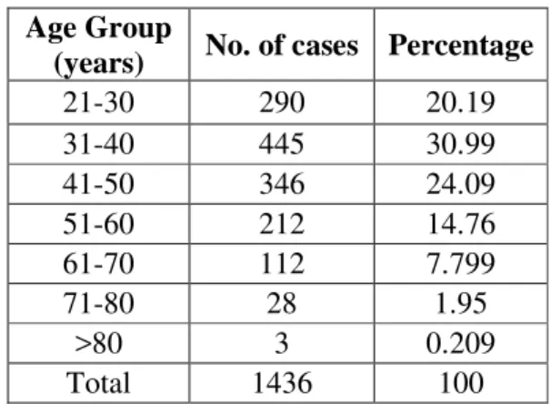
A total of 1436 cases were analyzed during a period of one year. Most of the women were in age group 31 – 40 years (Table 1)
Table 1: Age wise distribution of total number of patients
Age Group
(years) No. of cases Percentage
21-30 290 20.19
31-40 445 30.99
41-50 346 24.09
51-60 212 14.76
61-70 112 7.799
71-80 28 1.95
>80 3 0.209
Total 1436 100
As per as the patients presenting complain was concerned, vaginal discharge was commonest followed by lower abdominal pain and postmenopausal bleeding. The Pap smears were adequate and there was no other non-neoplastic or glandular cell abnormality noted apart from epithelial cell abnormality in 116 (8.0%) of the cases.
Morphology of epithelial cell abnormalities:
LSIL: Low Grade Squamous intraepithelial Lesion
Intermediate sized cells Nuclear atypia:
o Enlargement
o Irregular contour
o Hyperchromasia
o Slight chromatin coarseness Koilocytes
HSIL: High Grade Squamous intraepithelial Lesion
Parabasal sized cells
Discrete cells or syncytial groups (hyperchromatic crowded groups) Nuclear atypia
o Enlargement
o Markedly Irregular contour
36
p-ISSN:2231-6140,e-ISSN:2395-7859 Original Articleo Marked chromatin coarseness
SCC: Squamous cell carcinoma
HSIL features plus,
o Macronuclei
o Irregular chromatin distribution
o Tumor diathesis
‘Tadpoles’, Caudate and ‘fiber cells’ (Keratinizing type)
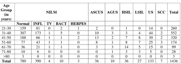
All other smears were either within normal limit or mild acute inflammatory cell infiltration. 133(9.2%) smears were found to be unsatisfactory for evaluation. A total of 1187 smears were reported as Negative for Intraepithelial Lesion or Malignancy (NILM) and 390 (22.7%) were inflammatory. Trichomonas infection 4 (0.2%) cases. The 116 abnormal cases comprised of 36 (2.5%) cases with ASC-US, 27 (1.8%) cases of LSIL, 36 (2.5%) cases of HSIL, 7 (0.4%) cases of invasive squamous cell carcinoma.
Table 2: Findings of pap smear cytology
Age Group
(in years)
NILM ASCUS AGUS HSIL LSIL US SCC Total
Normal INFL TV BACT HERPES
21-30 159 81 0 2 1 2 0 1 0 14 0 260
31-40 307 173 1 5 0 10 3 3 4 44 2 552
41-50 188 66 1 1 2 13 2 7 8 30 2 320
51-60 77 43 1 1 0 8 1 8 7 25 3 174
61-70 36 21 1 1 0 3 3 14 5 15 0 99
71-80 10 6 0 0 0 0 1 3 3 5 0 28
>80 3 0 0 0 0 0 0 0 0 0 0 3
Total 780 390 4 10 3 36 10 36 27 133 7 1436
INFL-Inflammatory smear, TV- Trichomonas Vaginalis, BACT-Bacterial Vaginosis, US-Unsatisfactory, SCC-Squamous cell carcinoma
Image 1: Squamous cell carcinoma [40X], Right lower inset - Caudate cell variant
37
p-ISSN:2231-6140,e-ISSN:2395-7859 Original Article
Discussion:
This study determines 1187 cases (82.6%) of negative for intraepithelial lesion or malignancy with non-specific inflammation 390 cases (27.1%) and 50% of NILM cases. Other studies revealed 95% and 74.3% cases of NILM respectively7.The Epithelial Cell Abnormality (ECA) rate, that is the total of ASCUS, LSIL, HSIL, and carcinoma diagnosis varied between 1.5 and 12.60% in various studies. The ECA rate of 8.0 % in our study was comparable to those reported in literature. Out of 116 neoplastic cases, 32 cases (27%) were in 41-50 years age group. In our study, the age group with most patients with abnormal smears was 41-50 years. Similar finding was detected by other studies8. A recent study conducted in Ningen Dock, Japan aimed to determine the gynaecological status of asymptomatic women who attended the hospital for health check-up, showed low prevalence cervical cell abnormalities of 1.2% 9. The explanation behind this result is mostly because of their cultural traditions and great concern regarding their health check-ups and less likelihood of having multiple sexual partners.
Our study revealed ASCUS 36(2.5%) and HSIL 36(2.5%) to be the most common epithelial cell abnormality. Similar results were obtained in other studies which also concluded ASCUS to be the most common epithelial cell abnormality10. In our study
Image 3 Low grade Squamous Intraepithelial Lesion [40X]
Image 4 Bacterial Vaginosis Showing Clue cells [40X]
Image 5 Bacterial Vaginosis Showing Acuteinflammatory cells [40X]
38
p-ISSN:2231-6140,e-ISSN:2395-7859 Original Article 1.88%had Low-grade Squamous Intraepithelial Lesion (LSIL), and2.5% had high-grade Squamous Intraepithelial Lesions (HSIL).Study from Saudi Arabia had 4.9% of cases were diagnosed with SIL11. In our study, the age group with most patients with abnormal smears was 41-50 years. Similar finding was detected by other studies8. Our study thus elucidates the importance of cervical screening test. Community health awareness campaigns and large scale Pap screening programs for women should be undertaken.Conclusion:
Cervical cancer is one of the most common malignancies in women of developing country like India. Pap smear is a simple, cheap, safe and practical diagnostic tool for early detection of cervical cancer in high risk group population; so it should be established as a routine screening procedure. It also has a greater role in diagnosis of inflammatory lesions including the identification of causative organism, atrophic changes, changes of radiation therapy and some rare tumors. As the most number of neoplastic lesions were observed in41-50 years of age (27% of all neoplastic lesions), It is recommended that at least a single life-time pap screening cytology of uterine cervix of all the women aged40 to 50 years.
References:
1. National Cancer Registry Program. Annual Report. IC New Delhi; 1990-1996.
2. Papanicolaou, GN, Traut, HF. The diagnostic value of vaginal smears in carcinoma of the uterus. Am Jr ObstetGynecol1941; 42:193-205.
3. Mintzer M, Curtis P, Resnick JC, Morrell D. The effect of the quality of Papanicolaou smears on the detection of cytological abnormalities Cancer 1999; 87:113-7.
4. Anderson and Jones: false positive cervico-vaginal cytology. acta cytol 41(6): 267, 1997
5. George Burkadze, Gulisa Turashvili: Cytology interpretations of cervical Pap smears in Georgia. The Internet Journal of Gynecology and Obstetrics. 2004; 3:2.
6. Banik U, Bhattacharjee P, Ahamad SU, Rahman Z. Pattern of epithelial cell abnormality in Pap smear: A clinic-pathological and demographic correlation. Cytojournal 2011; 8:8.
7. Solomon D, Davey D, Kurman R, Moriarty A, O'Connor D, Prey M, et al.The2001 Bethesda System: terminology for reporting results of cervical cytology. JAMA 2002; 287:2114-9.
8. Turkish Cervical Cancer And Cervical Cytology Research Group: Prevalence of cervical cytological abnormalities in Turkey. Int J Gynaecol Obstet. 2009; 106:206-9.
9. Imai A, Matsunami K, Takagi H, Ichigo S. Trend of Incidence in Positive Cervical Smears from 2002 - 2010 in Ningen Dock, a Special Japanese Health Checkup System. Ningen Dock 2012; 26:923-6.