ABSTRACT
Case R
epor
t
Rita Fontão-WendelSilvano Wendel
Vicente Odone
Jorge David Carneiro
Leandra Silva
Eduardo Isfer
A case report of neonatal
alloimmune thrombocytopenic
purpura: the importance of correct
diagnosis for future pregnancies
Blood Bank of Hospital Sírio-Libanês, São Paulo, Brazil
CONTEXT: Neonatal alloimmune thrombocyto-penic purpura (NAITP) is a neonatal disorder characterized by maternal alloimmunization against fetal platelet antigens inherited from the father. Intracranial hemorrhage leading to death or permanent neurological disability may occur in the fetus.
CASE REPORT: A healthy 30-year-old woman gave birth to her first baby by cesarean after an uneventful 36-week pregnancy. Ten hours after birth, the infant presented severe petechiae, with platelet count of 8 x 10³/µl. The mother’s platelet count was normal (180 x 103/µl). The infant re-ceived intravenous immunoglobulin and was dis-charged 18 days later, with platelet count of 100 x 103/µl. The cause of thrombocytopenia was not elucidated at that time. One year later, the infant died of neuroblastoma. Since the parents wanted another child, they were referred for investigation of this thrombocytopenia. Platelet genotyping and platelet antibody screening were performed, showing total HPA-1 system mismatch between mother (HPA-1b1b) and father (HPA-1a1a), with anti-HPA-1a antibodies in the mother’s serum. We concluded that the first baby was born with NAITP. Thus, in the second pregnancy, the mother was treated with several infusions of intravenous immunoglobulin. Careful ultrasound monitoring was performed, with normal results for mother and fetus throughout the pregnancy. The second baby was born by cesarean at 39 weeks, presenting 92x103 platelets/µl six hours after birth. The baby’s platelets were genotyped as HPA-1a1b and the mother’s serum again showed anti-HPA-1a antibodies. No clinical bleeding was observed. Intravenous immunoglobulin therapy was an effective treatment for preventing NAITP in the second baby.
KEY WORDS: Platelet antibody. Alloimmuniza-tion. Thrombocytopenia. Immunoglobulin. Hu-man platelet antigens.
INTRODUCTION
Neonatal alloimmune thrombocytope-nic purpura (NAITP) is a neonatal disorder characterized by maternal alloimmunization against fetal platelet antigens inherited from the father, which are not present in the mother. Its incidence is estimated to be around one in 350 to one in 2000 live births.1
NAITP is considered to be the counter-part of Rh hemolytic disease in newborns, but it involves maternal alloantibodies against platelet antigen systems and may affect the first child (around 30% of cases). Usually, the mother has a healthy and normal pregnancy with a normal platelet count. Nevertheless, severe thrombocytopenia in the fetus or newborn may be observed and intracranial hemorrhage may occur, leading to death or permanent neurological disabil-ity.1 Therefore, the prenatal management of
fetuses at risk is very important. However, there are still a lot of questions about what would be the best treatment.2 Some experts
have suggested fetal blood sampling, to check the fetal platelet count, but this procedure is very dangerous for the fetus and mother. Others have suggested intravenous injection of immunoglobulin, with or without cortico-steroids, for the mother. The diagnosing of NAITP is also difficult as it is initially based on clinical information and depends on rul-ing out other causes of thrombocytopenia. Laboratory testing is needed to confirm the presence of circulating maternal antibodies against fetal platelets or the presence of plate-let antigen incompatibility between father and mother, especially if platelet transfusion is needed to correct the thrombocytopenia. If platelet transfusion is required, it is manda-tory to investigate the compatibility between the patient and donor, as these antibodies are clinically important and may cause platelet destruction.
Platelet immunohematology has un-dergone major development over recent decades, and more sensitive techniques have become available for correctly iden-tifying platelet antibodies and/or platelet antigens.3 The most important platelet
antigen system involved in NAITP is HPA-1 (human platelet antigen 1), which accounts for 75-85% of cases, followed by HPA-5 (10-20% of cases).1 Both are
bial-lelic systems formed by a single nucleotide substitution in the DNA sequence, cor-responding to a single amino acid substi-tution in the respective protein. Usually the a allele has a high frequency in
Cau-casian populations, and the b allele has a
lower frequency. The phenotype frequency of these antigens in the blood donor population at our institution are: 1a1a: 70.5%; 1a1b: 27.5%; 1b1b: 2.0%; 5a5a: 80.4%; HPA-5a5b: 18.9%; and HPA-5b5b: 0.8% (personal observations).
We report on the case of a mother typed as HPA-1b1b, with anti-HPA-1a antibodies detected in her serum, whose first baby (probably HPA-1a1b) was born with NAITP. Because of correct clinical and laboratory diagnosis of the first baby’s condition, successful prenatal management for the second child was possible and no NAITP was observed.
CASE REPORT
A healthy 30-year-old woman (pri-mipara and primigravida) gave birth to her first baby by cesarean section after an uneventful 36-week pregnancy. Ten hours after birth, the infant presented severe pe-techiae, with a platelet count of 8 x 10³/µl.
The mother’s platelet count was normal. The infant received intravenous immu-noglobulin and was discharged 18 days
199
Genotype of platelets tested
Mother (auto)
1b1b;2a2a;3a3b;4a4a;5a5b;6a6a
1b1b;2a2b;3a3a;4a4a;5a5b;6a6a
1a1b;2a2a;3b3b;4a4a;5a5b;6a6a
1a1a;2a2b;3b3b;4a4a;5a5a;6a6a
1a1a;2a2a;3a3b;4a4a;5a5b;6a6a
later with a platelet count of 100 x 103/µl.
One year later, the infant died of another complication (neuroblastoma).
Since the parents wanted another child in the near future, it was suggested that immunohematological evaluation of platelets in the family should be un-dertaken, because of the unexplained thrombocytopenia presented by the first baby. For this purpose, the couple went to our service for platelet genotyping and platelet antibody screening. Whole blood samples were collected from the mother and father for HPA-1, HPA-2, HPA-3, HPA-4, HPA-5 and HPA-6 genotyping. The genomic DNA was extracted using a commercially available DNA isolation kit (DNAzol BD reagent - Life Technologies). HPA genotyping was performed using the polymerase chain reaction technique with sequence specific primers (PCR-SSP), as described by Meyer et al.4 Human
leukocyte antigen (HLA) typing was also performed in the mother’s sample. HLA class I typing was performed by serological tests (microlymphotoxicity) and HLA class II typing by PCR-SSP.
For platelet antibody screening, the mother’s serum was tested against a panel of genotyped platelets from known blood donors, using two different techniques: monoclonal antibody immobilization of platelet antigens (MAIPA)3 and the platelet
immunofluorescence test (PIFT) by flow cytometry.3
The mother’s HPA genotype was found to be: HPA-1b1b; HPA-2a2a; HPA-3a3b;
HPA-4a4a; HPA-5a5a; HPA-6a6a. The father’s HPA genotype was: HPA-1a1a;
2a2a; 3b3b: 4a4a; HPA-5a5b; HPA-6a6a. The mother’s HLA genotype was: HLA: A2, A-; B7, B8, Bw6; DRB1*0301, *1302; DRB3*0101, 0301; DQB1*0201, *0609.
The mother showed complete mismatch with the father in the HPA-1 system (moth-er HPA-1b1b; fath(moth-er HPA-1a1a) and partial mismatch in HPA-5 (mother HPA-5a5a; father HPA-5a5b). The mother also dem-onstrated the antigen HLA-DRB3*0101, which has a high prevalence among HPA-1b1b mothers with anti-HPA-1a5. The
platelet antibody screening by MAIPA (Figure 1a) and PIFT (Figure 1b) showed strong anti-HPA-1a alloantibodies in the mother’s serum. No HLA or autoantibodies were detected.
Figure 1. a) Absorbance values ob-tained by MAIPA technique, testing the mother’s serum against a panel of gen-otyped platelets in a case of neonatal alloimune thrombocytopenic purpura. The mother’s serum was strongly reac-tive against HPA-1a1a and HPA-1a1b donor platelets (bars 4, 5, 6) but was negative against HPA-1b1b donor platelets (bars 2, 3), thus showing pres-ence of anti-HPA-1a alloantibodies. No autoantibodies were detected (bar 1).
Cut-off
0,00 1,00 2,00 3,00 4,00 5,00
Absorbance
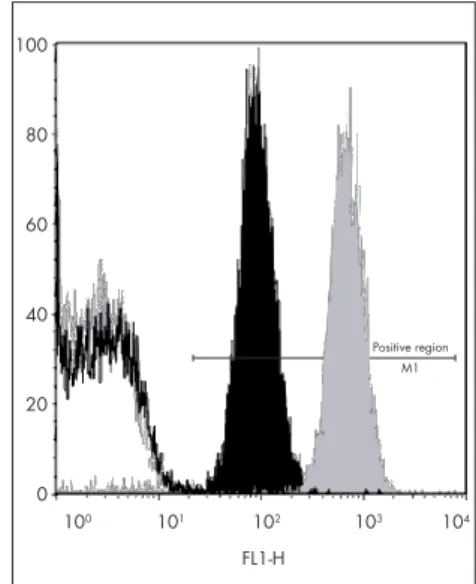
Figure 1. b) Flow cytometry results. The gray histogram is a positive control; the gray line histogram is a negative control; the positive region is where all the positive histograms must be; the black histogram is the mother’s serum against platelets typed HPA-1a1a, showing positive result; the black continuous line (overlapping with negative control) is the mother’s serum against platelets typed HPA-1b1b, showing negative result.
100
80
60
40
20
0
100 101 102 103 104
FL1-H
Positive region M1
Although we had no access to test the baby’s platelets (because he died due to neu-roblastoma one year after birth), the mother and father’s platelet genotyping showed total mismatch in the HPA-1 system, with the presence of anti-HPA-1a in the mother’s serum. Since HPA-1 is a biallelic autosomal codominant system, the offspring receives one allele from the father and another from the mother. Because in this case the mother was homozygous for HPA-1b (HPA-1b1b) and the father homozygous for HPA-1a (HPA-1a1a), all their descendents must be HPA-1a1b (heterozygous for HPA-1). From all this evidence, we concluded that the first baby was born with NAITP.
With these findings at hand for the sec-ond pregnancy, the mother received intrave-nous immunoglobulin infusion (400 mg/kg of weight) divided over three consecutive days, starting at the 19th week of gestation.
This treatment was repeated at the 24th
and 32nd weeks. All ultrasound tests (24th,
32nd and 34th weeks) revealed normal fetal
evolution. The mother’s platelet counts were normal throughout the pregnancy and her
second baby was born by cesarean section at the 39th week, with 92 x 103 platelets/µl
six hours after birth. The mother’s serum again showed anti-HPA-1a antibodies (a titer that was similar to the findings from the first pregnancy) and the baby’s platelet genotyping was HPA-1a1b. Although the infant was born with thrombocytopenia (the normal range for platelet counts is 150-400 x 103 platelets/µl), no clinical bleeding
was observed. Consequently, the mother and child were discharged 72 hours after the birth. The maternal alloimmunization against the infant’s platelets was evident, but the intravenous immunoglobulin therapy was an effective treatment for preventing any signs of bleeding in the second baby. Moreover, the fetus was successfully moni-tored during the pregnancy by ultrasound examinations. The correct diagnosing of the first baby’s condition was very important for achieving a good clinical approach for the second child.
Obstetricians should be aware of this disease and the importance of good follow-up for future pregnancies.
Sao Paulo Med J. 2005;123(4):198-200.
(1)
(2)
(3)
(4)
(5)
200
Resumo
Descrição de caso de púrpura trombocitopênica aloimune: importância do correto diagnóstico para futuras gestações
CONTEXTO: Púrpura trombocitopênica neonatal aloimune (PTNA) é uma doença neonatal caracterizada por aloimunização materna contra as plaquetas fetais, que apresentam antígenos herdados do pai. Podem ocorrer hemorragias cerebrais, levando à morte ou a anomalias neurológicas permanentes.
RELATO DE CASO: Mulher saudável, de 30 anos, deu à luz, por parto cesariano na 36a semana de gesta-ção, seu primeiro filho. Com 10 horas de vida, o recém-nascido apresentou petéquias e contagem de 8 x 10³ plaquetas/µl no sangue periférico; foi medicado com imunoglobulina e recebeu alta após 18 dias deµl no sangue periférico; foi medicado com imunoglobulina e recebeu alta após 18 dias del no sangue periférico; foi medicado com imunoglobulina e recebeu alta após 18 dias de internação, com 100 x 103 plaquetas/µl. A causa da trombocitopenia não foi elucidada na época. Um anoµl. A causa da trombocitopenia não foi elucidada na época. Um anol. A causa da trombocitopenia não foi elucidada na época. Um ano depois, a criança morreu de neuroblastoma. Como os pais desejavam outro filho, foram encaminhados para investigação da trombocitopenia. Genotipagem plaquetária e pesquisa de anticorpos antiplaquetários foram realizadas, mostrando total falta de concordância entre os sistemas HPA-1 do pai (HPA-1a1a) e da mãe (HPA-1b1b) e anticorpos anti-HPA-1a no soro da mãe. Concluímos que o primeiro bebê nasceu com PTNA. Por isso, na segunda gravidez, a mãe foi tratada com diversas infusões de imunoglobulina intravenosa. Foi realizado cuidadoso monitoramento por ultra-som, com resultados normais para mãe e feto durante a gravi-dez. O segundo bebê nasceu por cesárea às 39 semanas, apresentando 92 x 103 plaquetas/µl seis horasµl seis horasl seis horas após o nascimento. As plaquetas do recém-nascido foram genotipadas como HPA-1a1b e o soro da mãe novamente mostrou anticorpos anti-HPA-1a. Não houve hemorragia. A terapia de infusão de imunoglobulina foi efetiva na prevenção da PTNA no segundo filho.
PALAVRAS-CHAVE: Aloanticorpos. Alo-antígenos plaquetários. Trombocitopenia. Imunoglobulina G. Antí-genos de plaquetas humanas.
Author Information
Rita Fontão-Wendel. Pharmacist; supervisor of the Blood BankPharmacist; supervisor of the Blood Bank of Hospital Sírio-Libanês, São Paulo, Brazil.
Silvano Wendel, MD. Medical director of the Blood Bank of Hospital Sírio-Libanês, São Paulo, Brazil.
Vicente Odone, MD, PhD. Associate professor of the Depart-ment of Pediatrics, Universidade de São Paulo; oncologist at Hospital Sírio-Libanês, São Paulo, Brazil.
Jorge David Carneiro, MD. Department of Pediatrics, Univer-sidade de São Paulo, São Paulo, Brazil.
Leandra Silva, MD.Biomedic of the Blood Bank of Hospital Sírio-Libanês, São Paulo, Brazil.
Eduardo Isfer, MD. Medical director of “Fetus” prenatal diag-nostic and fetal medicine center, São Paulo, Brazil.
Address for correspondence:
Rita Fontão-Wendel
Banco de Sangue — Hospital Sírio Libanês Rua Dona Adma Jafet, 91
São Paulo (SP) — Brasil — CEP 01308-050 Tel.(+55 11) 3155-0350 — Fax (+5511) 3257-1290 E-mail: [email protected]
Copyright © 2005, Associação Paulista de Medicina
1. Forestier F, Hohlfeld P. Management of fetal and neonatal alloim-mune thrombocytopenia. Biol Neonate. 1998;74(6):395-401. 2. Engelfriet CP, Reesink HW, Kroll H, et al. Prenatal
manage-ment of alloimmune thrombocytopenia of the fetus. Vox Sang. 2003;84(2):142-9.
3. Goldman M, Trudel E, Richard L. Report on the Eleventh
International Society of Blood Transfusion Platelet Genotyping and Serology Workshop. Vox Sang. 2003;85(2):149-55. 4. Meyer O, Hildebrandt M, Schulz B, Blasczyk R, Salama A.
Simultaneous genotyping of human platelet antigens (HPA) 1 through 6 using new sequence-specific primers for HPA-5. Transfusion. 1999;39(11-12):1256-8.
5. Kaplan C. Immune thrombocytopenia in the foetus and the newborn: diagnosis and therapy. Transfus Clin Biol. 2001;8(3):311-4.
Sources of funding: None
Conflict of interest: None
Date of first submission: January 29, 2004
Last received: June 10, 2005
Accepted: June 17, 2005
References