Adipose Tissue Expansion and Weight Gain
Marcella K. Vaicik1.", Jill Thyboll Kortesmaa2.", Sofia Move´rare-Skrtic3
, Jarkko Kortesmaa2, Raija Soininen2, Go¨ran Bergstro¨m4, Claes Ohlsson3, Li Yen Chong5, Bjo¨rn Rozell6, Margo Emont7, Ronald N. Cohen7, Eric M. Brey1,8*`, Karl Tryggvason2,5`
1Department of Biomedical Engineering, Illinois Institute of Technology, Chicago, Illinois, United States of America,2Division of Matrix Biology, Department of Medical Biochemistry and Biophysics, Karolinska Institutet, Stockholm, Sweden,3Center for Bone and Arthritis Research, Institute of Medicine, The Sahlgrenska Academy, Gothenburg University, Go¨teborg, Sweden,4Department of Molecular and Clinical Medicine, Sahlgrenska University Hospital, Go¨teborg, Sweden,5Cardiovascular and Metabolic Disorders Program, Duke-NUS Graduate Medical School, Singapore, Singapore,6Clinical Research Center and Division of Pathology, Infectious Diseases, Department of Immunology, Microbiology, Pathology, and Infectious Diseases, Karolinska Institutet, Huddinge University Hospital, Stockholm, Sweden,7Section of Endocrinology, Department of Medicine, University of Chicago, Chicago, Illinois, United States of America,8Research Service, Hines Veteran Affairs Hospital, Hines, Illinois, United States of America
Abstract
Obesity is a global epidemic that contributes to the increasing medical burdens related to type 2 diabetes, cardiovascular disease and cancer. A better understanding of the mechanisms regulating adipose tissue expansion could lead to therapeutics that eliminate or reduce obesity-associated morbidity and mortality. The extracellular matrix (ECM) has been shown to regulate the development and function of numerous tissues and organs. However, there is little understanding of its function in adipose tissue. In this manuscript we describe the role of laminina4, a specialized ECM protein surrounding adipocytes, on weight gain and adipose tissue function. Adipose tissue accumulation, lipogenesis, and structure were examined in mice with a null mutation of the laminina4 gene (Lama42/2
) and compared to wild-type (Lama4+/+) control
animals.Lama42/2
mice exhibited reduced weight gain in response to both age and high fat diet. Interestingly, the mice had decreased adipose tissue mass and altered lipogenesis in a depot-specific manner. In particular, epididymal adipose tissue mass was specifically decreased in knock-out mice, and there was also a defect in lipogenesis in this depot as well. In contrast, no such differences were observed in subcutaneous adipose tissue at 14 weeks. The results suggest that laminin
a4 influences adipose tissue structure and function in a depot-specific manner. Alterations in laminin composition offers insight into the roll the ECM potentially plays in modulating cellular behavior in adipose tissue expansion.
Citation:Vaicik MK, Thyboll Kortesmaa J, Move´rare-Skrtic S, Kortesmaa J, Soininen R, et al. (2014) Laminina4 Deficient Mice Exhibit Decreased Capacity for Adipose Tissue Expansion and Weight Gain. PLoS ONE 9(10): e109854. doi:10.1371/journal.pone.0109854
Editor:Alessandro Bartolomucci, University of Minnesota, United States of America ReceivedJune 6, 2014;AcceptedSeptember 4, 2014;PublishedOctober 13, 2014
Copyright:ß2014 Vaicik et al. This is an open-access article distributed under the terms of the Creative Commons Attribution License, which permits unrestricted use, distribution, and reproduction in any medium, provided the original author and source are credited.
Data Availability:The authors confirm that all data underlying the findings are fully available without restriction. All relevant data are within the paper. Funding:Funding support from University of Chicago Diabetes Research and Training Center (DRTC) Pilot and Feasibility Grant Program DRTC grant P30 DK020595. The funders had no role in study design, data collection and analysis, decision to publish, or preparation of the manuscript.
Competing Interests:The authors have declared that no competing interests exist. * Email: brey@iit.edu
.These authors contributed equally to this work.
"These authors are first authors on this work.
`These authors also contributed equally to this work and are senior authors on this work.
Introduction
Obesity continues to increase globally in both industrialized and developing countries, with the World Health Organization reporting that worldwide obesity rates have doubled since 1980. Over 1 billion adults 20 years of age or older are overweight with an estimated 500 million of these defined as obese. Obesity is a major risk factor for several chronic diseases, contributing to a dramatic increase in morbidity and mortality due to type 2 diabetes, hypertension, heart disease, and other malignancies [1]. These issues with weight control result in a greater than $190 billion annual burden on US healthcare, and it has been suggested that the negative effects of obesity outweighs the positive effects of smoking cessation on the overall health of the population [2].
adipocytes invade the tissue stroma during adipose tissue expansion. In other tissues, it is well-known that interactions with the ECM can regulate cell growth, differentiation, and migration, and influence tissue development and repair [3–7]. However, little is known about the influence of the ECM on adipose tissue.
While adipose tissue-ECM interactions have not been studied extensively, there is some evidence that the ECM plays an important role in regulating adipogenesis and adipocyte function [8]. The synthesis and remodeling of ECM molecules is enhanced during adipogenesis [9,10]. Differentiating preadipocytes degrade the local matrix, invade the surrounding stroma, and then synthesize new ECM components as they mature [11]. The ECM surrounding preadipocytes transitions from fibronectin-rich to laminin-rich during differentiations primarily through an increase in ana4 chain containing BM protein laminin [12]. In
cell culture models, a preadipocyte cell line has been shown to express laminin, entactin and collagen IV during differentiation [13]. Laminins LN-411 and LN-421, are laminin isoforms consisting of a triple helix of the a4, b1 or b2 and c1 chains, are expressed in excess to other isoforms at this time [14]. Thea4 chain of laminin is present in the BM surrounding fully differentiated adipocytes and is upregulated during differentiation. However, the potential importance of thea4 chain laminins in the BM surrounding adipocytes has not been elucidated.
In this study, mice with a null mutation of the laminina4 gene (Lama42/2) were used to examine a potential role fora4 chain
laminins in adipose tissue expansion and function. Weight gain, adipose tissue function and adipose tissue structure were examined inLama42/2 mice and compared to wild-type control animals.
TheLama42/2mice were found to be resistant to age-related and
diet-induced obesity, and exhibited a depot-specific change in adipose tissue structure, volume and function.
Material and Methods
Animals, diets and housing
The generation of laminin a4 null mice (Lama42/2) was
previously described [15]. The mice were backcrossed to C57 BL/ 6 mice (Charles River) for more than 10 generations. Mice were fed a standard diet, or a high-fat diet containing 45 energy % fat (D12451, Research Diets), beginning at 4 weeks of age. The animals were fedad libitumand their food was weighed weekly. The mice were given a food refill up to 500 g after each weighing. The amount of food consumed was divided by the number of animals in a cage as an estimate of intake.
All animal procedures were approved by the IACUC at Karolinska Institutet or the University of Chicago. The animals were housed either in mixed cages (twoLama42/2and two
wild-type control animals) or in cages with onlyLama42/2 mice or
wild-types, in order to rule out the possibility that the weight differences observed were due to differences in dominance behavior. No differences were observed due to housing conditions.
Immunostaining
For immunostaining in mouse tissues, animals at 16 weeks of age were sacrificed and tissue harvested. Samples were placed in TissueTek (Sakura) in plastic molds and frozen in isopentane cooled to its freezing point. Cryosections of 8–12mm in thickness were made at238uC. The sections were allowed to dry for 1 hour at room temperature and then fixed in acetone for 10 minutes before staining, except for antibody to laminin a4, where the sections were additionally treated for 5 minutes in boiling 1 M Urea and washed in distilled water.
The antibodies used anti-nidogen/entactin (MAB 1946, Che-micon), anti-collagen type IV (polyclonal#AB756P, Chemicon), anti-perlecan (clone HK-102, Seikagaku Corp), anti-laminin a1 (clone 198 (35)), anti-laminina2 (clone 4H8-2), anti-laminina4 (polyclonal S8 (36)), anti-laminin a5 (serum 405). Secondary antibodies were FITC- or Cy3- conjugated and purchased from Jackson ImmunoResearch Laboratories, Inc. Tissue sections were examined with a Leica MDRB microscope (Leica) and pictures were taken with a Hamamatsu digital camera with Openlab (Improvision) software. Digital images were further processed with Photoshop 5.0 (Adobe).
Liver histopathology
Livers were harvested from 40 week old Lama42/2 mice for
histopathological evaluation (10 Lama42/2 mice on both diets).
For histological staining the tissue samples were fixed in 10%
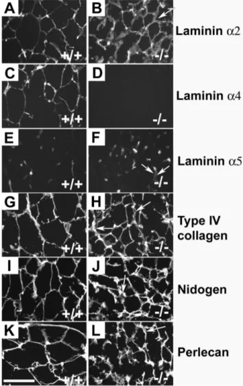
Figure 1. Immunostaining of WAT. The capillaries and BMs of
adipocytes of wild-type control and Lama4Lama42/2 mice were
positive with antibodies against laminina2 (A, B). Laminina4 antibodies stained both adipocytic BMs and capillaries in wild-type controls (C), while no staining was seen in mutants (D). The laminina5 antibody (E, F) stained capillaries inLama42/2and wild-type controls, but not the
adipocytic BM. Antibodies against type IV collagen (G; H), nidogen (I, J) and perlecan (K, L) all stained both capillaries and adipocytic BMs. Laminin a1 staining was completely negative (not shown). Arrows indicate a few locations where the adipocytic BM of theLama42/2
mice appeared slightly thickened. Bar 100mm.
neutral buffered formalin, paraffin-embedded and stained accord-ing to standard protocols. Tissue sections were examined with a Leica MDRB microscope (Leica) and pictures were taken with a Hamamatsu digital camera with Openlab (Improvision) software. Digital images were further processed with Photoshop 5.0 (Adobe).
Adipose tissue depot structure Lama4+/+
andLama42/2mice were fed a standard diet. At 14
weeks of age mice were sacrificed. Epididymal and subcutaneous fat depots were harvested, and the mass assessed. Mass of adipose tissues from each depot was normalized to the total individual animal weight the depot was harvested from using equation (1). The normalized % fat pad weight takes into account variation introduced from individual total animal weights.
fat pad weight
total animal weight100~
Normalized % fat pad weight ðequation1Þ
A portion of each fat pad type was then placed in formaldehyde and paraffin embedded. Samples were sectioned and stained with hematoxylin and eosin. Five images were taken with an Axiovert
200 inverted microscope using a 56objective (1.3mm/pixel) (Carl Zeiss MicroImaging, Inc., Thornwood, NY) for each fat pad. The images were used to manually measure the diameters of individual adipocytes using AxioVision (Carl Zeiss MicroImaging).
Lipogenesis. Lama4+/+ and Lama42/2 mice were fed a
standard diet. At 14 weeks of age mice were sacrificed. Fat pads were harvested and weighed prior to functional analysis with a lipogenesis assay. The assay was performed as described previously [16]. Briefly, adipocytes were isolated from the harvested fat pads by collagenase digestion and centrifugation. Isolated adipocytes were incubated with radioactive glucose in Krebs-Ringer bicar-bonate containing 10 nm insulin and 1% (w/v) BSA. The lipid fraction was extracted and radioactivity in the triglyceride fraction measured.
Statistics
The repeated measures of animal weights over time and food consumption over time were analyzed with ANOVA followed with Tukey test. Student’sttest was used for all other statistical analysis (two-tailed), p,0.05 was considered significant.
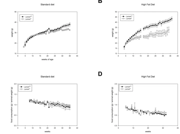
Figure 2. The weight difference of 16 maleLama42/2mice and 12 wild-type controls were statistically significantly (p = 0.002).(A).
When fed a high-fat diet (B), 8 male wild-type control mice gained statistically significant (p,0.001) more weight than the 8Lama42/2
mice. The weekly intake of standard food (C) and high fat diet (D) related to the weight of the animals were not significantly different.
Results
Adipose tissue composition of Lama42/2 mice
Immunofluorescence staining was first performed to compare BM composition surrounding adipocytes in Lama42/2 and
Lama4+/+
mice. Staining was performed for known adipose tissue BM proteins, including thea1,a2, a4 anda5 chains of laminin, type IV collagen, nidogen and perlecan. In wild-type control mice the a2 and a4 chains of laminin were present in the BM surrounding mature adipocytes (Figure 1). Laminin a5 was not observed in mouse adipocyte BM. Type IV collagen, perlecan and nidogen were present the murine pericellular adipocyte BM. When examiningLama42/2BM, the only difference in
compo-sition was the complete absence of laminin a4. All other BM proteins present in the adipose tissue BM of wild-type control mice were also observed in the pericellular BM ofLama42/2mice. The
adipocyte BM appeared somewhat thicker in theLama42/2mice.
Lama42/2mice are resistant to age and diet induced
obesity. The birth weight of laminin a4 deficient mice was ,10% lower than wild-type control littermates as previously reported [15]. At the time of weaning (3–4 weeks), no difference in weight was observed between Lama42/2 or wild-type control
animals (Lama42/2 20.8
60.26 g, n = 16;Lama+/+
18.261.05 g, n = 11). Animal weights were monitored first on a standard diet. A statistical difference in weight was observed between wild-type control andLama42/2animals (Figure 2A; p = 0.002).Lama42/2
weighed less than wild-type control animals from 16 weeks on and the difference between knockout and wild-type control animal weights increased steadily over time.
To further investigate weight gain in these animals, mice were supplied a high-fat diet starting at four weeks of age.Lama42/2
animals gained weight at a much lower rate and differences were observed from wild-type control animal weights within 4 weeks on this diet (Lama42/222.4560.73 g, n = 8;Lama+/+24.69
60.61 g, n = 8; Figure 2B). This difference continued to increase over time
Figure 3. TheLama42/2mice had reduced fat depots.The epididymal fat is delineated in an opened 40 week old male wild-type control mice
(A) and a maleLama42/2mice is shown in (B). Normalized fat pad depot mass as a % of total animal mass (C). Epididymal fat pad mass inLama42/2
mice was significantly lower thanLama4+/+mice (p,0.05). Age matched animalLama42/2
andLama4+/+, n = 4 for epididymal and subcutaneous. Error bars represent standard error.
withLama42/2exhibiting slow weight gain even on the high-fat
diet. In fact, at 34 weeks of age, there was no difference in the average weight ofLama42/2mice whether they were on standard
or high-fat diets (standard diet 32.660.5 n = 12 vs high-fat diet 32.862.57 n = 3). Wild-typetype control animals rapidly gained weight on the high fat diet resulting in dramatic differences from
Lama42/2 at 13 weeks (Lama42/2 25.460.89 g, n = 8;Lama+/+
30.761.27 g, n = 8;). A statistical difference in weight was observed between wild-type control and Lama42/2 animals on
high fat diet (Figure 2B; p,0.001).
The livers of the Lama42/2 mice on standard diet did not
display signs of liver steatosis after 40 weeks (File S1) suggesting that the decreased body fat inLama42/2mice does not represent
a form of lipodystrophy. On a high-fat dietLama42/2mice only
had very mild steatosis (File S1).
Food consumption
In order to evaluate whether the observed differences in weight and body composition resulted from lower food consumption, food intake was quantified weekly. There was no difference in the total amount of food consumed between Lama42/2 and wild-type
control animals on either standard (P = 0.418) or high fat diet (p = 0.098) (Figure 2C, D). These results suggest that the weight differences observed did not result from hypophagia in the
Lama42/2mice.
Adipose Tissue Structure
Lama42/2and age matched wild-type control animals 13 to 15
week on a standard diet were used to further examine adipose tissue structure and function. This time range was selected because it is prior to any statistically significant weight differences between
Lama42/2 and control animals, allowing for the examination of
adipose tissue function without confounding results due to obesity. At the time of sacrifice there were no differences in total animal mass between Lama42/2 and control animals (Lama42/2
25.9360.77 g, n = 4;Lama+/+
26.6062.35 g, n = 4;p= 0.80). Epididymal and subcutaneous adipose tissue were harvested from the mice (Figure 3A, B). The normalized percentage mass of
Lama42/2mice’s epididymal adipose tissue was significantly less
than control mice (Lama42/2 0.8060.09%, n = 4; Lama+/+
1.3260.14%, n = 4; p = 0.022) (Figure 3C). The mass of La-ma42/2 mice’s epididymal adipose tissue was less than control
mice (Lama42/20.2160.03 g, n = 4;Lama+/+
0.3660.07 g, n = 4;
p= 0.10). Interestingly, no differences in mass were observed between subcutaneous adipose tissue mass (Lama42/2
0.1860.04 g, n = 4;Lama+/+
0.2160.06 g, n = 4;p= 0.69). These results indicate that prior to any phenotypic observations in total animal weight gain, epididymal volume was reduced inLama42/2
mice.
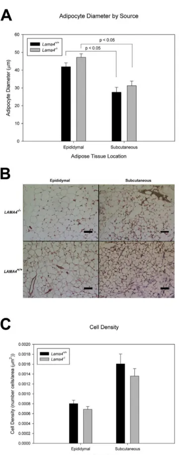
Histomorphometrix analysis was used to further analyze adipose tissue in the different depots. Mean adipocyte size determined from histological stains was greater in epididymal adipose tissue than in the subcutaneous adipose tissue depots in both types of mice (Figure 4B). In both subcutaneous and epididymal depots the mean adipocyte size was greater in the Lama42/2 mice
(Figure 4A). Cell density (number of cells per area) was lower in both epididymal and subcutaneous adipose tissue depots for
Lama42/2mice relative to wild-type controls (Figure 4C).
Figure 4. Adipocyte diameter in subcutaneous and epididymal
adipose tissue depots for age matched Lama42/2 and Lama
4+/+mice (A).
For epididymal n = 4 and subcutaneous n = 5, standard error shown as the error bars. There is a statistical difference between epididymal and subcutaneous (P,0.05). Histology images of
epididy-mal and subcutaneous Lama42/2 and Lama4+/+
, scale bar equals 300mm (B). Adipocyte cell density in subcutaneous and epididymal
adipose tissue depots for age matchedLama42/2andLama4+/+ mice (C). For epididymal n = 4 and subcutaneous n = 5, standard error shown as error bars.
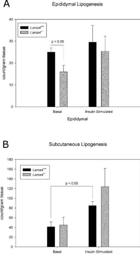
Figure 5. Lipogenesis levels in adipocytes from epididymal (A) and subcutaneous (B) adipose tissue in age matchedLama42/2and
Lama4+/+mice.
Epididymal fat pad basal lipogenesis is statistically significant betweenLama42/2andLama4+/+
mice (p,0.05). Subcutaneous fat pad inLama4+/+mice had statistically significant increase in lipogenesis between basal and insulin stimulated (p,0.05). The epididymal and subcutaneous n = 4. Error bars represent standard error.
Adipose Tissue Function
The reduced adiposity may be an indication of altered metabolic function. Adipose tissue function was examined by quantifying basal and insulin-stimulated lipogenesis in adipose tissue isolated from Lama42/2 and wild-type control mice
(Figure 5A, B). The epididymal depot ofLama42/2mice exhibited
impaired basal lipogenesis levels (Figure 5A). Interestingly, there were no differences in lipogenesis between subcutaneous adipo-cytes isolated from Lama42/2 and wild-type control mice
(Figure 5B).
Discussion
Differentiated adipocytes are surrounded by a thin BM layer. ECM in general, and BM specifically, have been shown to regulate cell behavior in a number of tissues and organs [3–7]. However, there is little knowledge of its role in adipocyte behavior. In this study, we used mice absent in a specific BM protein, laminina4, to investigate its role in adipose tissue. Using these mice we found a profound influence of laminina4 on the expansion and function of adipose tissue
Lama42/2 mice were found to gain weight at a much slower
rate than wild-type control mice. This occurred during normal aging and was more pronounced on a high fat diet. In fact,
Lama42/2mice did not exhibit any differences in weight whether
on normal chow or a high fat diet. Differences from wild type animals did not result from reduced food intake, and a reduction in adipose tissue was observed grossly in the mice deficient in laminin a4. While these results clearly showed that Lama42/2
mice have reduced adiposity, it was initially not clear if this resulted in normal or impaired adipose tissue function. Under both normal and high fat diets, livers in theLama42/2mice exhibited
little steatosis, suggesting that the decreased body fat did not represent a form of lipodystrophy.
Laminina4 is present around cells in the kidney, vasculature and muscle and has been shown to regulate different cell behaviors [17–26]. In order to further evaluate the role of laminin a4 specifically we isolated adipocytes from these tissues and analyzed their metabolic function. Surprisingly, we found that decreased laminin a4 leads to impaired adipocyte lipogenesis. Insulin stimulated lipogenesis resulted in increased lipogenesis in adipo-cytes from both Lama42/2 and wild-type control animals
indicating that insulin responsiveness appears to be intact, but basal lipogenesis levels were lower in Lama42/2 mice.
Interest-ingly, this difference was depot specific. Adipocytes isolated from epididymal adipose tissue exhibited impaired lipogenesis com-pared to wild-type controls while the subcutaneous adipose tissue depot had similar lipogenesis to wild-type control. This depot specific impairment is interesting and is further supported by the fact that epididymal adipose tissue depots in Lama42/2 mice
significantly are smaller in volume than wild-type control animals at 14 weeks. This difference was observed prior to any measurable differences in total body weight. While the total amount of epididymal adipose tissue inLama42/2mice was lower than
wild-type controls, the individual adipocytes were larger inLama42/2
mice relative to wild-type controls. The subcutaneous depots were similar in both total volume and cell size in comparisons between
Lama42/2 and wild-type control mice. Overall, these results
suggest an impaired function in adipose tissue from mice absent in laminina4 and that this impairment is manifested primarily in the epididymal depot.
This study shows a profound role for laminina4 on adipose tissue expansion and function. However, the mechanism under-lying this influence is unclear. There is very little knowledge of the
role of the ECM on adipocyte behavior. Cell culture and tissue engineering studies have shown that ECM substrata influence adipocyte behavior. In 2D culture ‘‘laminin’’ was found to be more potent than type IV collagen, fibronectin or type I collagen at promoting differentiation of preadipocytes [29]. This study did not distinguish which the specific laminin isoform used, but it is likely that this commercial laminin was purified from a mouse soft tissue tumor that has been shown to contain laminin 111 (a1:b1:c1). Studies with laminina4 are less common due to it only recently becoming available commercially. Preadipocytes have been shown to produce LN-411 (a4:b1:c1) during induction to an adipocyte phenotype [14] and laminin a4-rich ECM mixtures isolated promote greater adipogenesis than ECM lacking
a4 [30–32]. The a4 chain laminin could directly regulate adipocyte function through modulation of cell receptor signaling. Laminins containing the a4 chain could also influence adipocyte behavior based on indirect effects on the local cell microenvironment. The BM structure appeared thicker in immunohistochemical stains and previous studies have shown that the vascular BM in Lama42/2 mice is altered structurally
[15]. This structural change could reflect altered mechanical properties that may influence adipocyte function [33,34]. In addition, LN-411, which contains thea4 chain, has a chondroitin sulfate chain [35,36] which may function to sequester growth factors in the vicinity of a cell. This contributes to the regulation of growth factor availability and signaling near cell surface receptors [37]. Alterations in the laminin composition could alter the ability of the BM to bind growth factors and wild-type control the local concentration of regulatory factors. Laminina5 has had the gene variants shown to contribute to human body composition and weight [27]. A small subset of extremely obese humans were found to have variants in the laminin genes [28]. Future studies are needed that can address the mechanism including alteration in energy expenditure for the lean phenotype observed inLama42/2
mice. Additionally, genetic studies investigating laminin a4 in humans could lead to improved understanding of obesity.
Conclusion
In summary,Lama42/2mice exhibited reduced weight gain in
response to both age and high fat diet. The mass and function of the adipose tissue in Lama42/2 mice were altered in a
depot-specific manner. In particular, epididymal adipose tissue exhibited decreased mice and altered lipogenesis inLama42/2mice, but no
differences were observed in the subcutaneous depot. The results suggest that: (1) impaired lipogenesis leads to diminished fat mass inLama42/2mice; (2) alterations in lipogenesis are adipose tissue
depot-specific; and (3) specific ECM components dramatically influence adipose tissue function.
Supporting Information
File S1 Histology of livers of 10 months old male animals. In Lama42/2 animals the histological picture was
without signs of steatosis on standard diet, and on high-fat diet it was very mild.
(TIF)
Acknowledgment
We also would like to express our gratitude to Annika Bra¨nnstro¨m for critical reading of the manuscript and to Ha˚kan Westerblad for valuable discussions.
Author Contributions
Conceived and designed the experiments: MKV JTK SM JK RS GB CO BR RNC EMB KT. Performed the experiments: MKV JTK SM JK RS
GB CO LYC BR ME. Analyzed the data: MKV JTK SM JK RS GB CO LYC BR ME RNC EMB KT. Contributed reagents/materials/analysis tools: MKV JTK SM JK RS GB CO LYC BR ME RNC EMB KT. Wrote the paper: MKV JTK RNC EMB KT.
References
1. Frayn KN (2000) Visceral fat and insulin resistance - causative or correlative? British Journal of Nutrition 83: S71–S77.
2. Stewart ST, Cutler DM, Rosen AB (2009) Forecasting the effects of obesity and smoking on U.S. life expectancy. N Engl J Med 361: 2252–2260.
3. Aumailley M, Gayraud B (1998) Structure and biological activity of the extracellular matrix. Journal of Molecular Medicine-Jmm 76: 253–265. 4. Paulsson M (1992) BASEMENT-MEMBRANE PROTEINS - STRUCTURE,
ASSEMBLY, AND CELLULAR INTERACTIONS. Critical Reviews in Biochemistry and Molecular Biology 27: 93–127.
5. Kalluri R (2003) Basement membranes: Structure, assembly and role in tumour angiogenesis. Nature Reviews Cancer 3.
6. Lin CQ, Bissell MJ (1993) MULTIFACETED REGULATION OF CELL-DIFFERENTIATION BY EXTRACELLULAR-MATRIX. Faseb Journal 7: 737–743.
7. Domogatskaya A, Rodin S, Tryggvason K (2012) Functional Diversity of Laminins. Annual Review of Cell and Developmental Biology, Vol 28 28: 523– 553.
8. Kawaguchi N, Toriyama K, Nicodemou-Lena E, Inou K, Torii S, et al. (1998) De novo adipogenesis in mice at the site of injection of basement membrane and basic fibroblast growth factor. Proceedings of the National Academy of Sciences of the United States of America 95: 1062–1066.
9. Kawaguchi N, Toriyama K, Nicodemou-Lena E, Inou K, Torii S, et al. (1999) Reconstituted basement membrane potentiates in vivo adipogenesis of 3T3-F442A cells. Cytotechnology 31: 215–220.
10. Chavey C, Mari B, Monthouel MN, Bonnafous S, Anglard P, et al. (2003) Matrix metalloproteinases are differentially expressed in adipose tissue during obesity and modulate adipocyte differentiation. Journal of Biological Chemistry 278.
11. Kubo Y, Kaidzu S, Nakajima I, Takenouchi K, Nakamura F (2000) Organization of extracellular matrix components during differentiation of adipocytes in long-term culture. In Vitro Cellular & Developmental Biology-Animal 36.
12. Nie J, Sage EH (2009) SPARC Inhibits Adipogenesis by Its Enhancement of beta-Catenin Signaling. Journal of Biological Chemistry 284: 1279–1290. 13. Aratani Y, Kitagawa Y (1988) ENHANCED SYNTHESIS AND SECRETION
OF TYPE-IV COLLAGEN AND ENTACTIN DURING ADIPOSE CON-VERSION OF 3T3-L1 CELLS AND PRODUCTION OF UNORTHODOX LAMININ COMPLEX. Journal of Biological Chemistry 263: 16163–16169. 14. Niimi T, Kumagai C, Okano M, Kitagawa Y (1997) Differentiation-dependent
expression of laminin-8 (alpha 4 beta 1 gamma 1) mRNAs in mouse 3T3-L1 adipocytes. Matrix Biology 16: 223–230.
15. Thyboll J, Kortesmaa J, Cao RH, Soininen R, Wang L, et al. (2002) Deletion of the laminin alpha 4 chain leads to impaired microvessel maturation. Molecular and Cellular Biology 22: 1194–1202.
16. Neel BA, Brady MJ, Sargis RM (2013) The Endocrine Disrupting Chemical Tolylfluanid Alters Adipocyte Metabolism via Glucocorticoid Receptor Activation. Molecular Endocrinology 27: 394–406.
17. Sorokin LM, Pausch F, Durbeej M, Ekblom P (1997) Differential expression of five laminin alpha (1–5) chains in developing and adult mouse kidney. Developmental Dynamics 210: 446–462.
18. Ringelmann B, Roder C, Hallmann R, Maley M, Davies M, et al. (1999) Expression of laminin alpha 1, alpha 2, alpha 4, and alpha 5 chains, fibronectin, and tenascin-C in skeletal muscle of dystrophic 129ReJ dy/dy mice. Experimental Cell Research 246: 165–182.
19. Petajaniemi N, Korhonen M, Kortesmaa J, Tryggvason K, Sekiguchi K, et al. (2002) Localization of laminin alpha 4-chain in developing and adult human tissues. Journal of Histochemistry & Cytochemistry 50: 1113–1130.
20. Patton BL, Miner JH, Chiu AY, Sanes JR (1997) Distribution and function of laminins in the neuromuscular system of developing, adult, and mutant mice. Journal of Cell Biology 139: 1507–1521.
21. Miner JH, Patton BL, Lentz SI, Gilbert DJ, Jenkins NA, et al. (1997) The laminin alpha chains: Expression, developmental transitions, and chromosomal locations of alpha 1–5, identification of heterotrimeric laminins 8–11, and cloning of a novel alpha 3 isoform. Journal of Cell Biology 137: 685–701. 22. Gu YC, Sorokin L, Durbeej M, Hjalt T, Jonsson JI, et al. (1999)
Characterization of bone marrow laminins and identification of alpha 5-containing laminins as adhesive proteins for multipotent hematopoietic FDCP-mix cells. Blood 93: 2533–2542.
23. Lefebvre O, Sorokin L, Kedinger M, Simon-Assmann P (1999) Developmental expression and cellular origin of the laminin alpha 2, alpha 4, and alpha 5 chains in the intestine. Developmental Biology 210: 135–150.
24. Geberhiwot T, Ingerpuu S, Pedraza C, Neira M, Lehto U, et al. (1999) Blood platelets contain and secrete laminin-8 (alpha 4 beta 1 gamma 1) and adhere to laminin-8 via alpha 6 beta 1 integrin. Experimental Cell Research 253: 723–732. 25. Frieser M, Nockel H, Pausch F, Roder C, Hahn A, et al. (1997) Cloning of the mouse laminin alpha 4 cDNA. Expression in a subset of endothelium. European Journal of Biochemistry 246: 727–735.
26. Sorokin LM, Maley MAL, Moch H, von der Mark H, von der Mark K, et al. (2000) Laminin alpha 4 and integrin alpha 6 are upregulated in regenerating dy/ dy skeletal muscle: Comparative expression of laminin and integrin isoforms in muscles regenerating after crush injury. Experimental Cell Research 256: 500– 514.
27. De Luca M, Chambers MM, Casazza K, Lok KH, Hunter GR, et al. (2008) Genetic variation in a member of the laminin gene family affects variation in body composition in Drosophila and humans. Bmc Genetics 9: 11.
28. Mariman ECM, Bouwman FG, Aller E, van Baak MA, Wang P (2014) High frequency of rare variants with a moderate-to-high predicted biological effect in protocadherin genes of extremely obese. Genes and Nutrition 9: 9.
29. Hausman GJ, Wright JT, Richardson RL (1996) The influence of extracellular matrix substrata on preadipocyte development in serum-free cultures of stromal-vascular cells. Journal of Animal Science 74.
30. Uriel S, Huang J-J, Moya ML, Francis ME, Wang R, et al. (2008) The role of adipose protein derived hydrogels in adipogenesis. Biomaterials 29: 3712–3719. 31. Uriel S, Labay E, Francis-Sedlak M, Moya M, Weichselbaum R, et al. (2009) Extraction and Assembly of Tissue-Derived Gels for Cell Culture and Tissue Engineering. Tissue Engineering Part C-Methods 15: 309–321.
32. Abberton KM, Bortolotto SK, Woods AA, Findlay M, Morrison WA, et al. (2008) Myogel, a novel, basement membrane-rich, extracellular matrix derived from skeletal muscle, is highly adipogenic in vivo and in vitro. Cells Tissues Organs 188.
33. Shoham N, Gottlieb R, Sharabani-Yosef O, Zaretsky U, Benayahu D, et al. (2012) Static mechanical stretching accelerates lipid production in 3T3-L1 adipocytes by activating the MEK signaling pathway. American Journal of Physiology-Cell Physiology 302: C429–C441.
34. Shoham N, Gefen A (2012) Mechanotransduction in adipocytes. Journal of Biomechanics 45.
35. Sasaki T, Mann K, Timpl R (2001) Modification of the laminin alpha 4 chain by chondroitin sulfate attachment to its N-terminal domain. Febs Letters 505: 173– 178.
36. Kortesmaa J, Doi M, Patarroyo M, Tryggvason K (2002) Chondroitin sulphate modification in the alpha 4 chain of human recombinant laminin-8 (alpha 4 beta 1 gamma 1). Matrix Biology 21: 483–486.