MEASUREMENT OF EVOLUTION THERAPY
USING A DIGITAL CALIPER IN BELL’S PALSY
Mensuração da evolução terapêutica com paquímetro digital
na paralisia facial periférica de Bell
Claudia Hosana da Maceno Salvador(1),Adriana Tessitore (2), Leopoldo Nisan Pfeilsticker (3), Jorge Rizzato Paschoal(4), Kátia Nemr (5)
(1) Phonoaudiologist; Specialization Student in Orofacial Motri-city at Cefac.
(2) Phonoaudiologist, Master and PhD in Medical Sciences from the College of Medical Sciences from the State Uni-versity of Campinas.
(3) Otolaryngologist Doctor; Responsible Assistant for the Ambulatories of Surgery and Maxillo-Facial Traumatology and Ambulatory of the Skull Base and Facial Paralysis of Otolaryngology from the College of Medical Sciences of the Clinical Hospital from Unicamp.
(4) Otolaryngologist Doctor, Responsible Professor for the Ambulatory of Skull Base and Facial Paralysis of the Oto-laryngology course from the College of Medical Sciences from the Hospital of Unicamp, PhD in Medical Sciences, UNICAMP.
(5) Phonoaudiologist, Assistant Professor of the College of
Medicine from the University of São Paulo; Ph.D. in Social Psychology from the University of São Paulo.
Conlict of interest: non-existent
INTRODUCTION
Facial paralysis has progressively been the subject of the phonoaudiologists who work with motor and orofacial rehabilitation1-6.
The facial palsy (PFP), also described as pathology of disiguring symptoms of the biopsychic-social senses, is triggered by a partial blockage of the facial nerve, VII cranial nerve7. The
PFP has sudden onset and unilateral beginning8,9
and compromises the harmony and symmetry of facial movements, causing strong impact disigu -rement, which causes functional and psychological impairment10,11.
The absence of movements of facial muscles results in disigurement and impairment of facial expression, fundamental to the process of human ABSTRACT
Purpose: to assess the use of the digital caliper in the measurement of the facial mimic movements in different moments of the speech therapy. Method: prospective longitudinal study, with 20 subjects between 7 and 70 years-old, 13 females and 7 males, all diagnosed with Bell’s Palsy, attended in the Facial Paralysis Ambulatory, of the otorhinolaryngology subject of a University Public Hospital. The use of a Digimess 100,174BL digital measuring caliper was adopted for this study. The measurements were carried out in the facial mimic movement, always starting from a ixed point to a mobile point in the structures: the tragus and the labial commissure, external corner of the eye and labial commissure and also internal corner of the eye and the nasal ala. All measurements were carried out both prior and after the treatment. The quantiication of the incompetence of the movement was measured by simple percentage. The Wilcoxon signed rank test was applied to check for possible differences between both moments considered (with and without movements), as the study variables. Results: the measurements had a statistically signiicant result (p<0.05) in all the proposed measured structures (tragus and labial commissure, external corner of the eye and labial commissure, and internal corner of the eye and nasal ala), showing that there are possibilities of measuring of movement and absence of movement using a digital caliper. Conclusion: the caliper has demonstrated to be a useful device which has permitted to objectively compare the evolution of the rehabilitation of facial mimic in Bell’s Palsy in the sample studied here.
eficacy results in the use of caliper. It was done the same with a small change in the methodology removing one of the measures that demonstrated that it was unnecessary to measure the inner corner of the eye to the corner of the lips, this measure was not relevant, so the current research used only three steps.
In this study we evaluated the use of digital calipers to measure the movements of facial mimics at different times of the phonoaudiological treatment.
METHOD
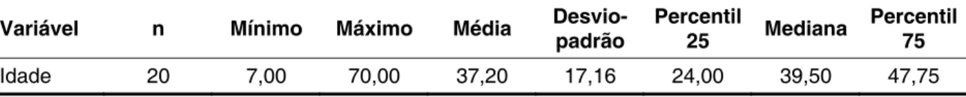
This is a prospective longitudinal study, which evaluated 20 cases of individuals diagnosed with Bell’s Palsy, in the Facial Paralysis Ambulatory from the Otolaryngology course from the College of Medical Sciences Hospital of Unicamp, aged from 07 to 70, who were an average age of 37,20 and standard deviation (dv) of 17.16 years (Table 1). communication (facial mimics). The speech is
hampered by the deviation of the nasal-labial ilter and the inadequate articulation of labial-dental and bilabial phonemes due to laccidity occurred at the beginning of facial paralysis, the muscles of the face, both in the buccinator muscle and in the orbicularis oris, which causes an articulation impairment12.
To clinically quantify the impact of facial paralysis in the facial mimics as well as the result of inter-vention processes were developed subjective and objective methods, among which one can point out: the graduation of House Brackman13, Yanagihara14,
as well as methods such as facial function Index15,
Moiré topography16, three-dimensional analysis of
facial movements17, EMG18,19 and anthropometric
measurements20, 21.
Among the developed methods, we studied the results of manual measurements of the movement of the paralyzed face, in order to develop an objective system which would not be compute-rized of PFP quantiication, easily done with the use of digital caliper22. This study demonstrated the
Table 1 – Description of the age variable
Variável n Mínimo Máximo Média Desvio-padrão Percentil 25 Mediana Percentil 75
Idade 20 7,00 70,00 37,20 17,16 24,00 39,50 47,75
Inclusion criteria were: (a) aged from 07 to 70 years old, (b) patients with Bell’s facial paralysis for a fortnight.
Exclusion criteria were the other causes of facial paralysis.
After evaluation and medical diagnosis, the patients were referred to the Department of Orofacial Rehabilitation – Phonoaudiology, where the integrative process of orientation and measu-rement began.
Proposal for evaluation in this study was adopted using a Digimess 100.174BL digital caliper gauge, instrument with a resolution of 0.00 mm/152, 78mm, properly secured with tape.
Measurements were performed in the following structures in movement of the facial mimics, always starting from a ixed point to a mobile point:
The caliper was initially placed of the tragus ixed point with opening up to the structure of the labial commissure, in the sequence of the measured ixed point was from the outer corner of the eye to the labial structure and to inalize put in the structure of the inner corner of the eye with opening up the structure of the wing of the nose.
The patient sat on proper and comfortable posture with both feet propped, keeping his gaze straight ahead during measurement. To measure the smile it was made two measurements TR (Tragus) – LC (Labial commissure) and CEO (Outside the Corner of the Eye) – CL, while for nasal contraction it was considered as CIO (Inside the Corner of the Eye) and AN (Wing nose).
Patient and therapist sat facing each other to perform the measurement. First, the side of the paralyzed face and subsequently the non paralyzed side.
Measurements were performed before and after speech therapy at the clinic where most cases were treated at an average time of up to 05 weeks. To quantify the incompetence of movement (IM), which represents how much the paralyzed side (LP) is more laccid than the normal side (LN) in simple percentage, we used the following formula21:
IM = LP-LN x 100
possible differences between movements of the facial muscles, always starting from a ixed point to a mobile point, which are considered for the variables of interest. We adopted a signiicance level of 5% (0,050).
RESULTS
The results can be seen and analyzed more speciically in detail by the following tables:
Table 2 is a characterization of the sample, thus enabling the identiication of the evolution time in weeks, as well as the initial and the end of each measured structure by n, from each studied subject. The applied therapeutic protocol consisted of:
1) Initial guidelines as to the care with the affected eye, and the use of inducing maneuvers of the facial movement, twice a day after meals; 2) protocol for the initial phase of PF with orofacial
maneuvers and functional orientation;
3) protocol for the recovery phase, using orofacial isotonic and isometric exercises23.
This study was fully approved by the Ethics Committee of the College of Medical Sciences from the State University of Campinas-UNICAMP (1104/2008).
For data analysis we used Wilcoxon test of Signed Posts, with the purpose of investigating
Table 2 – Characteristics of the sample
TR X CL CEO x CL CIO X NA
N Idade PM UM TES IM
Inicial
IM Final
IM
Inicial IM Final
IM Inicial
IM Final
1 46 30/01/09 20/02/09 3 6,60 0,12 6,44 4,55 9,39 6,17
2 22 26/10/07 09/11/07 2 21,46 5,43 32,54 11,07 11,85 6,52
3 11 19/10/07 25/10/07 2 6,96 6,08 4,68 3,82 0,24 2,33
4 13 09/11/07 30/11/07 2 12,83 5,93 18,67 -0,16 11,63 0,97
5 47 04/03/08 31/03/08 5 12,54 -4,73 23,63 2,81 24,28 1,39
6 28 26/06/08 19/09/08 5 2,60 0,83 8,98 7,44 5,84 5,22
7 27 01/08/08 08/08/08 2 15,30 -3,52 13,67 3,83 3,67 -0,61
8 23 01/08/08 22/08/08 4 33,27 31,58 24,90 12,64 34,51 18,16
9 42 01/08/08 08/08/08 2 11,82 14,16 15,22 13,03 2,80 0,64
10 46 06/06/08 18/07/08 5 11,56 4,17 15,89 -2,56 11,60 17,01
11 7 20/06/08 01/08/08 3 96,04 4,59 31,32 3,21 8,72 3,70
12 33 04/07/08 26/09/08 5 27,03 18,44 53,41 22,60 42,09 16,03
13 29 14/02/08 28/02/08 3 0,68 1,75 2,73 2,12 9,66 8,16
14 70 22/11/08 11/12/08 3 8,92 5,03 -0,43 4,34 3,82 3,79
15 61 15/11/08 29/11/08 3 10,21 56,05 12,22 12,93 11,98 11,98
16 52 21/02/09 05/03/09 3 -0,06 -0,06 -0,55 -0,22 20,85 15,36
17 48 06/12/08 13/12/08 2 1,20 2,58 6,13 6,13 8,31 8,28
18 45 15/05/09 25/05/09 2 10,77 2,58 5,27 3,06 24,75 5,49
19 57 15/05/09 05/06/09 4 21,87 3,41 47,94 12,14 22,15 61,71
20 37 18/09/09 16/10/09 3 24,53 13,98 35,59 26,26 23,93 29,64
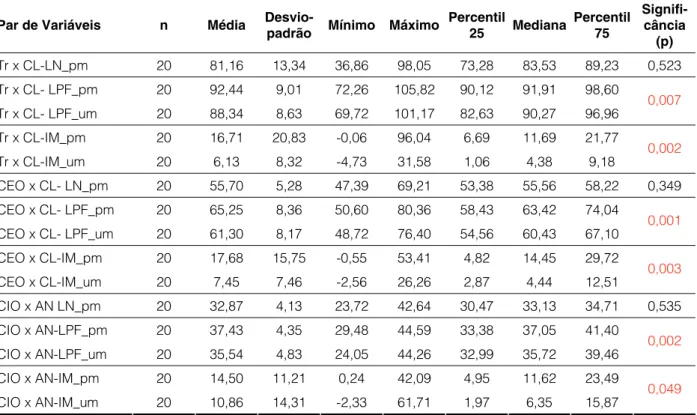
In Table 3 there are the description and compa-rison of the results obtained at the beginning and the end of the measurements of the total sample, as well as the standard deviation and signiicance level
The use of caliper was introduced into Phonoaudiology, that enables the possibility to perform static measurements of the human face studied by anthropometry21, 22, associated to
movements of the expression of the facial mimic11, 12 and the results of the movements. In orofacial
motricity, the caliper is also used to analyze movements in oral breathing patients22. Its appli-cation has been effective in related ields such as cosmetic dentistry26.
Independent of the patient’s age (children or adults), in facial paralysis, there is always a potential recovery. The use of the caliper facilitates the measurement of the results in those cases by not being invasive, since its use is preceded only by simple requests to the patients for them to perform movements associated to the expression of the facial mimics.
It can be noted, in this study, when comparing the initial and inal data of the process that measured measures emphasize the improvement of movement. We observe in the results a statistically signiicant improvement in the proposed movements of smiling and nasal contraction, as shown in Table 1, in 18 out of 20 evaluated patients.
In this table it is still possible to see that the request in the nose contraction (smelling something bad face) the motion is more limited because it is not as common as the movement of the smile.
DISCUSSION
Based on a recent study that showed that the muscles of the face have their own characte-ristics that must be respected in rehabilitation24,
and considering the characteristics of the muscle ibers, as well as the residual contractile capacity and the potential for resistance to muscle fatigue that orofacial muscles provide6, it emerged then the relection on the possibility of requesting the movements of contraction smile and nose, for this work, and the caliper as a measuring tool18.
In the literature there are several studies showing the importance of measuring the prognosis of facial paralysis13-21. An example is the use of an
objective and computerized approach for measu-rements of facial synkinesis as biofeedback25
surface electromyographic as an adjunct in muscle reeducation to enhance appreciation of the facial expression24.
Legend: Tr = tragus, CL= labial commissure, LN= normal side ,pm= irst measurement, um= last measurement, IM= Incompetence of
movement, LPF= Side of facial paralysis, CEO = outer corner of the eye, CIO= inner corner eye, AN= Wing Nose. Statistical Test: Wilcoxon Signed Posts
Table 3 – Description and comparison between the initial time and the end of the measurements
Par de Variáveis n Média Desvio-padrão Mínimo Máximo Percentil 25 Mediana Percentil 75 Signifi-cância (p)
Tr x CL-LN_pm 20 81,16 13,34 36,86 98,05 73,28 83,53 89,23 0,523
Tr x CL- LPF_pm 20 92,44 9,01 72,26 105,82 90,12 91,91 98,60
Tr x CL- LPF_um 20 88,34 8,63 69,72 101,17 82,63 90,27 96,96 0,007
Tr x CL-IM_pm 20 16,71 20,83 -0,06 96,04 6,69 11,69 21,77
Tr x CL-IM_um 20 6,13 8,32 -4,73 31,58 1,06 4,38 9,18 0,002
CEO x CL- LN_pm 20 55,70 5,28 47,39 69,21 53,38 55,56 58,22 0,349
CEO x CL- LPF_pm 20 65,25 8,36 50,60 80,36 58,43 63,42 74,04
CEO x CL- LPF_um 20 61,30 8,17 48,72 76,40 54,56 60,43 67,10
0,001
CEO x CL-IM_pm 20 17,68 15,75 -0,55 53,41 4,82 14,45 29,72
CEO x CL-IM_um 20 7,45 7,46 -2,56 26,26 2,87 4,44 12,51
0,003
CIO x AN LN_pm 20 32,87 4,13 23,72 42,64 30,47 33,13 34,71 0,535
CIO x AN-LPF_pm 20 37,43 4,35 29,48 44,59 33,38 37,05 41,40
CIO x AN-LPF_um 20 35,54 4,83 24,05 44,26 32,99 35,72 39,46 0,002
CIO x AN-IM_pm 20 14,50 11,21 0,24 42,09 4,95 11,62 23,49
the difference between the measurements of both hemifaces approach to the maximum of the normal range.
The caliper can be considered an important beacon when the evaluator objectively compares gain movement by paralysis of the affected side with the unaffected side in numerical values.
Moreover, the difference of the incompetency of the initial movement, in relation to the inal criterion can be a clearer prognosis of each patient, which can reverberate better control of the therapeutical progress of paralysis; both in terms of quantity and the quality of the adopted procedures and thus allowing organizing better the chosen treatment and therapy.
After the clinic, using the caliper as the instrument for quantifying the movement, it was noted that the inal measurement of the side affected by paralysis showed a value of improvement, which made it numerically closer to the reference value of the unaffected side.
The use of digital caliper takes the professional (phonoaudiologists and other health professionals) of empiricism and refers them to a new level of evaluation and objective quantiication.
CONCLUSION
The caliper has proved to be a useful tool that allowed us to compare, objectively, the evolution of therapeutic therapy in rehabilitation of Bell’s palsy in the sample.
At the same time it was also measured and calculated the limitation of the initial and inal movement suggested by the formula described in the methodology, thus conirm the importance of this calculation, which facilitates the visualization of the percentage gain of movement.
It is observed that the movements of the tragus ixed point to the movable point (labial commissure of the not paralyzed side) were unaltered, however, when comparing to the irst measurement of the involved side with the latter, with the corresponding measurements made on the paralyzed side, this is the irst with the last measurement, it can be noted that there was a statistically signiicant improvement (0.007), this evidence also obtained in other struc-tures measured from the ixed point to the movable point (Table 3).
The higher numeral reference, from one of the hemifaces, indicates the limitation of movement in comparison to the numeral reference of the side unaffected by paralysis, or it is due to the invol-vement of unilateral facial paralysis outline a limited movement on the opposite side there is no limitation of movement, so I have a reference number given by caliper initially greater than the end in the same hemiface. Therefore, the muscle has a gain of movement which initially it lacked, and the caliper is an instrument which facilitates this view.
JBP rec. Ibero-am. Odontopediatr. Odontol. 2005; 8(41):26-31.
8. Calais LL, Gofi-Gomez MVS, Bento RF, Comerlatti LR. Avaliação funcional da mímica na paralisia facial central por acidente cerebrovascular. Pró-Fono. 2005; 17(2):213-22.
9. Freitas KCS, Gofi-Gomez MV. Degree of perception and discomfort regarding facial condition in subjects with peripheral facial paralysis in sequele stage. Rev Soc Bras Fonoaudiol. 2008; 13(2):113-8. 10. Liriano RYG, Magalhães SLB, Barros F, Testa JRG, Fukuda Y. Relação da presença de hiperacusia em pacientes com paralisia facial periférica de Bell. Rev Bras Otorrinolaringol. 2004;70(6):776-9.
11. Gonzáles HJM. Estúdio epidemiológico de la páralisis de Bell o paralísis facial idiopática, realizando em el servicio de isiatria del Hospital Clínico Universitário de la Universidade Central de Venezuela, noviembro 2003, marzo 2004: resultados preliminares. Acta odonto. 2007;45(3): 384-7.
12. Tessitore A, Pfelsticker LN, Paschoal JR. Aspectos neuroisiológicos da musculature facial, visando a reabilitação na paralisia fácil. Rev CEFAC. 2008;10(1):68-75.
13. House JW, Brackmann DE. Facial nerve grading system. Otolaryngol Head Neck Surg. 1985;93(2):146-7.
REFERENCES
1. Atolini JN, Jorge JJJ, Gignon VF, Kitice T, Prado LSA, Santos VGW. Paralisia facial periférica/ incidência das várias etiologias num ambulatório de atendimento terciário/ Facial nerve palsy: incidence of different ethiologies in a tertiary ambulatory; Arq. Int. otorrinolaringol. ( impr), 2009;13(2):167-71. 2. Batista KT, Cauhi AF. Resultados da reabilitação cirúrgica da face paralisada;Rev. Bras. Cir. Plast. 2008; 23(3):149-52.
3. Brosens C, Botargues M. Are steroids and antivirals useful in the treatment of idiopathic facial paralysis. Evid actual práct ambul. Jul-ago 2008;11(4):124-5.
4. Bauso D. Paralysus facial idiopatica o parálisis de Bell. Evid.actual. práct. Ambul. 2006;9(1): 22-5. 5. Hernández PL, García AS,Pieruzzini OR, Mora CA, Ledezma RJG. Parálisis facial periférica em el departamento de otorrinolaringologia Hospital Militar Dr Carlos Azevedo. Acta otorrinolaringol. 2007;19(1):31-3.
6. Lazarini PR, Fernandez AMF, Brasileiro VSB, Custódio SEV. Paralisia facial periférica por comprometimento de tronco cerebral: a propósito de um caso clínico. Rev Bras Otorrinolaringol. 2002;68(1):140-4.
7. Leal RB, Rodrigues MJ. Reabilitação bucal em pacientes portadores de paralisia facial periférica.
RESUMO
Objetivo: avaliar o uso do paquímetro digital na mensuração dos movimentos da mímica facial em diferentes momentos do tratamento fonoaudiológico. Método: estudo longitudinal prospectivo, em 20 sujeitos com idade entre 07 e 70 anos, sendo 13 do genero feminino e 07 masculino, com diag-nóstico de paralisia facial periférica de Bell, atendidos no Ambulatório de Paralisia Facial, da disci-plina de otorrinolaringologia de um Hospital Público Universitário. Neste estudo foi adotado o uso de um medidor paquímetro digital da marca Digimess 100.174BL, instrumento com resolução de 0,00mm/152,78mm. As medições foram realizadas no movimento da mímica facial, sempre partindo de um ponto ixo para o ponto móvel nas estruturas: tragus e comissura labial, canto externo do olho e comissura labial e também canto interno do olho e asa do nariz, sendo realizadas pré e pós trata-mento fonoaudiológico. A quantiicação da incompetência do movitrata-mento foi mensurada por meio de porcentagem simples. Foi aplicado teste dos Postos Sinalizados de Wilcoxon, para veriicar possíveis diferenças entre ambos os momentos considerados (com e sem movimentos), como as variáveis de interesse. Resultados: as mensurações tiveram um resultado estatisticamente signiicante (p<0,05) em todas as estruturas medidas propostas (tragus e comissura labial, canto externo do olho e comis-sura labial e canto interno do olho e asa do nariz), demonstrando que há possibilidades de se fazer medições de movimento e de ausência de movimento utilizando o paquímetro digital. Conclusão: o paquímetro mostrou ser um instrumento útil que permitiu comparar, de forma objetiva, a evolução da reabilitação da mímica facial na Paralisia Facial Periférica de Bell na amostra estudada.
system of month breathing children:anthroposcopic approach. Pro Fono. 2007; 19(4):347-51.
21. Cattoni DM, Fernandes FD. Anthropometric orofacial measurements of children from São Paulo and from North America: comparative study. Pro Fono. 2009; 21(1):25-9.
22. Quintal M, Tessitore A, Paschoal JR, Pfeilsticker LN. Quantiicação da Paralisia Facial com Paquímetro Digital. Rev CEFAC. 2004;6(.2):170-6. 23. Tessitore A. Avaliação do angulo da comissura labial na reabilitação na Paralisia Facial. Tese apresentada a Faculdade de Ciências Médicas da Unicamp.2010;47p.
24. Vanswearingen J. Facial Reabilitation: a neuromuscular reeducation,patient-centered approach. Facial Plast Surg. 2008;24(2):250-9.
25. Wu ZB, Silverman CA, Listron CJ, Tessema B, Cosetti MK. Objective computerized versus subjective analysis of facial synkinesis. Laryngoscope. 2005; 115(12):2118-22.
26. Farias RL. Interpretação e conceituação dos tipos de peris faciais por meio de paquimetro do peril facial do comitê de avaliadores utilizando fotograias faciais. Tese apresentada a Universidade Estadual Paulista. 2006;129p.Disponível http:// bases.bireme.br/cgi-bin/wxislind.exe/iah/online. 14. Satoh Y, Kanzankij J, Yoshiharas. A
comparision and conversion table of”the House-Brackmann Facial nerve grading system” and “Yanagihara grading system”. Auris Nasus Larynx. 2002;27(3);207-12.
15. Johnson PJ, Bajai-Luthra A, Llull R, Johnson PC. Quantitative facial motion analysis after fucunctional free muscle reanimation producers. Plast Reconstr Surg. 1997;100(7):1710-9.
16. Saito N, Nishijima T, Fujimura T, Moriwaki S, Takema Y. Development of a new evaluation method for cheek sagging using a Moire 3D analysis system, Skin Res Technol. 2008;14(3): 287-92. 17. Listrom CJ, Silverman CA, Susman WM. Facial-motion analysis with a video and computer system: a preliminary report. Am J Otol. 2002;21(1):123-9. 18. Kasse CA, Cruz OLM, Leonhardt FD, Testa JRG, Ferri RG, Vierther EY.Valor prognóstico de dados clínicos em paralisia de Bell. Rev Bras otorrinolaringol.2005;71(4):454-8.
19. Rahal A, Gofi-Gomes MVS. Avaliação eletromiográica do músculo masseter em pessoas com paralisia facial periférica de longa duração. Rev CEFAC. 2007; 9(2):207-12.
20. Cattoni DM, Fernandes FD, Di Francesco RC, Latorre M do R. Characteristics of the stomatognathic
http://dx.doi.org/10.1590/S1516-18462012005000085
Received on: September 14, 2011 Accepted on: December 29, 2011 Mailing Address:
Claudia Hosana da Maceno Salvador
Rua Alvaro Ribeiro nº 15 Apto 18 – Ponte Preta Campinas – SP
CEP: 13041-730