(1) Universidade Federal de Ciências da Saúde de Porto Alegre, Porto Alegre, Rio Grande do Sul, Brasil.
Source: Coordenação de Aperfeiçoamento de Pessoal de Nível Superior.
Conlict of interest: non-existent
Animal models for hearing evaluations: a literature review
Modelos animais para avaliação auditiva: revisão de literatura
Aléxia dos Reis(1) Suelen Pizzolatto Dalmolin(1) Eliane Dallegrave(1)
Received on: February 09, 2017 Accepted on: May 31, 2017
Mailing adress:
Eliane Dallegrave
Rua Sarmento Leite, 245 - Porto Alegre Rio Grande do Sul, Brasil
CEP: 90050-170
E-mail: [email protected]
ABSTRACT
This review aims to outline which animal models are viable for preclinical hearing research, conside -ring their anatomical and physiological characteristics, and their advantages and disadvantages of use. PubMed, Scielo, and Portal Periodicos Capes were consulted, using descriptors concerning hearing, hea -ring tests and animal species, individually and crossed with each other. The abstracts of the articles found in the databases were read, with subsequent selection based on the following criteria: free articles, use of animal models in audiological procedures that included the description of the evaluation methods, the advantages and/or disadvantages of using the species, and published between 1995 and 2016. Despite
the existence of alternative models, mammals are still widely used in research. It has been found that rats,
mice and guinea pigs are frequently used, and, in addition to these, sheep, rabbits and chinchillas. The methods for auditory evaluation mainly comprise distortion product otoacoustic emissions, brainstem
auditory evoked potential and histological evaluation, especially in rodents. Choosing the experimental
ani-mal to evaluate the auditory system depends on anatomical, physiological, economic, spatial and psycho -social factors, and on the evaluation’s objective.
Keywords: Models, Animal; Hearing; Speech, Language and Hearing Sciences
RESUMO
O objetivo dessa revisão é delinear os modelos animais viáveis para a pesquisa pré-clínica auditiva, con-siderando suas características anatômicas, isiológicas, vantagens e desvantagens. Foram consultadas as bases de dados Scielo, Pubmed e Periódicos Capes, utilizando descritores envolvendo audição, testes auditivos e espécies animais, individualmente e cruzados entre si. Foram lidos os resumos dos artigos
encontrados nas bases de dados, com posterior seleção baseada nos critérios: artigos disponíveis em
sua integridade, uso de modelos animais em procedimentos audiológicos que incluísse a descrição dos
métodos de avaliação, as vantagens e/ou desvantagens do uso da espécie, publicados entre 1995 e
2016. Apesar da existência de modelos alternativos, os mamíferos são ainda amplamente utilizados em pesquisa. Constatou-se que os ratos, camundongos e cobaios são frequentemente utilizados e, além
destes, ovelhas, coelhos e chinchilas. Os métodos para avaliação auditiva contemplam principalmente emissões otoacústicas por produto de distorção, potencial evocado auditivo de tronco encefálico e ava
-liação histológica, principalmente em roedores. A escolha do animal de experimentação para ava-liação do sistema auditivo depende de fatores anatômicos, isiológicos, econômicos, espaciais, psicossociais e do
objetivo da avaliação.
INTRODUCTION
In the present scenario, many scientiic tests are carried out by in vitro methods in a controlled laboratory environment, or in silico, mimicking biological processes with computer assistance. Neither of them use animals, but they have restrictions, as some research can only be performed in vivo.
The importance of animal use in research for scien-tiic advancement and improvement of the knowledge of the physiological mechanisms of diseases is highlighted in several studies, showing the importance of in vivo evaluation techniques that can be applied in humans in the future1.
The basis of the use of animal models permeates many aspects for its justiication. To be able to use living beings in research, it is essential to know the charac-teristics of their anatomy and physiology, which tests are appropriate for the correct interpretation of results, and the advantages and disadvantages of using each species.
Thus, this review aims to delineate viable animal models for preclinical hearing research – that is, performed on animals to predict possible effects in humans –, considering their anatomical characteristics, and advantages and disadvantages of use.
METHODS
Scielo, PubMed and Portal Periodicos Capes were consulted, using descriptors in English concerning hearing, hearing tests and animal species, individually and crossed with each other. The descriptors used for hearing were hearing, ear, auditory, hair cell, ear anatomy, anatomy hearing and hearing advantages, while those related to hearing tests were distortion product and streams processing auditory cortex. As for the animals, the descriptors used to cross the terms were animal, animal model, cat, dog, chinchilla, Rhesus,
zebraish and rabbits.
The abstracts of the articles found in the databases were read, with subsequent selection based on the following inclusion criteria: free articles, use of animal models in audiological procedures that included the
description of the evaluation methods, the advan-tages and/or disadvanadvan-tages of use of the species, and published between 1995 and 2016.
The exclusion criterion was use of animal models in audiological procedures without the description of the evaluation method.
Other sources, such as the Alternative Methods Network (RENAMA)2 and the National Council for Animal Experimentation Control3 (CONCEA), were consulted in order to standardize the concept of the conscious use of animals and examine other sources, duly cited, outside the described databases.
LITERATURE REVIEW – RESULTS
Table 1 shows the research results, according to the descriptors and consulted databases.
The search for alternative methods for auditory evaluation in animal models that are adequate to predict the possible effects in humans of the exposure to ototoxic agents reveals that, among the speciic methods, distortion products and auditory evoked potential were the most used (Table 2).
Regarding animal models in hearing research, rodents are the most commonly used animals, often in the investigation of the pathophysiological mecha-nisms of hearing damage and their possible reversal. In addition to these animals, ish, sheep, dogs, cats, monkeys and other alternative models were also cited.
After evaluating the abstracts of the studies selected by the inclusion criteria, the anatomical, histological and audiological characteristics, which will base the classiication of the models regarding the advantages and disadvantages, will be presented and discussed.
Anatomical studies
Table 1. Research results according to the databases consulted, descriptors, evaluated articles and referenced articles
Database Descriptor Evaluated Referenced
Periódicos CAPES Animal hearing 3 1
Scielo Auditory and dogs 10 2
Animal model anatomy 2 1
Pubmed
Chinchilla distorction product 10 6
Rabbit model hearing 13 5
Hair cell zebraish 178 3
Alternative methods animal ear 72 2
Alternative methods animal hearing 27 2
Hearing mice advantages 13 2
Rabbit ear anatomy hearing 10 2
Streams processing auditory cortex 82 2
Auditory evaluation methods in animal models 18 1
Alternative methods model insects hearing 2 1
Cat eat anatomy 101 1
Dogs ear anatomy 87 1
Rabbit distorction product 12 1
Rhesus hearing 34 1
Table 2. Studies containing hearing evaluations in animal models
Evaluation Species Number of
studies
Distortion Product Otoacoustic Emissions (DPOAEs) Paca12, Chinchilla18,31,32,33, Guinea Pig26, Rabbit28,29,34,40,
Rabbit/Rat/Chinchilla30 11
Brainstem Auditory Evoked Potential (BAEP) Paca12, Rhesus Monkey (Macaca mulata)15, Chinchilla18,32, Rabbit25, Guinea Pig26, Mouse27, Dog36,37 9
Tympanometry Chinchilla18, Mouse27, Rabbit28,29 4
Cochlear microphonic Chinchilla32,33 2
Summating potentials; Compound action potentials Chinchilla32,33 2
Auditory Evoked Potential (AEPs) Zebraish19, Turtle (Caretta caretta)38 2
Evoked Potentials from a Inferior Colliculus Chinchilla31,33 2
Compound action potentials; Optically evoked
compound action potentials Cat
35 1
Transient otoacoustic emissions Paca12 1
Pure Tone Responses Locust (Locusta migratoria)39 1
Behavioral methods Turtle (Caretta caretta)38 1
Otoscopy Mouse27 1
Otologic surgery Pig8 1
Histology by optical microscopy
Cat6,35, Chinchilla7,31,32, Guinea Pig9, Sheep11, Rhesus Monkey (Macaca mulata)15, Guinea Pig/Rat17, Zebraish19, Mouse27, Rabbit28
12
Histology by electron microscopy Paca12, Guinea Pig/Rat17, Rabbit25, Guinea Pig26 4
Cytocochleogram/cochleogram Chinchilla31,32 2
Time-Lapse Imaging Zebraish22 1
inferiorly to the bulla11. Moreover, the otoacoustic emissions in animals are different from the results found in humans, and these can be correlated with the differences already known in hair cell patterns12.
The use of monkeys as animal models for auditory evaluation is especially recommended in the analysis of cerebral cortex function in central processing deicits, because this area inds more similarities between monkeys and humans than between humans and rodents13-15. Besides that, there are similarities between monkeys and humans in progressive hearing damage, which increase in severity with aging15.
The function of the Eustachian tube in humans is smaller when compared to monkeys, probably due to differences between the two species in the anatomy of the tensor veli palatini muscles and the levator veli palatini muscles. In addition, it is highlighted that the occurrence of otitis media in humans, especially in children, is remarkably higher than in laboratory monkeys10.
There are similarities between the anatomy of human and sheep ears, and the access paths used for surgery are preserved in the procedure. An important similarity between the auditory system of humans and sheep is the size relationship between the structures16.
Rats have a fragile junction of the tympanic bulla and two and a half turns of the cochlea. They are not as easy to handle as guinea pigs and often present otitis media, because the tympanic membrane does not seal around the external auditory canal and the Eustachian tube is inherently horizontal in the anatomy of rats1.
Guinea pigs have full bullas, fused malleus and incus, and three and a half turns of the cochlea17. In Guinea pigs, the anatomy of the temporal bone, the cochlea and its components, and the vestibulocochlear nerve resembles the humans’, which makes them excellent models for comparative studies to the human ear. This animal does not have internal auditory meatus, only external, and the Eustachian tube is cartilaginous9.
Authors claim that, in research on drugs that have an effect on the cochlea, guinea pigs are a better model than rats due to the greater number of cochlear turns17. Furthermore, guinea pigs were easily handled in surgical experiments concerning stapes, the tympanic membrane and the oval window, as well as in micro-dissections, by reason of the size and strength of the temporal bone17.
Pacas are also animals used in hearing research. Anatomically, their cochlea has a spiral structure constituted by three and a half turns, called: basal turn The tympanic membrane separates the external
from the middle ear, being supported by a tympanic ring, which is dorsally interrupted by a notch. The tympanic ring has the coniguration of an inclined semi-transparent blade, being oval in dogs, pointed in cats, circular in swine, and oval in equines and bovines4. In
cats, it spans the width of the ibrocartilaginous rings that form the entrance to the external ear canal, and is ine, semitransluscent, and of white-gray color6.
The middle ear consists of tympanic cavity, hearing ossicles and auditory tube. The latter connects the tympanic cavity to the nasal pharynx and has the function of equalizing the air pressure on both sides of the tympanic membrane4. The tensor tympani muscle provides greater sensitivity to the transmission system, and the stapes muscle has a mitigating effect on the transmission4.
The inner ear consists of membranous chambers and ducts illed with endolymph – the membranous labyrinth –, and the endolymph movement stimulates the inner ear’s sensory cells. The membranous labyrinth comprises the vestibular labyrinth, including sacculus, utricle, cochlear labyrinth and union duct, where the spiral organ of Corti and the cochlea are situated4.
The most important differences in relation to human anatomy include, in rats, the facial nerve, which emerges more supericially and in the antero-rostral temporal bone, the thickness of the ossicles in the middle ear, which are almost entirely hidden on the epitympanum, and the carotid artery, which passes between the crura of the stapes1. Among the
differ-ences identiied in chinchillas, we highlight the fusion of the malleus and the incus ossicles7; and, in pigs, analysis revealed that the external appearance of the temporal bone shows discrepancy when compared to humans8.
Anatomically, there is a difference between the VIII nerve of guinea pigs and men, due to the fact that guinea pigs have the cochlear component involved by the vestibular component until both fascicles join so it is not possible to distinguish the cochlear vestibular component9. In monkeys, the Eustachian tube is shorter
and lexible, especially in the irst years of life, and the physiological function is lower due to the paratubal muscles’ anatomy10.
transgenic quails, focusing on two brain regions fundamental for the sound localization circuit in the auditory brainstem. The results demonstrated that there are structural and functional similarities between the neurons of the regions analyzed in transgenic quails and chickens; quails can be a great model24.
Histological studies
The use of histological methods allows the evalu-ation of the pathophysiology of hearing loss. In this sense, one study analyzed cochlear lesion induced by experimental bacterial meningitis in rabbits, observing structures such as the organ of Corti, hair cells, support cells and stria vascularis, scala tympani, basilar membrane, scala media, spiral ligament and tectorial membrane through electronic microscopy25.
Scanning electron microscopy was used in guinea pigs to evaluate the acute organophosphorus toxicity in the auditory system26. The presence, in this animal, of three and a half turns of the cochlea, Hensen’s cells, tectonic membrane, Reissner’s membrane and organ of Corti was evidenced by the same method17. In rats, it was possible to demonstrate the presence of tectonic membrane, Reissner’s membrane and organ of Corti1. This analysis also allowed the electrophysiological, functional and ultrastructural characterization of the paca’s inner ear12.
Another possibility is the histological evaluation by optical microscopy, which evaluated the operation of the middle ear of genetic strains of 61 mice27. The external, middle and inner ears of cats were also analyzed by this method6, as well as the cochlea of rabbits exposed to vibration28. In addition, character-istics of the sheep’s temporal bone allow the visual-ization, through optical microscopy, of cellular aspects, ear architecture, intracavitary spaces and anatomy.11
One less-used analysis is time-lapse imaging. A study with zebraish analyzed, using this imaging test, the death of ciliated cells in the lateral line induced by cisplatin22. It should be emphasized that this animal is considered a good model for an evaluation of hair cell loss19.
Studies on audiological evaluation
Distortion product otoacoustic emissions (DPOAE) are widely cited in the literature in studies with animal models. Pre- and post-noise exposure analyses in rabbits have shown that DPOAE can be used to assess (1 turn), turn 2 (1 turn), turn 3 (1 turn) and apical turn
(1/2 turn)12.
One advantage of using chinchillas in auditory system research is easy access to surgery of middle ear structures, as these animals have large tympanic bullas18. Their ear has similar structures to the human ear, such as stapes, cochlea, distribution of hair cells and vestibular system7. However, it has anatomical differences when compared to the human ear, such as a fusion of the malleus and the incus ossicles, which has also been identiied in guinea pigs7.
Zebraish (Danio rerio) do not have an auditory
organ, such as the cochlea, but they have vestibular otolith organs similar to those of mammals, such as the saccule and the utricle19. In addition, these ish have an accessible set of hair cells in the lateral line, neuro-masts, similarly to other cold-blooded vertebrates, such as salamanders, newts and tadpoles20. They also have small organs, which require fewer cells to perform body functions21, and the access to hair cells in the zebraf-ish’s body surface is a factor that allows the precise determination of the time of exposure to an agent, in this study, the cisplatin22.
Yet, despite the advantages of the zebraish model over the mammal model as to the high throughput and easy access to the sensory hair cells, some data on ish cannot be applied to mammals, by virtue of the cell differences, molecular characteristics of the teleost and auditory cells of mammals23.
The pig is an alternative model for otologic surgery because, anatomically, the temporal bone is in the same position as in humans, and the tympanic membrane, middle ear and ossicular chain have similar-ities regarding structure dimensions. The temporal line, the spina suprameatum, the external auditory canal and the mastoid cells are considered classic landmarks found in humans; however, these structures were not identiied in pigs. In addition, it has advantages over stapes surgery, such as easy vision and manipulation of the incudostapedial joint8. The disadvantage of using
them as a model is the access dificulty to the middle ear as the temporal bone is covered with soft tissue. The pneumatized cell system is located inferiorly to the anterior tympanic cavity and not posteriorly – as in humans –, does not have a den, and, for viewing the side channel, it is necessary to remove part of the external auditory canal8.
and diagnose initial noise-induced hearing loss, even when the result of tonal audiometry is normal29.
DPOAE similarities between humans, rabbits, chinchillas and rats have been demonstrated30,and this evaluation was used to determine if the application of buthionine sulfoximine by infusion directly into the cochlea improved the ototoxicity of carboplatin in the chinchilla cochlea31. The severity of ototoxicity caused by this drug was evaluated not only by DPOAE, but also by evoked potentials from the inferior colliculus and, anatomically, by cytochocleograms31. In a research whose objective was to differentiate the inhibitory mechanisms related to tympanic tensor muscles and stapes in chinchillas, the analyses occurred through real-time DPOAE, which allowed the comparison of components of the medial olivocochlear system and of the middle ear muscle relex18.
In an analysis, the carboplatin was applied in chinchillas’ ears to verify if selective lesions in inner hair cells and in auditory nerve ibers would generate results of electrophysiological tests similar to those presented in cases of auditory neuropathy. The authors evaluated the animals by cochlear microphonic, DPOAE, summating potential, compound action potential and BAEP32.
Another analysis with the application of carboplatin in chinchillas’ ears aimed to investigate the effects of initial morphological damage on the cochlea and the auditory nerve in the central and peripheral auditory system. Cochlear microphonic and DPOAE were used for the evaluation of external ciliary cells, and the summating potentials, to evaluate the inner ciliary cells. The components of action potentials were measured to evaluate the function and integrity of the inner hair cells and the afferent synaptic ibers in the auditory nerve. The midbrain evoked potentials were measured in the inferior colliculus to evaluate the functioning of the central auditory system. The results indicated that the measures of thresholds and amplitudes failed to detect peripheral pathologies until relatively high damage was achieved33.
DPOAE and tympanometry were used in rabbits in research on the effects of vibration on hearing. The protocol consisted of baseline audiometry, rest periods, exposure periods, rest periods28. This exam was also chosen in a study on the acute toxicity of organophos-phorus in the auditory system of guinea pigs, together with BAEP26.
Still using rabbits, an experiment performed topical application of Papaverine directly to the internal auditory
artery and the cochleovestibular nerve, comparing cochlear blood low and DPOAE between the control group and the treated group, showing the functional loss of cochlear activity34.
In cats, the tests, found in the literature, to assess the researched cochlear function were composed of auditory evoked potentials and optically evoked action potentials recorded in the round window. The results demonstrated the effectiveness of pulsed infrared radiation stimulating auditory neurons without causing detectable injury, but a limitation on the effectiveness of the stimulation of spiral ganglion cells by pulsed infrared radiation may be the presence of a signiicant amount of bone in front of the optical iber, which would cause light diffraction and scattering35.
Brainstem auditory evoked potentials (BAEP) are also widely used in hearing research. Besides the advantage related to the functional assessment from the cochlea to the brainstem, it is a noninvasive test. In a study on dogs, the authors propose the test’s wave latency values as a reference for comparison with Boxer dogs with different diseases, as well as for evaluation in dogs of different ages, in this case, without sedation36. However, one of the examination’s disadvantages is the possible use of sedation in case of need for chemical restraint, which, in another study, was done through intramuscular administration of morphine and acepromazine. This did not affect the interpretation of the evoked potential, although it caused prolongation in latencies of waves II, III and intervals I-III and IV, without interfering with their identiications37.
Brainstem auditory evoked potentials (BAEP) were used in Rhesus monkeys in a research that investi-gated presbycusis15. There was a connection between age, threshold increase in ABR and decrease in cochlear histopathology of this primate. The animals studied were between 10 years and three months to 35 years and three months-old, equivalent to 30 to 105 human years. Anesthetic ketamine and medetomidine were used to perform the functional test, in order to provide the appropriate position of the animals for the exam; this is one of the test’s disadvantages, as there is evidence that their administration may increase wave latency.
The function of the middle ear of genetic strains of 61 mice was evaluated through tympanometry, otoscopy and analysis of brainstem auditory responses. The combination of these evaluations allows not only the morphological analysis of the middle ear, but also the evaluation of inlammation27.
An alternative experimental model for hearing evaluation is the Caretta caretta turtle. Auditory evoked potentials and behavioral methods were chosen for measuring audiograms of a captiveadult female turtle completely submerged. It was evidenced that the audiograms collected through the behavioral testing and auditory evoked potentials are similar, being the auditory evoked potentials advantageous as they can be conducted in a few hours and with untrained animals38. Another alternative model, the zebraish, has already been evaluated by auditory evoked potentials to analyze the death of hair cells induced by the admin-istration of aminoglycosides19.
There is also research in the literature using Locusta migratory locusts for auditory analyses. These animals are characterized by the responses of the auditory receptor cells in pure tones and by having the tympanal auditory organ located in the irst abdominal segment39. Besides these, rabbits were models in the testing of the effects of mannitol administered topically in the round window after the induction of episodes of repeated ischemia by compression of the internal auditory artery40.
Other features
In the literature, there are auditory studies using alternative models. Birds, such as quails, are often used, since they have advantages such as small size, great egg production and early sexual maturity, being possible to develop transgenic lines of quails in the laboratory24.
Birds and chickens are cited as a study model for the evaluation of ciliary cells, being the birds previously characterized as to the time of regeneration, identii -cation of such precursor cells and cellular processes20.
Zebraish have been widely used as a model in biological research because of their tolerance to temperature variations, ease of reproduction, identi-ication of genes through mutations, and excellent embryology. The embryos are large and transparent and can be seen through the chorion during the irst twenty-four hours post-fertilization21.
In a descriptive analysis of the ovine ear anatomy, which, among its purposes, was to identify a suitable
animal for experimentation and training in otologic surgery, advantages were reported, such as docile behavior, and no need to keep the animal conined in the laboratory. Thus, in long observational periods, the sheep could be kept in farms, other than in the laboratory, increasing the animals’ comfort and reducing the susceptibility to infection by diseases. In addition, sheep are widely available due to the economic activities related to meat consumption and wool use16.
The easy manipulation of the guinea pig, by reason of its small size and for being a docile animal, has also been highlighted in the literature9. Animals such as cats, dogs and monkeys – in addition to having a different body size from humans – can be dificult to handle in the laboratory, because they are aggressive and susceptible to diseases, can be costly, be less available, and, for being pets, can cause a negative psychosocial effect and meet objection from animal rights agencies16.
DISCUSSION
Table 3 shows the advantages and disadvantages of using animal models in audiological assessments, by species.
In relation to the anatomical characteristics of mammals as animal models, there are several advan-tages regarding their use in audiological evaluations, mainly with reference to anatomical similarities to humans, especially monkeys, although some structures are different or absent. However, in some cases, these animal models require sedation for the tests, bring a negative psychosocial relex and are more expensive.
Economically, small animals are more advanta-geous because of the smaller volume of food ingested and less space required in the laboratory to maintain animal comfort. Animal size does not seem to be a completely dependent factor in the manipulation of the model, since sheep – although they have a signiicantly larger size than rats – are easily manipulated in audio-logical studies, unlike reports related to the possible dificulty in manipulating rats.
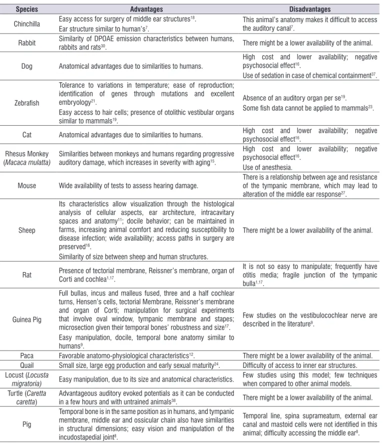
Table 3. Advantages and disadvantages of the use of animal models, by species
Species Advantages Disadvantages
Chinchilla Easy access for surgery of middle ear structures 18.
Ear structure similar to human’s7.
This animal’s anatomy makes it dificult to access
the auditory canal7.
Rabbit Similarity of DPOAE emission characteristics between humans,
rabbits and rats30. There might be a lower availability of the animal.
Dog Anatomical advantages due to similarities to humans.
High cost and lower availability; negative psychosocial effect16.
Use of sedation in case of chemical containment37.
Zebraish
Tolerance to variations in temperature; ease of reproduction;
identiication of genes through mutations and excellent
embryology21.
Easy access to hair cells; presence of otolithic vestibular organs similar to mammals19.
Absence of an auditory organ per se19.
Some ish data cannot be applied to mammals23.
Cat Anatomical advantages due to similarities to humans. High cost and lower availability; negative psychosocial effect16.
Rhesus Monkey (Macaca mulatta)
Similarities between monkeys and humans regarding progressive auditory damage, which increases in severity with aging15.
High cost and lower availability; negative psychosocial effect16.
Use of anesthesia.
Mouse Wide availability of tests to assess hearing damage.
There is a relationship between age and resistance of the tympanic membrane, which may lead to alteration of the middle ear response27.
Sheep
Its characteristics allow visualization through the histological
analysis of cellular aspects, ear architecture, intracavitary spaces and anatomy11; docile behavior; can be maintained in farms, increasing animal comfort and reducing susceptibility to disease infection; wide availability; access paths in surgery are preserved16.
Similarity of size between sheep and human structures.
There might be a lower availability of the animal.
Rat Presence of tectorial membrane, Reissner’s membrane, organ of Corti and cochlea1,17.
It is not so easy to manipulate; frequently have
otitis media; fragile junction of the tympanic bulla1,17.
Guinea Pig
Full bullas, incus and malleus fused, three and a half cochlear turns, Hensen’s cells, tectorial Membrane, Reissner’s membrane
and organ of Corti; manipulation for surgical experiments
that involve oval window, tympanic membrane and stapes;
microsection given their temporal bones’ robustness and size17.
Easy manipulation, docile, temporal bone anatomy similar to humans9.
Few studies on the vestibulocochlear nerve are described in the literature9.
Paca Favorable anatomo-physiological characteristics12. There might be a lower availability of the animal. Quail Small size, large egg production and early sexual maturity24. Dificulty of access to inner ear structures. Locust (Locusta
migratoria) Easy manipulation, due to its size and anatomical characteristics.
Few studies using this model; few techniques when compared to other animal models.
Turtle (Caretta caretta)
Advantageous auditory evoked potentials as it can be conducted
in a few hours and with untrained animals38. There might be a lower availability of the animal.
Pig
Temporal bone is in the same position as in humans, and tympanic membrane, middle ear and ossicular chain also have similarities in structural dimensions; easy vision and manipulation of the incudostapedial joint8.
Temporal line, spina suprameatum, external ear canal and mastoid cells were not identiied in this
With respect to the difference between the number of turns in rats and guinea pigs, the authors of the study analyzed suggest that, in research using drugs that inluence the cochlea, it would be better to use guinea pigs, since it is the model with a larger number of turns17. This study does not justify such an assertion, but there may be a relationship between the greater number of turns and a larger basilar membrane size, which would generate a larger spectrum of frequencies and more hair cells.
Despite the absence of an auditory organ and some results that are not applicable in mammalian animal models, zebraish are widely cited in auditory evalu -ation analyses, mainly in the assessment of hair cells in the lateral line of this animal model, demonstrating that anatomical characteristics, reproducibility and habitat are relevant for their choice in different assessment methods.
As an option to the animal models already estab-lished in the literature and commonly used, the occur-rence of alternative models was veriied. Some animals have anatomical similarities, others have been chosen for practicality, size, reproducibility and even for the possibility of transgenic reproduction.
The Rede de Métodos Alternativos ao Uso de Animais2 (RENAMA) highlights the application of the 3R principles: reduction: the use of the smallest possible number of animals to obtain the necessary information to the experiment; reinement: the pain, suffering or stress of the animal used in the experiment should be minimized; and replacement: when the required level of information is acquired without the use of live verte-brate animals.
In accordance with the normative resolutions of Conselho Nacional de Controle de Experimentação Animal3 (CONCEA), it is essential to aim for the possi-bility of alternatives to the use of animals. If they do not exist, the best techniques proposed should be considered, in order to reine the study and reduce the number of animals used.
With regard to anatomy, isolated attributes may not be decisive for the determination of the animal model used; the advantages and disadvantages of the animals’ auditory system characteristics should be evaluated for the appropriate choice.
In order to achieve the objectives proposed in the research, the ideal is that there is a balance between the auditory evaluation method, its feasibility in relation to access to equipment, the presence of a trained professional to evaluate the animal model, and a
considerable number of advantages in anatomical and structural terms and greater possibility of generalization for the human auditory system.
It was found, in the literature, a great availability of alternatives for conducting auditory evaluations, such as DPOAE, auditory evoked potentials, scanning electron microscopy (SEM) and cytocochleograms.
In relation to the methods of hearing evaluation, DPOAE and BAEP were the most used, showing themselves to be important research tools. These tests, being objective and non-invasive methods, allow the characterization of hearing damage more reliably than behavioral techniques, especially in research with animal models. However, they are limited to the possible use of sedation or anesthesia, which may interfere with wave latency.
Histologically, cellular morphology characterization may facilitate the analysis of the pathophysiology of hearing loss. However, the choice of the animal model interferes with the type of evaluation to be performed. The counting of hair cells in alternative animal models, such as ish, has been performed by methods such as time-lapse imaging, a technique less widespread than optical or electron microscopy, common in mammalian and avian studies.
Variations in studies may be justiied by the research objective, as well as by the animal model chosen and the access to equipment. Therefore, it is essential to know the characteristics of the auditory system of the chosen model, its advantages, disadvantages and limitations in experimental practice. Considering all these aspects, the determination of the number of animals should be as small as possible, respecting, in particular, the standards proposed by RENAMA.
CONCLUSION
The choice of the experimental animal to evaluate the auditory system depends on anatomical, physi-ological, economic, spatial, psychosocial factors and the evaluation’s objective. Rodents are still the most commonly used animal models, and the most frequently cited hearing evaluations are distortion product otoacoustic emissions and brainstem auditory evoked potential.
ACKNOWLEDGEMENTS
REFERENCES
1. Albuquerque AAS. Estudo comparativo da estrutura da orelha interna de ratos e cobaias através da microscopia eletrônica de varredura [monograia]. Jaboticabal (SP): Universidade de São Paulo; 2006. 2. Ministério da Ciência, Tecnologia e Inovação. 2014.
Rede Nacional de Métodos Alternativos (RENAMA). [acesso em 17 de maio 2016]. Disponível em: http:// renama.org.br/
3. Conselho Nacional de Controle de Experimentação Animal (CONCEA). Normativas do CONCEA. Para produção, manutenção ou utilização de animais em atividades de ensino ou pesquisa cientíica. Lei, decreto, portarias, resoluções normativas, orientações técnicas. [acesso em 20 de março 2016]. Disponível em: http://www.mct.gov.br/upd_ blob/0238/238343.pdf
4. König HE, Liebich HG. Órgão Vestibulococlear. In: König HE, Liebich HG. Anatomia dos animais domésticos: texto e atlas colorido. 4ª ed. Porto Alegre (RS): Artmed; 2011. p. 613-28
5. König HE, Liebich HG. Orelha. In: König HE, Liebich HG. Anatomia dos animais domésticos: texto e atlas colorido. 6ª ed. Porto Alegre (RS): Artmed; 2016. p. 601-14.
6. Sula MM, Njaa BL, Payton ME. Histologic Characterization of the Cat Middle Ear: In Sickness and in Health. Vet Pathol. 2014;51(5):951-67.
7. Carrasco ML, Cristóbal Maass OJ, Dentone SL, Miranda GG, Kakuljan PM. Estudio morfológico del oído medio e interno de la Chinchilla laniger. Rev. Otorrinolaringol. Cir. Cabeza Cuello. 2008;68(3):263-74.
8. Garcia LB, Andrade JSC, Testa JRG. Anatomical study of the pigs temporal bone by microdissection. Acta Cir. Bras. 2014;29(3):77-80. 9. Vasconcelos CAC. Aspectos descritivos e
quantitativos da anatomia macroscópica e microscópica do nervo vestíbulo-coclear de cobaias. [Dissertação]. Ribeirão Preto (SP) Faculdade de Medicina de Ribeirão Preto, 2005. 10. Bluestone CD, Swarts JD. Human evolutionary
history: Consequences for the pathogenesis of otitis media. Otolaryngol Head Neck Surg. 2010;143(6):739-44.
11. Soares HB. O estudo histológico do osso temporal do ovino- uma contribuição para a caracterização da ovelha como modelo animal para treinamento e investigação experimental em otologia [Dissertação]. Porto Alegre (RS): Universidade
Federal do Rio Grande do Sul, Faculdade de Medicina, 2004.
12. Silva IA. Caracterização eletroisiológica, funcional e ultraestrutural por microscopia eletrônica de varredura da orelha interna da Cuniculus paca: um novo modelo experimental [dissertação]. Ribeirão Preto (SP): Universidade Federal de São Paulo. Faculdade de Medicina de Ribeirão Preto, 2012. 13. Rauschecker JP, Tian B. Mechanisms and
streams for processing of “what” and “where” in auditory cortex. Proc Natl Acad Sci U S A. 2000;97(22):11800-6.
14. Kaas JH, Hackett TA. Subdivisions of auditory cortex and processing streams in primates. Proc Natl Acad Sci U S A. 2000;97(22):11793-9.
15. Engle JR, Tinling S, Recanzone GH. Age-Related Hearing Loss in Rhesus Monkeys Is Correlated with Cochlear Histopathologies. Snyder J, ed. PLoS ONE. 2013;8(2):e55092.
16. Lavinsky L, Goycoolea M. In search of a teaching, training and experimental model for otological surgery. In: Tos M, Thompson J, editors. Otitis Media Today. Copenhagen (Dinamarca): Kugler Publications; 1997. p. 341-8.
17. Albuquerque AAS, Rossato M, De Oliveira JAA, Hyppolito MA. Conhecimento da anatomia da orelha de cobaias e ratos e sua aplicação na pesquisa otológica básica. Rev Bras Otorrinolaringol. 2009;75(1):43-9.
18. Wolter NE, Harrison RV, James AL. Separating the contributions of olivocochlear and middle ear muscle relexes in modulation of distortion product otoacoustic emission levels. Audiol Neurootol. 2014;19(1):41-8.
19. Uribe PM, Sun H, Wang K, Asuncion JD, Wang Q, Chen CW et al. Aminoglycoside-Induced Hair Cell Death of Inner Ear Organs Causes Functional Deicits in Adult Zebraish (Danio rerio). PLoS One. 2013;8(3):e58755.
20. Brignull HR, Raible DW, Stone JS. Feathers and Fins: Non-mammalian models for hair cell regeneration. Brain Res. 2009;1277:12-23
22. Ou HC, Raible DW, Rubel EW. Cisplatin induced hair cell loss in zebraish (Danio rerio) lateral line. Hear Res. 2007;233(1-2):46-53.
23. Monroe JD, Rajadinakaran G, Smith ME. Sensory hair cell death and regeneration in ishes. Frontiers in Cellular Neuroscience. 2015;9:131.
24. Seidl AH, Sanchez JT, Schecterson L, Tabor KM, Wang Y, Kashima DT et al., Transgenic Quail as a Model for Research in the Avian Nervous System – A Comparative Study of the Auditory Brainstem. J Comp Neurol. 2013;521(1):5-23.
25. Osborne MP, Comis SD, Tarlow MJ, Stephen J. The cochlear lesion in experimental bacterial meningitis of the rabbit. Int J Exp Pathol. 1995;76(5):317-30. 26. Körbes D. Toxicidade de agrotóxico
organofosforado no sistema auditivo periférico de cobaias: estudo anatômico e funcional [dissertação]. Santa Maria (RS): Universidade Federal de Santa Maria; 2009.
27. Zheng QY, Tong YCI, Alagramam KN, Yu H. Tympanometry Assessment of 61 Inbred Strains of Mice. Hear Res. 2007;231(1-2):23-31.
28. Moussavi Najarkola SA, Khavanin A, Mirzaei R, Salehnia M, Muhammadnejad A. Cochlear Damages Caused by Vibration Exposure. Iranian Red Crescent Medical Journal. 2013;15(9):771-4. 29. Moussavi-Najarkola SA, Khavanin A, Mirzaei
R, Salehnia M, Muhammadnejad A, Akbari M. Temporary and permanent level shifts in distortion product otoacoustic emissions following noise exposure in an animal model. Int J Occup Environ Med. 2012;3(3):145-52.
30. Martin GK, Stagner BB, Chung YS, Lonsbury-Martin BL. Characterizing distortion-product otoacoustic emission components across four species. J Acoust Soc Am. 2011;129(5):3090-103.
31. Henderson D, Hu BH, MCFadden SL, Zheng XY, Ding D. The role of glutathione in carboplatin ototoxicity in the chinchilla. Noise Health. 2000;3(9):1-10.
32. El-Badry MM, McFadden SL. Evaluation of Inner Hair Cell and Nerve Fiber Loss as Suficient Pathologies Underlying Auditory Neuropathy. Hear Res. 2009;255(1-2):84-90.
33. El-Badry MM, McFadden SL. Electrophysiological Correlates of Progressive Sensorineural Pathology in Carboplatin-Treated Chinchillas. Brain research. 2007;1134(1):122-130.
34. Morawski K, Telischi FF, Merchant F, Namyslowski G, Lisowska G, Lonsbury-Martin BL. Preventing
Internal Auditory Artery Vasospasm Using Topical Papaverine: An Animal Study. Otol Neurotol. 2003b;24(6):918-26.
35. Rajguru SM, Matic AI, Robinson AM, Fishman AJ, Moreno LE, Bradley A, et al.; Optical cochlear implants: evaluation of surgical approach and laser parameters in cats. Hear Res. 2010;269(1-2):102-11. 36. Palumbo MIP, Resende LAL, Pantoja JCF,
Mayhew IG, Borges AS. Brainstem auditory-evoked potential in Boxer dogs. Pesq. Vet. Bras. 2014a;34(10): 1007-10.
37. Palumbo MIP, Ramos MC, Outeda NC, Resende LAL, Pantoja JCF, Borges AS. A sedação sobre os potenciais evocados auditivos em cães. Cien. Rural. 2014b;44(5):891-6.
38. Martin KJ, Sarah CA, Joseph CG, Anton DT, Gordon BB, David AM. Underwater hearing in the loggerhead turtle (Caretta caretta): a comparison of behavioral and auditory evoked potential audiograms. J Exp Biol. 2012;215(Pt 17):3001-9. 39. Gollisch T, Schütze H, Benda J, Herz AV. Energy
Integration Describes Sound-Intensity Coding in an Insect Auditory System. J Neurosci. 2002;22(23):10434-48.