Tinnitus Neural Mechanisms and Structural
Changes in the Brain: The Contribution of
Neuroimaging Research
Patricia Simonetti
1Jeanne Oiticica
11Department of Otolaryngology, School of Medicine, University of São
Paulo, Brazil
Int Arch Otorhinolaryngol 2015;19:259–265.
Address for correspondence Patricia Simonetti, MSc, Department of Otorhinolaringology, FMUSP, Av Eneas de Carvalo Aguiar 255, São Paulo, Sao Paulo 05403000, Brazil (e-mail: [email protected]).
Introduction
Tinnitus can be defined as the perception of sound (noise, pure tone, among others) in the absence of external sound stimulus. From clinical observation of patients with this symptom, we can see that tinnitus is not only a sound sensation, but a whole sound experience based on an acoustic signal, which can cause many different reactions. Stress, sleep disturbance, and difficulties concentrating are among the changes that affect the quality of life of these patients. We will use the termperceptionto explain the different reactions to the symptom. The key point behind those differences lies exactly in the central nervous system.
For a long time, the peripheral auditory system was assumed, from a psychoacoustic point of view, to explain the symptoms presented by patients, and the cochlea was
considered the main “generator” of tinnitus. This model, however, did not explain, for example, how patients still presented the symptom after surgical removal of a schwan-noma and the auditory nerve section.1The neurophysiologi-cal model suggested that, besides the peripheral auditory system, other systems also appear to be involved in the perception of chronic tinnitus.2Currently, it is clear to the scientific community that central mechanisms contribute crucially to tinnitus generation as well to its persistence, because:
1. Tinnitus persists in most cases, even after complete section of the eighth cranial nerve (auditory nerve).1
2. Many patients with hearing loss simply do not suffer from chronic tinnitus.
3. Tinnitus bothers only a small portion of patients.
Keywords
►
tinnitus
►
functional
neuroimaging
►
auditory cortex
►
neural networks
►
limbic system
Abstract
Introduction
Tinnitus is an abnormal perception of sound in the absence of an external
stimulus. Chronic tinnitus usually has a high impact in many aspects of patients
’
lives,
such as emotional stress, sleep disturbance, concentration dif
fi
culties, and so on. These
strong reactions are usually attributed to central nervous system involvement.
Neuro-imaging has revealed the implication of brain structures in the auditory system.
Objective
This systematic review points out neuroimaging studies that contribute to
identifying the structures involved in the pathophysiological mechanism of generation
and persistence of various forms of tinnitus.
Data Synthesis
Functional imaging research reveals that tinnitus perception is
associated with the involvement of the nonauditory brain areas, including the front
parietal area; the limbic system, which consists of the anterior cingulate cortex, anterior
insula, and amygdala; and the hippocampal and parahippocampal area.
Conclusion
The neuroimaging research con
fi
rms the involvement of the mechanisms
of memory and cognition in the persistence of perception, anxiety, distress, and
suffering associated with tinnitus.
received
November 18, 2014
accepted
February 9, 2015
published online
March 30, 2015
DOI http://dx.doi.org/ 10.1055/s-0035-1548671.
ISSN 1809-9777.
Copyright © 2015 by Thieme Publicações Ltda, Rio de Janeiro, Brazil
4. Psychoacoustic characteristics of tinnitus, such as frequen-cy and loudness, hardly reflect or correlate to the degree of annoyance reported by the patient or even treatment outcomes.
5. Perception of tinnitus does not only occur after damage to the auditory system; it can also be triggered in situations of complete and utter silence.
Those observations have radically changed the previous view of the cochlear tinnitus. Therefore, it is assumed that in cases of persistent and chronic tinnitus, the brain’s tonotopic maps in the auditory cortex are reorganized, with growth and overrepresentation of tinnitus-related frequencies. The straight enrollment of the limbic system is also well known. However, the exact location of the brain areas affected by these changes, and how this justifies and clarifies the set of aberrant and negative reactions implicated in chronic tinni-tus, is still quite controversial.
The main objective of this systematic review is to identify, based on articles published in the literature, the areas of the brain that are actually involved in the pathophysiological mechanisms of chronic tinnitus and the contribution of neu-roimaging research. The U.S. National Library of Medicine (PubMed), Lilacs database, Scielo database, Cochrane database, and Academic Google were used to search for works published in the previous 20 years. The following descriptors were used: tinnitus AND functional neuroimaging; Tinnitus AND PET; Tinnitus AND fRMI; Tinnitus AND neural network. The re-search was limited to articles written in English. We found 1,233 publications, but a few other filters were used: we selected clinical trials in humans as the study design, and only 68 publications matched the criteria. We chose to analyze studies that used positron emission tomography (PET) and functional magnetic resonance imaging (fMRI) as methodo-logical procedures, and we ended with 31 papers to review.
Review of the Literature
According to a few authors,3,4 cochlear injury induced by overexposure to noise or caused by ototoxic agents leads to an enhanced“firing rate,”or rather“spontaneous neuralfiring rate” in various structures, including the dorsal cochlear nucleus, ventral cochlear nucleus, central nucleus of the inferior colliculus, and secondary auditory cortex. The authors correlated the three types of neural activity with tinnitus perception: (1) increased rate of spontaneous neuralfiring; (2) increased neural synchrony; (3) increased“bursting” activ-ity. The increased spontaneous neuralfiring rate in the dorsal cochlear nucleus is observed in cells that persist after cochlear injury, the fusiform cells. This fact shifts cortical balance (excitationversus inhibition), and this inhibitory regulation is diminished by deafferentation of central structures. Among these three mechanisms, the neural synchrony is the compo-nent most related to tinnitus because it produces a high impact on postsynaptic targets and ends up recruiting cortical neu-rons and the perception of tinnitus downstream.
The functional organization of cortical and subcortical neural maps can be altered by sensory experience. Sensory
deprivation destabilizes neural maps, resulting in increased excitability, neural synchronization, and increased spontane-ousfiring in cortical and subcortical neurons.5–7Not surpris-ingly, another author showed that tinnitus is not eliminated after ablation of the dorsal cochlear nucleus.8
Some other types of chronic tinnitus seems to be depen-dent on changes in systems other than the auditory system—
for example, changes in the somatosensory pathway.9,10 Technological advances in neuroimaging and electrophys-iology promoted newfindings in tinnitus research in the last decade. We could see a growing number of studies using hemodynamic techniques such as PET, computed tomogra-phy, single-photon emission computed tomogratomogra-phy, and fMRI. These techniques allow us to measure and track cere-bral bloodflow and metabolic activity of specific regions.
Magnetoencephalography (MEG) and electroencephalog-raphy, which are neuromagnetic techniques, directly mea-sure neural synchrony and also appear in the literature in a complementary form. These techniques allow us to“map”
(i.e., identify) the structures involved in the pathophysiologi-cal mechanism of generation and persistence of various forms of tinnitus. The technology mentioned above contributed to the knowledge we have today: the perception of tinnitus can be the result of associated or overlapped neural maps.
The literature shows great variability in the methodology used for research in neuroimaging. Some studies were done under functional paradigms and focused on the research of anatomical differences in brain structure. Most studies sur-veyed used two basic paradigms—(1) evoked by sound stim-ulation, or (2) controlled stimulus that induces tinnitus (orofacial movements, drug administration, for example)—
and allow intrasubject comparison, trying to identify the neural activity that can be correlated to tinnitus.11The study of the brain at rest might be a more interesting paradigm to demonstrate the“typical” neural activity of tinnitus.12 We can use PET, fMRI, MEG, and electroencephalography to reliably quantify interrelationships of different brain regions that are connected and that constantly exchange information by analyzing the brain activity at rest. Those are called“maps of resting state.”These maps show us the“functional connec-tivity”(operational interactions of multiple and distinct brain regions engaged and quantified).13
PET studies have demonstrated an enhanced metabolic activity in various structures of the auditory system of tinni-tus patients when compared with their controls without tinnitus: medial geniculate nucleus, primary and secondary auditory cortex, and associative temporal-parietal areas.1,11,14,15
Functional Studies
Our research revealed neuroimaging studies that used differ-ent paradigms, either PET or fMRI, ranked in differdiffer-ent tasks: evoked by sound, somatosensory modulation,16 eye move-ment,17,18and administration of drugs such as lidocaine that partially or completely suppress the perception of tinnitus, as we could see in►Table 1.
without complaint and revealed increased activity in the left primary auditory cortex (area 41 Broadmann).19
In 1998, a study of a group of individuals who perceived change in sensation of tinnitus by means of orofacial move-ments and a control group underwent PET examinations.16 The subjects received sound stimuli unilaterally, and those individuals who“modulated”the tinnitus by jaw movement produced unilateral, rather than bilateral, response as expected.
One of the major difficulties in researching chronic tinni-tus is to separate the effects of hearing loss and tinnitinni-tus, because the vast majority of patients with tinnitus have some degree of hearing loss. Hyperacusis, which affects 40% of these patients, is also an item of confusion in the interpreta-tion of research results. Few studies showed that increased activation of the inferior colliculus after sound stimulation in patients with tinnitus was correlated to the perception of tinnitus, but hyperacusis and hearing loss were not controlled in the study.9,14
Researchers studying the involvement of the cognitive system compared three groups (individuals with normal hearing versus subjects with bilateral hearing loss, and those with bilateral hearing loss and chronic tinnitus) in two different situations: passive and active listening.20The sub-jects were asked to perform auditory discrimination tasks of nonverbal sounds. The author sought to test the distract effect (attention) of tinnitus in multisensory activities. MRI for functional mapping was used. In the passive listening activi-ties, there was no significant difference between groups. All showed greater activation in the superior temporal cortex, including medial temporal and superior gyrus and superior temporal sulcus. During the activities of active listening, differences were evident, but differences were not statisti-cally significant. The study showed the differential involve-ment of the neural map of auditory attention and short-term
memory network, encompassing cortex regions in frontal, parietal, temporal, and anterior cingulate.
A considerable number of studies uses fMRI BLOD (blood oxygen level dependent) technique (depending on blood oxygen level) to investigate the neural correlates of tinnitus. This technique, however, is not able to register sustained increases in spontaneous activity. Consequently, studies have used sound stimulus to verify the abnormal auditory proc-essing in patients with tinnitus and hearing loss.21There was greater activation in the inferior colliculus, and we could see that the greater the stimuli, the greater the response in these regions (peripheral auditory tract), except in the dorsal cochlear nucleus. The correlation between cortical and sub-cortical groups was also negative for those individuals, and once these connections occur in the thalamus, this correlation may be interpreted as a thalamic dysfunction.
Stress and negative emotions may actually increase the perception of tinnitus as the neurophysiological model de-termined.2That model primarily included the limbic system as an integral part in the pathophysiology of chronic tinnitus. Other authors have attempted to establish the involvement of this system in the regulation of aberrant auditory sys-tem.22–24In an attempt to identify structural changes,2228 patients with chronic tinnitus and normal hearing were compared with a control group matched for sex and age. Analyzing the region of interest, the researchers found de-creased gray matter in regions responsible for environmental stimuli adaptation and inhibitory function of unpleasant stimuli: nucleus acumens and thalamic reticular nucleus. Using fMRI and presenting stimulus of various frequencies, one coincident with the perception of tinnitus (obtained through psychoacoustic measurements), the researchers compared corticostriatal limbic-evoked, auditory cortex and thalamus responses of subjects with and without tinnitus. In addition to the altered auditory areas (medial geniculate body Table 1 Summary of PET and FMRI studies and neural structures involved in tinnitus perception
Authors n Method Paradigm Results (activated areas/neuroanatomic alterations)
Lockwood et al16 4/6 PET Orofacial movements Temporal gyrus/hippocampus
Giraud et al18 4 PET Eye movements Associated areas/auditory/temporal-parietal Mirz et al24 12 PET Lidocaine/suppression Right temporal-front gyrus
Andersson et al41 1 PET Lidocaine/suppression Left temporal lobe
Mirz et al41 8 PET Sound/lidocaine Amygdala
Muhlau et al22 28/28 fMRI Sound Nucleus acumbens/thalamic /reticular nucleus (↓volume) Plewnia et al36 9 PET Lidocaine/rest Postcingulum, temp-par cortex
Melcher et al14 fMRI Sound Inferior colliculus
Van Gendt et al42 18/9 fMRI Gaze evoked/sound "IC and CN ;↓AC /medial genicular body Rauschecker et al26 23/21 fMRI Sound ↓Gray matter vmPFC/nucleus accumbens;
"gyrification dmPFC
Husain et al20 8/7/11 fMRI Sound ↓Front/parietal lobes Schecklmann et al27 91 PET Rest Left Heschl gyrus
and Heschl gyrus), significant differences were observed in the subcallosal area in the ventromedial prefrontal cortex and nucleus acumens. Continuing this research, the authors also show anatomical differences in the ventral prefrontal cortex (reduction in gray matter) of subjects with chronic tinnitus, responsible for suppressing the aberrant auditory system activity in the thalamic structure. These differences were correlated positively to perceptual factors such as loudness and awareness (percentage of time that the individual per-ceives the tinnitus), and the correlation with other variables such as depression and anxiety were weak or absent.26
Ninety-one individuals with chronic tinnitus underwent an F-deoxyglucose PET study and had clinical characteristics as duration and distress correlated to neuronal activation patterns.27Tinnitus duration correlated positively with right inferior frontal, right ventromedial prefrontal, and right posterior cingulated cortex. Parahippocampal and hippocam-pal areas and posterior inferior temporal gyrus were corre-lated to tinnitus distress, rated by tinnitus questionnaire score. The study also revealed the overactivation of the left Heschl gyrus.
Resting State/Functional Connectivity Studies
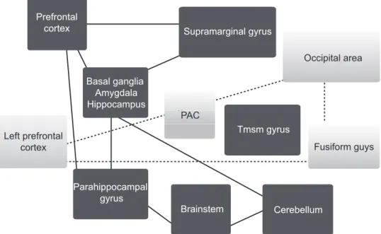
As mentioned earlier, study of the brain at rest has been increasingly used as an alternative to functional“mapping”of tinnitus, as the results show consistent and reliable patterns of functional connectivity, which in turn reflect the percep-tual and cognitive processes present in patients with tinnitus. Among the studies surveyed, as seen in►Fig. 1, several areas showed greater connectivity: the brainstem, basal ganglia, hippocampus and parahippocampus areas, and cerebellum. Likewise, some areas showed less connectivity: the primary
auditory cortex, left prefrontal cortex, left fusiform gyrus, and occipital regions of both hemispheres.
In a pilot study,28researchers evaluated the spontaneous activity of auditory areas between the right and left brain hemispheres with and without tinnitus: the scores of func-tional connectivity between two groups were measured. The
findings (mean connectivity scores) were significantly lower in the auditory areas of the left and right hemispheres for individuals with tinnitus. Individuals with tinnitus also showed increased connectivity in the left amygdala and medial prefrontal cortex.
Another study,12 mentioned previously, using fMRI and analyzing functional connectivity of the brain at rest of 12 tinnitus patients and comparing with controls matched for gender and hearing loss, revealed robust bilateral activity between auditory cortical areas in both hemispheres (proba-bly due to symmetrical hearing loss in both groups) and increased functional connectivity in the right supra marginal gyrus and middle temporal gyrus for the tinnitus group.
A randomized clinical trial compared 17 subjects with chronic tinnitus with age-matched controls using fMRI and analyzing maps of functional connectivity of these individu-als as they performed cognitive tasks.29 We know that cognitive distraction tasks decreases the perception of tinni-tus and how tinnitinni-tus influences the ability to concentrate for assignments. The study revealed differences between audito-ry/visual/occipital cortical maps between the tinnitus group and the control group.
A group of researchers performed fMRI to verify functional connectivity at rest into three groups: individuals with hearing loss and mild tinnitus, individuals with hearing loss, and a third control group of normal-hearing subjects
Prefrontal
cortex Supramarginal gyrus
Basal ganglia Amygdala Hippocampus
Parahippocampal gyrus
Brainstem
Tmsm gyrus
Cerebellum
Occipital area
Fusiform guys Left prefrontal
cortex
PAC
without tinnitus, matched for age.30After primary analysis, three areas of connectivity were identified: areas of resting state auditory state (primary and secondary auditory cortex), default mode (default mode network: medial prefrontal cortex, posterior cingulate, precuneus, bilateral superior frontal gyrus and bilateral inferior parietal lobes), and atten-tion maps (dorsal attenatten-tion network: bilateral posterior intraparietal sulcus and visual field). The results showed a strong correlation between the functional regions of the limbic system (specifically in the left amygdala and dorso-medial prefrontal cortex) in the parahippocampus left area and attention network in subjects with tinnitus when com-pared with their peers without tinnitus and without hearing loss.
Another study compared the Tinnitus Handicap Inventory, tinnitus questionnaire, and tinnitus characteristics such as loudness and duration during resting-state fMRI to investi-gate possible correlations in functional connectivity.31 Re-sults showed a modified functional connectivity pattern in tinnitus sufferers’parahippocampal region in addition to the posterior cingulate/precuneus region and a correlation was found with the Tinnitus Handicap Inventory score.
In general, tinnitus research using neuroimaging still produced divergent results, with poor reproducibility. This statement is made based on studies done with different techniques, different methodologies, and few patients.32
From the current functional imaging techniques studies we have so far, new models and new therapies have been proposed, contributing to a more contemporary view of chronic tinnitus.
Discussion
All the studies mentioned previously showed an evident and reciprocal enrollment of emotional and sensory areas, reveal-ing increased cerebral gray matter in the central auditory pathways, located only in the thalamus, and in direct opposi-tion, a decrease in cerebral gray matter outside the central auditory pathway in the subcallosal area, specifically in the nucleus acumens.22,26 The reduction in subcallosal gray matter (including the nucleus acumens) is intriguing for several reasons:
1. This area is directly related to unpleasant emotions trig-gered by musical dissonance.
2. Activation of this region is triggered by aversive sounds. 3. This area plays a crucial role in the generation of adaptive
behavioral responses to environmental stimuli.
4. In humans, this area is activated during Pavlovian conditioning.
5. In animals, it is implicated in phenomena called“targeted reward”and“avoidance learning.”
6. In animals with this area injured, the habituation of the trauma preceded by acoustic warning sound is diminished. 7. The nucleus acumens receives glutamatergic afferent in-puts from the amygdala afferent and serotonergic raphe nucleus of the brainstem structures involved in the regu-lation of sleep and the state of excitement.
8. Between the nucleus acumens and the thalamus, there are interconnected parallel circuits (reticular thalamic nucle-us) so that thefirst circuits inhibit the second.
Therefore, the reduced volume of gray matter in the nucleus acumens should decrease its inhibitory influence on the thalamus. But the increase of gray matter concentra-tion in the posterior thalamus suggests a new model of generation of tinnitus: (1) reorganization in the medial geniculate nucleus (possibly through corticofugal feedback) due to peripheral auditory deafferentation, which generates neuronal activity related to tinnitus in the central auditory pathways and leads to a permanent increase in the concen-tration of thalamic gray matter on thalamus; (2) the tinnitus-related activity in the medial geniculate nucleus is transmit-ted in parallel to the limbic structures through the amygdala, which in turn triggers negative emotional associations with the perception of tinnitus. The hypothesis is that permanent habituation mediated by the subcallosal area (nucleus acu-mens), which normally helps to cancel the tinnitus signal in the thalamus, prevents the signal being relayed to the audi-tory cortex. Thus, the reduction in volume of the subcallosal region results in chronic tinnitus.
In the Muhlau study,22these anatomical differences (de-crease of gray matter in the ventromedial prefrontal cortex and decreased volume in dorsomedial prefrontal cortex and supramarginal gyrus) were correlated with psychogenic and perceptual factors related to tinnitus. The difference in thick-ness of the anterior insula was positively correlated to factors such as anxiety and stress, and the thickness of the anterior subcallosal angle was related to factors such as depression and anxiety. Alterations found in the dorsomedial prefrontal cortex were positively correlated to the percentage of time that the individual was aware of tinnitus. This study confirms the alterations on prefrontal cortex are not associated with psychogenic factors but perceptual factors, confirming the role of this structure in regulating the perception of tinnitus. The subcallosal area contains dopaminergic and seroto-nergic neurons whose activity is modulated by stress and excitability, factors known to affect the perception of tinnitus. Depression, insomnia, and aging are associated with reduced levels of brain serotonin (including the nucleus acumens) and are also correlated to tinnitus. Therefore:
1. Neural activity related to tinnitus is perpetuated primarily in the medial geniculate nucleus, a result of reorganization after peripheral deafferentation.
2. The inhibitory feedback of the subcallosal area usually helps tune the neural activity related to tinnitus. 3. Reduction of gray matter in the subcallosal area reduces
feedback and increases risk of tinnitus in patients with hearing impairment. However, these results do not fully explain whether these structural changes are responsible for the onset of tinnitus or are consequential to tinnitus installation.
Some of these neural changes include: (1) the reorganization of the tonotopic map in the auditory cortex and thalamic struc-tures; (2) hyperactivity of these structures (not in the auditory nervefibers); (3) increasedfiring rate of neurons (burst type) in the dorsal cochlear nucleus; (4) increased synchronous neural activity particularly in the corresponding area of peripheral hearing loss regions.
Apparently, the reduction of peripheral afferent auditory triggers adaptive compensatory changes in the balance between excitation and inhibition (homeostatic plasticity), which can even occur within a physiological range of neuronalfiring (no tinnitus), or can be an unwanted side effect (in susceptible individuals) of the increased spontaneous neural activity, with phase locked in a synchronous pattern, leading to the perception of tinnitus. Stress is an important mechanism in the induction of neural plasticity. Despite the fact that stress has a protective effect against noise-induced trauma, the combination of stress and hearing loss may increase the likelihood of tinnitus.
Functional neuroimaging studies of the brain confirm that the brain regions affected by tinnitus extend beyond the auditory centers, including areas of the brain involved in cognitive processing in higher centers. The areas of the brain that differ in individuals with and without tinnitus have described in detail.20,33 Coincidentally, the affected brain regions (prefrontal cortex, parietal cortex, and insula and cingulate gyrus) in patients with tinnitus are the same regions that show increased activity during the performance of cogni-tive tasks that require attention in normal individuals. Neuro-cognitive research also shows that activation of this neural network is closely related to consciousness or awareness phenomena. Another interesting fact is that, restricted to the auditory pathways, aberrant neural activity is not sufficient to generate tinnitus. It is necessary that the aberrant activity is dispersed in the global neural network. The perception of a stimulus is given by the interconnection of systems.
Neuroimaging has shown that brain activity and function-al connectivity in patients in a neurovegetative state (without consciousness or awareness) is decreased in the anterior and posterior cingulate and front temporal parietal areas. In these patients, painful stimuli activate the primary somatosensory cortex and thalamus, which is disconnected from the second-ary cortex. Similarly, auditory stimuli activate the primsecond-ary cortex only bilaterally. Stimuli become conscious only when connected to the frontal and parietal areas (cingulate cortex, dorsal anterior cortex, and anterior insula). This network is important for the integration of sensory experience, and increased connectivity in these regions results in a state of sustained vigilance. In summary, deafferentation (of any type) results in an increase in neural activity in the primary cortex and reaches consciousness when connected to the associative coactivated (front-parietal, cingulate cortex, and anterior insula) areas.34Based on this concept, another study suggest that drugs that have multiple effects of low-level synaptic processes in highly specialized neural pathways (therapeutic rifles) may be more effective in breaking this behavior of the network and reduce tinnitus.35
The difficulty to treat and effectively control persistent and chronic tinnitus relies on the complexity and range of
aber-rantly activated neural networks. Thefirst step is to under-stand the factors behind the loudness of tinnitus (attention, context, and personality).
As we have seen, the brain changes in patients with tinnitus are not restricted to auditory regions. It is already reported that there is a functional increase in responses (activity) in various nonauditory structures, including the hippocampus,16 amygdala,24and cingulate gyrus36; decreased gray matter was reported in the hippocampus and subcallosal area,15including the nucleus acumens.22 There is also an increased activity coupled phase seen in MEG studies between the anterior cingulate and the right frontal lobe, which is more intense in patients with tinnitus compared with controls; this is directly correlated with the observed scores on bothersome scales. These results suggest that the thalamus is involved in the neural tinnitus network, which possibly is the prerequisite for the conscious perception of sound.37 The limbic and the prefrontal area are associated with emotion and attention and contribute to this anxiety in many patients with tinnitus. Recent theories indicate that nonauditory regions have direct implication on the onset of tinnitus perception. The observa-tions that approximately two thirds of patients are able to modulate the intensity and the frequency of their tinnitus by somatic maneuvers (jaw clenching, tension of the neck muscles) and that tinnitus can arise from somatosensory injuries led to the search of neural connections between the auditory and somatosensory systems that could explain these phenomena.9,38–40When electrical stimulation of somatosen-sory pathways precedes the acoustic stimulus, this may change both the peak time of the response evoked by sound and the synchrony offiring between neurons in the dorsal cochlear nucleus, which can be correlated to tinnitus.
Final Comments
cortex and subcortical structures, between the central infor-mation (predictive or effector, top-down) and the informa-tion from the periphery (or afferent obtained, bottom-up), can trigger auditory attention and induce the neural activity while the brain tries to build a more accurate representation of the actual hearing status.
References
1 House JW, Brackmann DE. Tinnitus: surgical treatment. Ciba Found Symp 1981;85:204–216
2 Jastreboff PJ. Phantom auditory perception (tinnitus): mechanisms of generation and perception. Neurosci Res 1990;8(4):221–254 3 Eggermont JJ, Roberts LE. The neuroscience of tinnitus. Trends
Neurosci 2004;27(11):676–682
4 Eggermont JJ, Roberts LE. The neuroscience of tinnitus: under-standing abnormal and normal auditory perception. Front Syst Neurosci 2012;6:53
5 Noreña AJ. An integrative model of tinnitus based on a central gain controlling neural sensitivity. Neurosci Biobehav Rev 2011;35(5): 1089–1109
6 Schaette R, McAlpine D. Tinnitus with a normal audiogram: physiological evidence for hidden hearing loss and computational model. J Neurosci 2011;31(38):13452–13457
7 Engineer ND, Møller AR, Kilgard MP. Directing neural plasticity to understand and treat tinnitus. Hear Res 2013;295:58–66 8 Roberts LE, Eggermont JJ, Caspary DM, Shore SE, Melcher JR,
Kaltenbach JA. Ringing ears: the neuroscience of tinnitus. J Neuro-sci 2010;30(45):14972–14979
9 Levine RA. Somatic (craniocervical) tinnitus and the dorsal cochlear nucleus hypothesis. Am J Otolaryngol 1999;20(6): 351–362
10 Shore SE. Multisensory integration in the dorsal cochlear nucleus: unit responses to acoustic and trigeminal ganglion stimulation. Eur J Neurosci 2005;21(12):3334–3348
11 Lanting CP, de Kleine E, van Dijk P. Neural activity underlying tinnitus generation: results from PET and fMRI. Hear Res 2009; 255(1–2):1–13
12 Davies J, Gander PE, Andrews M, Hall DA. Auditory network connectivity in tinnitus patients: a resting-state fMRI study. Int J Audiol 2014;53(3):192–198
13 Rogers BP, Morgan VL, Newton AT, Gore JC. Assessing functional connectivity in the human brain by fMRI. Magn Reson Imaging 2007;25(10):1347–1357
14 Melcher JR, Levine RA, Bergevin C, Norris B. The auditory midbrain of people with tinnitus: abnormal sound-evoked activity revis-ited. Hear Res 2009;257(1–2):63–74
15 Landgrebe M, Langguth B, Rosengarth K, et al. Structural brain changes in tinnitus: grey matter decrease in auditory and non-auditory brain areas. Neuroimage 2009;46(1):213–218
16 Lockwood AH, Salvi RJ, Coad ML, Towsley ML, Wack DS, Murphy BW. The functional neuroanatomy of tinnitus: evidence for limbic system links and neural plasticity. Neurology 1998;50(1):114–120 17 Simmons R, Dambra C, Lobarinas E, Stocking C, Salvi R. Head, Neck, and Eye Movements That Modulate Tinnitus. Semin Hear 2008; 29(4):361–370
18 Giraud AL, Chéry-Croze S, Fischer G, et al. A selective imaging of tinnitus. Neuroreport 1999;10(1):1–5
19 Arnold W, Bartenstein P, Oestreicher E, Römer W, Schwaiger M. Focal metabolic activation in the predominant left auditory cortex in patients suffering from tinnitus: a PET study with [18F]deoxyglucose. ORL J Otorhinolaryngol Relat Spec 1996; 58(4):195–199
20 Husain FT, Pajor NM, Smith JF, et al. Discrimination task reveals differences in neural bases of tinnitus and hearing impairment. PLoS ONE 2011;6(10):e26639
21 Boyen K, de Kleine E, van Dijk P, Langers DR. Tinnitus-related dissociation between cortical and subcortical neural activity in humans with mild to moderate sensorineural hearing loss. Hear Res 2014;312:48–59
22 Mühlau M, Rauschecker JP, Oestreicher E, et al. Structural brain changes in tinnitus. Cereb Cortex 2006;16(9):1283–1288 23 Leaver AM, Renier L, Chevillet MA, Morgan S, Kim HJ, Rauschecker
JP. Dysregulation of limbic and auditory networks in tinnitus. Neuron 2011;69(1):33–43
24 Mirz F, Pedersen B, Ishizu K, et al. Positron emission tomography of cortical centers of tinnitus. Hear Res 1999;134(1–2): 133–144 25 Seydell-Greenwald A, Leaver AM, Turesky TK, Morgan S, Kim HJ,
Rauschecker JP. Functional MRI evidence for a role of ventral prefrontal cortex in tinnitus. Brain Res 2012;1485:22–39 26 Leaver AM, Seydell-Greenwald A, Turesky TK, Morgan S, Kim HJ,
Rauschecker JP. Cortico-limbic morphology separates tinnitus from tinnitus distress. Front Syst Neurosci 2012;6:21
27 Schecklmann M, Landgrebe M, Poeppl TB, et al. Neural correlates of tinnitus duration and distress: a positron emission tomography study. Hum Brain Mapp 2013;34(1):233–240
28 Kim JY, Kim YH, Lee S, et al. Alteration of functional connectivity in tinnitus brain revealed by resting-state fMRI? A pilot study. Int J Audiol 2012;51(5):413–417
29 Burton H, Wineland A, Bhattacharya M, Nicklaus J, Garcia KS, Piccirillo JF. Altered networks in bothersome tinnitus: a functional connectivity study. BMC Neurosci 2012;13:3
30 Schmidt SA, AkrofiK, Carpenter-Thompson JR, Husain FT. Default mode, dorsal attention and auditory resting state networks exhibit differential functional connectivity in tinnitus and hearing loss. PLoS ONE 2013;8(10):e76488
31 Maudoux A, Lefebvre P, Cabay JE, et al. Connectivity graph analysis of the auditory resting state network in tinnitus. Brain Res 2012; 1485:10–21
32 Song JJ, De Ridder D, Van de Heyning P, Vanneste S. Mapping tinnitus-related brain activation: an activation-likelihood estima-tion metaanalysis of PET studies. J Nucl Med 2012;53(10): 1550–1557
33 Langguth B, Schecklmann M, Lehner A, et al. Neuroimaging and neuromodulation: complementary approaches for identifying the neuronal correlates of tinnitus. Front Syst Neurosci 2012;6:15 34 De Ridder D, Elgoyhen AB, Romo R, Langguth B. Phantom percepts:
tinnitus and pain as persisting aversive memory networks. Proc Natl Acad Sci U S A 2011;108(20):8075–8080
35 Elgoyhen AB, Langguth B, Vanneste S, De Ridder D. Tinnitus: network pathophysiology-network pharmacology. Front Syst Neurosci 2012;6:1
36 Plewnia C, Reimold M, Najib A, et al. Dose-dependent attenuation of auditory phantom perception (tinnitus) by PET-guided repeti-tive transcranial magnetic stimulation. Hum Brain Mapp 2007; 28(3):238–246
37 Schlee W, Weisz N, Bertrand O, Hartmann T, Elbert T. Using auditory steady state responses to outline the functional connec-tivity in the tinnitus brain. PLoS ONE 2008;3(11):e3720 38 Rubinstein B, Axelsson A, Carlsson GE. Prevalence of signs and
symptoms of craniomandibular disorders in tinnitus patients. J Craniomandib Disord 1990;4(3):186–192
39 Pinchoff RJ, Burkard RF, Salvi RJ, Coad ML, Lockwood AH. Modula-tion of tinnitus by voluntary jaw movements. Am J Otol 1998; 19(6):785–789
40 Shore S, Zhou J, Koehler S. Neural mechanisms underlying somatic tinnitus. Prog Brain Res 2007;166:107–123
41 Andersson G, Lyttkens L, Hirvelä C, Furmark T, Tillfors M, Fredrik-son M. Regional cerebral bloodflow during tinnitus: a PET case study with lidocaine and auditory stimulation. Acta Otolaryngol 2000;120(8):967–972