Use of intravitreal bevacizumab
or triamcinolone acetonide as
a preoperative adjunct to vitrectomy
for vitreous haemorrhage in diabetics
Injeção intravítrea de bevacizumabe ou triancinolona
como adjuvantes da vitrectomia posterior no tratamento
da hemorragia vítrea em diabéticos
Daniel Araújo Ferraz
1, Celso Morita
2, Rony Carlos Preti
1, Vinicius Paganini Nascimento
2,
Otacílio Oliveira Maia Junior
3, André Carvalho de Barros
2, Beatriz SayuriTakahashi
2, Walter Yukihiko Takahashi
41Postgraduate in Retina and Vitreous, Department of Ophthalmology, Universidade de São Paulo (USP) – São Paulo (SP), Brazil;
2Assistant of the Vitreoretinal Service, Department of Ophthalmology, Universidade de São Paulo (USP) – São Paulo (SP), Brazil;
3Vitreoretinal specialist of the Department of Ophthalmology, São Rafael Hospital, Monte Tabor Foundation – Salvador (BA), Brazil;
Postdoctoral of the Department of Ophthalmology, Universidade de São Paulo (USP) – São Paulo (SP), Brazil;
4Head of the Vitreoretinal Service, Department of Ophthalmology, Universidade de São Paulo (USP) – São Paulo (SP), Brazil.
The authors declare no conflicts of interest
A
BSTRACTPurpose: To evaluate the effect of preoperative intravitreal bevacizumab (IVB) or triamcinolone (IVT) on the rate of early postvitrectomy hemorrhage in proliferative diabetic retinopathy (PDR). Methods: Eligible eyes were assigned randomly to 1 of 3 groups: the IVB group received 1.25 mg bevacizumab, the IVT group received 4,0mg triamcinolone and the control group underwent a sham procedure. The primary outcome measure was the incidence of early postvitrectomy hemorrhage. Secondary outcome measures included changes in visual acuity (BCVA) and adverse events. Results: Twenty and seven eyes, 9 in each group were randomized. The incidence of vitreous hemorrhage was lower in the IVB group (p=0.18). Postoperative vitreous hemorrhage at 1 month also was less in the IVB group compared with the control group (p ≥ 0.05). The rate of bleeding immediately after surgery was higher in IVT group with 4 (44.4%) cases. The overall mean visual acuity was 1.72 ± 0.37 logMAR preoperatively and 1.32 ± 0.73 logMAR in 6 months after surgery. Accessing visual acuity by group evidenced that the IVB group had initial mean logMAR VA of 1.87 and 1.57 logMAR VA at the six months (p = 0.84). In IVT group, initial mean VA was 1.75 logMAR and 0.96 logMAR VA at six months (p ≤ 0.001). And in control group, the initial mean VA was 1.85 logMAR and 1.57 logMAR VA at six months (p= 0.34). Conclusion: Intravitreal injection of bevacizumab 1 week before vitrectomy seems to reduce the incidence of early postvitrectomy hemorrhage in diabetic patients. There was a better visual acuity outcome in the triamcinolone group.
Keywords: Diabetic retinopathy/surgery; Vitrectomy; Angiogenesis inhibitors/therapeutic use; Triamcinolone/therapeutic use; Visu-al acuity
I
NTRODUCTIONI
n spite of all the scientific advances in medicine and in our knowledge of the pathophysiology and treatment of diabe-tes over the past 25 years, diabetic retinopathy remains one of the leading causes of blindness in individuals between 20 to 64 years-old in industrialized countries(1,2).Proliferative diabetic retinopathy (PDR) is a major cause of visual loss in diabetic patients. In PDR, the growth of new vessels from the retina or optic nerve, is thought to occur as a result of vascular endothelial grow factor (VEGF) released into
the vitreous cavity as a response to ischaemia(3-5). Early
postvitrectomy hemorrhage in diabetic patients is a major concern for both surgeons and patients. On the patient’s side, expectations regarding visual improvement are not met, and this may be crucial in monocular patients. On the surgeon’s side, this complication interferes with fundus examination, detection of iatrogenic retinal breaks, and performing laser therapy.
Because VEGF has been shown to play a major role in retinal neovascularization(3,4), in conjunction with other factors(6,7)
anti-VEGF treatments have been hypothesized as an alternative adjunctive treatment for retinal neovascularization(8,9).
To date, pars plana vitrectomy (PPV) is the treatment of choice for PDR with vitreous hemorrhage and retinal detachment. There is a high risk of complication due to the bleeding from fibrovascular membrane(10).
Oral, topical and periocular steroids have been widely used in the past in many pathologic ocular conditions that have an underlying inflammatory basis. The retinal neovascular proliferation in PDR often has an accompanying inflammatory component. The intravitreal (IV) steroid injections, particularly triamcinolone acetonide, may potentially be important in quelling intraocular inflammation(11-13). It is therefore intuitive that IV
steroid injections could be beneficial to PDR.
Bevacizumab (Avastin Genetech Inc.,South San Francisco, California, USA) is a humanized anti-vascular endothelial growth factor (VEGF) antibody used for metastatic colorectal cancer. Recent reports have described the application of bevacizumab to treat ocular neovascular disorders including PDR(14-15). Adjunctive
use of intravitreal bevacizumab for severe PDR before vitrectomy has also been reported. However, the preferable timing from the injection to surgery has not been determined yet(16-18).
The purpose oh this study was to evaluate the efficacy of bevacizumab or triamcinolone acetonide intravitreal injections
R
ESUMOObjetivo: Avaliar o efeito no pré-operatório da injecao intravítrea de bevacizumab (IVB) ou triancinolona (IVT) sobre a taxa de hemorragia precoce pos-vitrectomia na retinopatia diabética proliferativa. Métodos: Os olhos foram distribuídos em três grupos: IVB - 1,25 mg bevacizumab, IVT - 4,0 mg de triancinolona e o grupo controle - simulação da injeção. O objetivo primário foi a avaliação da incidência da hemorragia precoce pós-vitrectomia. Os objetivos secundários incluíram mudanças na acuidade visual corrigida e eventos adversos relacionados à injeção. Resultados: Dos Vinte e sete olhos, 9 foram randomizados em cada grupo. A incidência de hemorragia
vítrea foi menor no grupo IVB (P=0,18). A hemorragia vítrea em 1 mês também foi menor no grupo IVB (P ≥ 0,05). A taxa de
sangramento pós-operatório imediato foi maior no grupo IVT com 4 (44,4%) dos casos. A média da acuidade visual (AV) foi de 1,72 ± 0,37 logMAR no pré-operatório e 1,32 ± 0,73 logMAR em 6 meses após a cirurgia. Analisando a AV por grupo evidenciamos que o grupo IVB tinha inicialmente AV média logMAR de 1,87 e AV logMAR de 1,57 em seis meses (p = 0,84). No grupo IVT, a média inicial de AV foi de 1,75 logMAR e 0,96 logMAR em seis meses (p ≤ 0,001). E no grupo controle, a média inicial foi de 1,85 logMAR e 1,57 logMAR no seis meses (p = 0,34). Conclusão: A injeção intravítrea de bevacizumab antes da vitrectomia parece diminuir a incidência de hemorragia vítrea precoce pós-vitrectomia em diabéticos. Houve um melhor resultado na acuidade visual no grupo da triancinolona.
Descritores: Retinopatia diabética/cirurgia; Vitrectomia; Inibidores da angiogênese/uso terapêutico; Bevacizumabe; Triancinolona; Acuidade visual
as an adjuvant before vitrectomy surgery for vitreous haemorrhage in diabetic patients.
M
ETHODSThe design of this study was an interventional consecutive, randomized prospective study. Twenty-seven patients (27 eyes) aged from 17 to 79 years old (mean age 52 years) with severe PDR and persistent Vitreous Hemorrhage (VH) for 6 months were enrolled and underwent PPV. Of the 27 enrolled patients, 9 underwent IV bevacizumab (1.25 mg in 0.05 ml) 5–7 days before PPV (IVB group ), 9 underwent IV triamcinolone acetonide (4.0 mg in 0.1 ml) 5-7 days before PPV (IVT group) and 9 had surgery alone (control group). The patientes were randomized in order to assign each study participant to group 1, 2 or 3. The main outcome measure was VH recurrences; a secondary goal was visual outcome at 6 months. The primary outcome measure was the incidence of early (<4 weeks) and late (>4 weeks) recurrent VH. Recurrent VH was defined as a new episode of grade 1 or more VH occurring 1 week after surgery.
Demographics and clinical findings, including age, gender, diabetes mellitus category and disease duration, blood glucose control, diabetes mellitus type, hypertension and renal disease anticoagulation were recorded. At baseline, each patient underwent detailed ophthalmologic examinations, including measurement of best-corrected visual acuity (BCVA) using EDTRS acuity test, slit-lamp biomicroscopic examination and indirect funduscopic examination when possible. Vitreous hemorrhage was evaluated by eye ultrasound. Post-surgery follow-up was scheduled at 1 week, 1 and 6 months. At each visit, complete ophthalmologic examinations were performed. The graduation of VH was done by indirect fundus examination using the classification above (clinical findings) and this findings evaluated by the score changes during the visits(19).
Exclusion criteria were: tractional retinal detachment, tractional-rhegmatogenous retinal detachment, previous
Score Description Clinical findings
1 Minimal Posterior pole clearly visible
2 Mild Posterior pole details slightly haze
3 Moderate Posterior pole details very haze
4 Marked Posterior pole details barely visible
vitrectomy in the study eye.
The procedure followed standard intravitreal injection protocol(19). The surgical technique was the same for the groups.
All patients underwent 20- gauge three-port PPV and further endolaser panretinal photocoagulation.
The study protocol adhered to the tenets of the Declaration of Helsinki. The off-label use of the drug and its potential risks and benefits were discussed extensively with all patients. All participants gave written informed consent before entering the study. Visual acuity measured in EDTRS was converted to logMAR for data analysis. Continuous variables were presented as mean and standard deviations (SD) and the Mann–Whitney test was performed. Non-parametric Friedman test was used to compare the evolution of mean VA. A significance level of 95% was considered statistically significant.
R
ESULTSOf the 27 cases, nine were randomized to IVB group, nine to IVT group and nine to control group. The mean age of patients was 52.2 ± 13.9 years, 17 males and 10 females. The clinical characteristics of patients are shown in Table 1. There was no statistically significant between the clinical findings and visual acuity or recurrent VH.
The incidence of vitreous hemorrhage was lower in the IVB group 1 week after the operation (p=0.18). Postoperative
Initiais Age Sex VA Pre VA pos Recurrent Time(days) Type of DM AAS Insuline Hb1ac Creatinine VH – (Score)
NT 82 F 1,9 1,2 - - 2 No No 6,1 1,7
OM 57 M 2,2 1,9 - - 2 Yes Yes 5,9 1,5
JRM 58 M 1,1 1,1 2 7 2 Yes Yes 7 1,8
VS 56 M 2,2 1,9 - - 2 Yes Yes 5,5 1,5
LCS 58 M 2,2 1,9 - - 2 No Yes 7,1 1
RM 67 M 1,9 1,2 - - 2 Yes No 5,8 1,7
ISS 42 M 2,2 2,2 2 7 2 No No 8,2 9
SB 48 F 1,6 1,6 - - 2 Yes Yes 6,8 2,3
MB 32 M 1,8 1,8 2 1 1 No Yes 6,3 0,9
RF 41 M 1,9 1,4 - - 2 No Yes 7,3 1,1
ST 53 F 1,6 1,8 3 1 2 No No 5,8 1,3
GA 27 F 1,8 1,4 1 7 1 Yes Yes 6,2 1,1
ES 26 M 1,9 1,8 2 7 1 No Yes 5,8 1,9
MMD 50 F 1,6 1,4 1 7 2 No Yes 8,3 1
ES 26 M 1,8 1,8 1 1 1 No Yes 5,8 1,9
GC 60 M 1,9 0,6 - - 2 No No 7 2
RF 41 M 1,6 1,6 3 14 2 No Yes 7,3 1,3
CAM 51 M 1 0,6 - - 1 Yes Yes 7,2 1,6
WF 54 M 1,8 1,3 - - 2 Yes Yes 7,8 1,4
EIM 54 F 1,8 1,2 - - 2 Yes Yes 6,8 1,5
AL 60 F 2,2 1,3 - - 2 No No 7,1 1,4
FGB 66 F 1,8 1,8 1 14 2 No No 6,7 2,3
MM 66 F 1,8 1,6 - - 2 No Yes 6,3 1,5
NGS 62 M 1,8 1,4 - - 2 No No 7,5 1,3
FDO 61 M 1 0,2 - - 2 No Yes 6,9 1,8
LF 67 M 0,9 0,9 1 7 2 Yes Yes 8,8 2,0
NGV 65 F 1,6 1,4 - - 2 Yes Yes 8,7 1
VA = Visual acuity; VH = Vitreous Haemorrhage; DM = Diabetic Melitus; AAS = Acidum acetylsalicylicum Table 1
Clinical characteristics of patients
vitreous hemorrhage at 1 month also was no statistically significantly less in the IVB group compared with the control group (p ≥ 0.05). The rate of bleeding immediately after surgery was higher in IVT group with 4 (44.4%) cases. There wasn’t possible to calculate the relative risk for occurrence of any gra-de of vitreous hemorrhage in this sample.
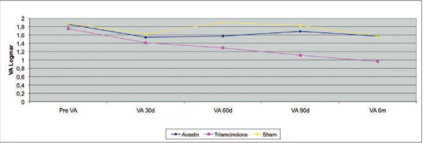
The overall mean visual acuity was 1.72 ± 0.37 logMAR preoperatively and 1.32 ± 0.73 logMAR in 6 months after surgery, Figure 1. Accessing visual acuity by group evidenced that the bevacizumab group had initial mean logMAR VA of 1.87 and 1.57 logMAR VA at the six months, which was not statistically significant (p = 0.84). In IVT group, initial mean VA was 1.75 logMAR and 0.96 logMAR VA at six months, and this result was statistically significant (p ≤ 0.001). And in control group, the initial mean VA was 1.85 logMAR and 1.57 logMAR VA at six months (p =0.34), which was also not statistically significant.
There were no local complications (uveitis, infection or elevation of IOP) or systemic side effects (hypertension and thromboembolic events) after the injections or the surgeries.
D
ISCUSSIONtreatment of cystoid macular edema, membrane choroidal neovascularization and retinal neovascularization. Bevacizumab Injection induces rapid regression of neovascularization of the retina and iris in proliferative diabetic retinopathy(15-17). Studies
showed that patients who underwent bevacizumab in the end of vitrectomy had a lower rate of recurrent bleeding and reoperation(15).
The preoperative injection of intravitreal bevacizumab may reduce intraoperative bleeding during dissection of membrane in PDR with tractional retinal detachment(17). Intravitreal
bevacizumab injection may not prevent postoperative VH, probably due to washing of bevacizumab during vitrectomy(20).
In this study, immediate postoperative VH occurred in 1 eyes in group 1, 4 eyes in group 2 and 8 eyes of the control group. But the rate of VH was not statistically significant between groups. The lower rate of re-bleeding in group 1 could be due to the theory in which bevacizumab helps to inhibit angiogenesis by reducing vascular endothelial growth factor (VEGF) (15-17).
The exact mechanism by which bevacizumab reduces the incidence of immediate postoperative VH is unclear. Vascular Endothelial Growth Factor, the main factor in PDR, decreases significantly after bevacizumab injection and there is a rapid resolution of the neovascularization of the retina(15,16) One possible
mechanism is that this regression of neovascularization improves the integrity of the vessels retinal blood and therefore reduces the risk of postoperative bleeding (21-23).
The visual acuity in all groups showed improvement with 6 months follow-up. There are few studies that assessed visual acuity later than 6 months for patients who underwent bevacizumab before PPV. In IVB group the initial mean VA was 1.87 logMAR and it became 0.96 logMAR postoperatively.
In this study all groups had improvement in the visual acuity within 6 months postoperatively, but only in the group that underwent triamcinolone acetonide injection, this improvement was statistically significant (p <0.05). Moreover, visual acuity improvement did not change between group control and IVB. These results are similar to that found by others authors(21-24). And there was a better visual acuity outcome
in the triamcinolone group. We propose that the improvement in visual acuity in this group may be due to a progressive resolution of macular edema as triamcinolona’s duration. It is worth noting that due to vitreous hemorrhage, macular edema was not evaluated preoperatively. Thus, there is the possibility of an underestimation of macular edema.
Intravitreal injection of bevacizumab 1 week before vitrectomy seems to reduce the incidence of early postvitrectomy hemorrhage in diabetic patients. The need for vitrectomy also may be decreased significantly in these cases.
The main weakness points in our study are the small sample size and the complexity of diabetic patients. Many of these patients had renal complications and uncontrolled diabetes that can contribute to an unfavorable outcome. Despite these limitations, the results observed in this series may encourage carrying out further studies to determine whether the use of bevacizumab or triamcinolone as a preoperative adjunctive to obtain success in vitrectomy for proliferative diabetic retinopathy.
R
EFERENCES1. Varma R, Torres M, Peña F, Klein R, Azen SP; Los Angeles Latino Eye Study Group.. Prevalence of diabetic retinopathy in adult Latinos: the Los Angeles Latino eye study. Ophthalmology. 2004; 111(7):1298–306. 2. Kempen JH, O’Colmain BJ, Leske MC, Haffner SM, Klein R, Moss SE, et al. The prevalence of diabetic retinopathy among adults in the United States. Arch Ophthalmol. 2004; 122(4):552–63
3. Adamis AP, Miller JW, Bernal MT, D’Amico DJ,Folkman J, Yeo TK, et al. Increased vascular endothelial growth factor levels in the vitreous of eyes withproliferative diabetic retinopathy. Am J Ophthalmol. 1994;118(4): 445–50.
4. Aiello LP, Avery RL, Arrigg PG, Keyt BA, Jampel HD, Shah ST, et al. Vascular endothelial growth factor in ocular fluid of patients with dia-betic retinopathy and other retinal disorders. N Engl J Med. 1994; 331(22): 1480–7.
5. Pe’er J, Shweiki D, Itin A, Hemo I, Gnessin H, Keshet E. Hypoxia-induced expression of vascular endothelial growth factor by retinal cells is a common factor in neovascularizing ocular diseases. Lab In-vest. 1995; 72(6):638–45.
6. Watanabe D, Suzuma K, Suzuma I, Ohashi H, Ojima T,Kurimoto M, et al. Vitreous levels of angiopoietin 2 and vascular endothelial growth factor in patients with proliferative diabetic retinopathy. Am J Ophthalmol. 2005; 139(3): 476–81.
7. Tolentino MJ, McLeod DS, Taomoto M, Otsuji T, Adamis AP, Lutty GA. Pathologic features of vascular endothelial growth factor-induced retinopathy in the nonhuman primate. Am J Ophthalmol. 2002; 133(3): 373–85.
8. Adamis AP, Altaweel M, Bressler NM, Cunningham ET Jr, Davis MD, Goldbaum M, et al. Changes in retinal neovascularization after pegaptanib (Macugen) therapy in diabetic individuals. Ophthalmol-ogy. 2006; 113(1):23–8.
9. Spaide RF, Fisher YL. Intravitreal bevacizumab (Avastin) treatment of proliferative diabetic retinopathy complicated by vitreous hemor-rhage. Retina 2006; 26(3): 275–8.
10. Helbig H, Kellner U, Bornfeld N, Foerster MH. [Vitrectomy in diabetic retinopathy: outcome, risk factors, complications]. Klin Monbl Augenheilkd. 1998; 212(5):339–342. German
11. Machemer R, Sugita G, Tano Y. Treatment of intraocular prolifera-tions with intravitreal steroids. Trans Am Ophthalmol Soc. 1979;77:171–80.
12. Tano Y, Sugita G, Abrams G, Machemer R. Inhibition of intraocular proliferations with intravitreal corticosteroids. Am J Ophthalmol. 1980;89(1):131–6.
13. Tano Y, Chandler D, Machemer R. Treatment of intraocular prolifera-tion with intravitreal injecprolifera-tion of triamcinolone acetonide. Am J Ophthalmol.1980;90(6):810–6.
14. Krämer I, Lipp HP. Bevacizumab, a humanized anti-angiogenic mono-clonal antibody for the treatment of colorectal cancer. J Clin Pharm Ther .2007; 32(1): 1–14.
15. Avery RL, Pearlman J, Pieramici DJ, Rabena MD, Castellarin AA, Nasir MA, et al. Intravitreal bevacizumab (Avastin) in the treatment of proliferative diabetic retinopathy. Ophthalmology. 2006; 113(10): 1695.e1-15.
16. Chen E, Park CH. Use of intravitreal bevacizumab as a preoperative adjunct for tractional retinal detachment repair in severe prolifera-tive diabetic retinopathy. Retina. 2006;26(6): 699–700.
17. Mason JO 3rd, Nixon PA, White MF. Intravitreal injection of bevacizumab (Avastin) as adjunctive treatment of proliferative dia-betic retinopathy. Am J Ophthalmol. 2006; 142(4): 685–8.
18. Haritoglou C, Kook D, Neubauer A, Wolf A, Priglinger S, Strauss R, et al. Intravitreal bevacizumab (Avastin) therapyfor persistent diffuse diabetic macular edema. Retina. 2006;26(9): 999–1005.
19. Davis JL, Madow B, Cornett J, Stratton R, Hess D, Porciatti V, et al. Scale for photographic grading of vitreous haze in uveitis. Am J Ophthalmol. 2010;150(5):637-41.
Correspondence to:
Daniel Ferraz, MD. Department of Ophthalmology,
University of São Paulo Medical School
Av. Dr. Enéas de Carvalho Aguiar, nº 255 - Cerqueira
César
CEP 05403-000, São Paulo, (SP) – Brasil
Phone: 55-11-30697871; fax: 55-11- 30697871
http://www.hcnet.usp.br.
20. Romano MR, Gibran SK, Marticorena J, Wong D, Heimann H. Can a preoperative bevacizumab injection prevent recurrent postvitrectomy diabetic vitreous haemorrhage? Eye (Lond). 2009; 23(8):1698-701.
21. Aiello LP, Aiello LP, Brucker AJ, Chang S, Cunningham ET Jr, D’Amico DJ, et al. Evolving guidelines for intravitreous injections. . Retina. 2004:24(5 Suppl): S3-19.
22. Moradian S, Ahmadieh H, Malihi M, Soheilian M, Dehghan MH, Azarmina M. . Intravitreal bevacizumab in active progressive prolif-erative diabetic retinopathy. Graefes Arch Clin Exp Ophthalmol. 2008; 246(12):1699–705.
23. Jorge R, Costa RA, Calucci D, Cintra LP, Scott IU. Intravitreal bevacizumab (Avastin) for persistent new vessels in diabetic retin-opathy (IBEPE study). Retina.2006;26(9):1006–13.