INTRODUCTION
Retinal vein occlusion (RVO) is an important cause of visual loss worldwide. It is the second-most common retinal vascular disorder, and epidemiologic studies reported prevalence rates of 0.7-1.6% in the general population(1-2). An estimated 520 new cases per 1 million people develop annually(3) and 15.3% of cases involve the central retinal vein.
Macular edema, ischemic maculopathy, anterior and posterior segment neovascularization, vitreous hemorrhage, and neovascular glaucoma (NVG) are possible complications associated with central retinal vein occlusion (CRVO). The anterior segment is the main site of neovascularization in CRVO. The risk of development increases with the degree of retinal ischemia, and it is most likely to develop during
the first 3 months after occlusion(4,5).The cumulative incidence of NVG in ischemic CRVO is approximately 40% over 1 year, compared with 10% in nonischemic eyes(6).
The Central Vein Occlusion Study (CVOS) reported that scatter panretinal laser photocoagulation (PRP) is recommended promptly after the development of neovascularization over 2 h or more in the iris or any angle neovascularization(7).
Recent prospective, randomized, controlled trials evaluated intravitreally injected drugs for treating CRVO and tried to deine treatment strategies for macular edema secondary to CRVO.
Steroids reduce vascular permeability and stabilize the blood-re-tina barrier(8). The mechanism involves inhibition of inlammatory me-diators and vascular permeability factors such as vascular endothelial
Incidence of anterior segment neovascularization during intravitreal treatment for
macular edema secondary to central retinal vein occlusion
Incidência de neovasos de segmento anterior durante o tratamento de edema macular secundário a
oclusão da veia central da retina
Luiz FiLipe AdAmi LucAtto1, octAviAno mAgALhães-Junior1, JuLiAnA m. B. prAzeres1, AdriAno m. FerreirA1, rAmon A. oLiveirA1, niLvA s. morAes1,
FLávio e. hirAi1, mAuricio mAiA1
Submitted for publication: August 30, 2016 Accepted for publication: November 7, 2016
1 Department of Ophthalmology, Universidade Federal de São Paulo, SP, Brazil.
Funding: No specific financial support was available for this study.
Disclosure of potential conflicts of interest: None of the authors has any potential conflict of interest to disclose.
Corresponding address: Luiz Filipe Adami Lucatto. Universidade Federal de São Paulo. Rua Bo tucatu, 821 São Paulo, SP 04023062 Brazil Email: [email protected]
Approved by the following Research Ethics Committee: Universidade Federal de São Paulo (CAAE: 18765113.1.0000.5505).
ABSTRACT
Purpose: To analyze the effects of injections of intravitreal triamcinolone aceto-nide (IVTA) and intravitreal bevacizumab (IVB) on the incidence rates of anterior segment neovascularization (ASN) and neovascular glaucoma (NVG) in patients with macular edema secondary to central retinal vein occlusion (CRVO).
Methods: In this prospective, randomized, double-masked, sham-controlled study, 35 patients with macular edema following CRVO were randomized to intravitreal bevacizumab, intravitreal triamcinolone acetonide, or sham injections during the first 6 months of the study. The primary outcome was the incidence rate of ASN at month 6. The secondary outcomes were the mean changes from baseline in best-corrected visual acuity (BCVA) and central foveal thickness (CFT ) on optical coherence tomography over time to month 12.
Results: ASN developed in 8 (22.86%) eyes, including 5 (62.50%) eyes in the sham group and 3 (37.50%) eyes in the IVTA group, during 12 months of fol low-up (p=0.009). BCVA difered signiicantly (p<0.05) among the groups only at month 1. CFT did not differ significantly (p<0.05) among the groups over 12 months. NVG required surgery and developed in one eye despite laser treatment.
Conclusion: Early treatment with intravitreal antivascular endothelial growth factor therapy decreases the rates of ASN and NVG after CRVO.
Keywords: Neovascularization; Pathologic; Bevacizumab; Retinal vein occlusion; Macular edema; Glaucoma; Neovascular
RESUMO
Objetivo: Analisar as taxas de incidência de neovascularização do segmento anterior (NSA) e de glaucoma neovascular (GNV), em pacientes com edema macular secundário a oclusão de veia central da retina (OVCR), em tratamento com injeções intravítreas de triamcinolona (IVTA) ou bevacizumab (IVB).
Métodos: Neste estudo prospectivo, randomizado, duplo mascarado e sham
con-trolado, 35 pacientes com edema macular secundário a OVCR foram randomizados para IVB, IVTA ou para o grupo controle (sham), durante os 6 primeiros meses do estudo. O desfecho primário foi a taxa de incidência de NSA no mês 6. Os desfechos secundários foram alterações médias da acuidade visual corrigida (BCVA) e espessura foveal central (EFC) ao exame de tomografia de coerência óptica, até o mês 12.
Resultados: NSA ocorreu em oito (22,86%) olhos, cinco (62,50%) olhos no grupo sham e três (37,50%) olhos no grupo tratado com injeções intravítreas de Triamcinolona, Não houve nenhum caso com NSA no grupo tratado com bevacizumab durante 12 meses de acompanhamento (p=0,009). A BCVA apresentou diferença estatisticamente significante (p<0,05) entre os grupos, somente no mês 1. A EFC não apresentou dife-renças estatisticamente significantes (p<0,05) entre os grupos ao longo dos 12 meses. GNV ocorreu em um olho apesar do tratamento com laser e este paciente necessitou de intervenção cirúrgica.
Conclusão: O tratamento precoce com injeções intravítreas de Anti VEGF podem
diminuir as taxas de neovascularização do segmento anterior e glaucoma neovascular após oclusão de veia central da retina.
growth factor (VEGF); thus, it may prevent neovascularization(9,10). The SCORE study compared the eicacy and safety of two doses (1 and 4 mg) of intravitreal triamcinolone acetonide (IVTA) for the treatment of macular edema after CRVO in 272 eyes(11). The study reported improved BCVA in 27% of eyes treated with 1 mg of IVTA and fewer ocular adverse events in this group. Neovascularization occurred in 9.8% of eyes treated with 1 mg of IVTA and 4.4% in those treated at a dose of 4 mg. Implantation of sustained corticosteroid delivery devices resulted in improved best-corrected visual acuity (BCVA) in other studies(12).
VEGF plays a key role in the pathophysiology of CRVO and its com-plications. Several studies proposed treatment with the anti-VEGF drugs bevacizumab (Avastin, Genentech Inc., South San Francisco, CA), ranibizumab (Lucentis, Genentech Inc.), and alibercept (Eylea, Regeneron Pharmaceuticals, Tarrytown, NY).
The CRUISE study illustrated that patients treated with monthly intravitreal ranibizumab (0.3 or 0.5 mg) achieved better results than controls(13).The improved BCVA was maintained at the 12-month endpoint.
The HORIZON trial followed the same patients who enrolled in the CRUISE study during the second year. At the end of 24 months, BCVA did not differ significantly among the three groups (0.3 mg, 0.5 mg, and sham)(14).
Bevacizumab is an anti-VEGF drug used to manage retinal vas-cular disorders such as age-related mavas-cular degeneration, diabetic ede ma, and retinal vein occlusions(15-18). Several retrospective and prospective studies reported decreased retinal thickness and impro-ved BCVA after intravitreal injections of the drug(19,20).
Although most recent studies suggested the therapeutic bene-its of intravitreal steroids and anti-VEGF for treating macular edema secondary to CRVO, none focused on the efects of such treatments for preventing anterior segment neovascularization (ASN) and NVG as a primary endpoint.
The purpose of the this prospective study was to analyze the efects of intravitreal bevacizumab (IVB) injections compared with IVTA or sham injections for preventing ASN and NVG in patients with macular edema due to CRVO.
METHODS
S
TUDYDESIGNThis 12-month randomized, double-masked, sham-controlled study was designed to evaluate the incidence rates of ASN and NVG in three groups treated for macular edema secondary to CRVO.
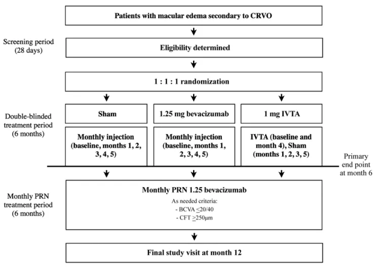
The study included a 28-day screening period, a 6-month treat-ment period (baseline to month 6) in which patients received monthly injections, and an additional 6-month, open-label PRN treatment period (month 6 to inal study visit).
The study was conducted according to the tenets of the Decla-ration of Helsinki and federal laws. All patients were informed about the purpose of the study, and they provided informed consent. The ethics committee of our institution approved the study. The primary outcome was the presence of ASN in the study eye, as determined by ophthalmologic examination at the 6-month follow-up visit.
S
CREENINGANDELIGIBILITYThe primary investigator (LFAL) determined patient eligibility at the Retina Division of the Department of Ophthalmology, Federal University of São Paulo, using the criteria in table 1.
During the screening visit, after providing informed consent, all participants provided a complete medical history and underwent an ophthalmologic examination that included measurements of BCVA using a Snellen chart, slit-lamp examination, gonioscopy, measure-ment of intraocular pressure (IOP), pupillary relex, binocular fun dus examination, optical coherence tomography (SD-OCT) (Spectralis OCT, Heidelberg Engineering, Heidelberg, Germany), and wide-angle luorescein angiography (FA) (HRA, Heidelberg Engineering).
R
ANDOMIZATIONIf the physician investigator judged a patient eligible for partici-pation in the study, then he or she was randomized to one of three treatment groups as follows (Figure 1): group 1, sham injections; group 2, 1.25-mg IVB injections; and group 3, 1-mg IVTA injections.
The patients in groups 1 and 2 received monthly sham and 1.25-mg IVB injections, respectively, at baseline and months 1, 2, 3,
Table 1. Inclusion and exclusion criteria
Inclusion criteria*
≥18 years of age with macular edema secondary to CRVO and less than 90 days since symptoms appeared
BCVA ≤20/40 according to the Snellen chart
Central foveal thickness ≥250 μm according to a central 1-mm diameter circle with a Spectralis OCT (Heidelberg Engineering, Heidelberg, Germany)
Exclusion criteria*
Any iris or angle neovascularization evident on slit lamp or gonioscopy examination without pupillary dilation
Presence of macular edema due to a cause other than CRVO
Prior episode of RVO
IOP ≥25 mmHg, open-angle glaucoma (either primary open-angle glaucoma or other cause), prior steroid-induced IOP elevation, or pseudoexfoliation
Evidence on examination of any diabetic retinopathy
History or presence of wet or dry AMD Any previous treatment for macular edema
Previous panretinal scatter photocoagulation or sector laser photocoagulation
Prior anti-VEGF treatment
Any ocular surgery within 6 months before baseline
Prior pars plana vitrectomy Intra or periocular acute infection
*= pertains to the study eye, except where noted otherwise.
CRVO= central retinal vein occlusion; BCVA= best-corrected visual acuity; BRVO= branch retinal vein occlusion; RVO= retinal vein occlusion; IOP= intraocular pressure; AMD= age-related macular degeneration; VEGF= vascular endothelial growth factor.
4, and 5. The patients in group 3 received IVTA injections at baseline and month 4; at months 1, 2, 3, and 5, the eyes of patients rando-mized to group 3 received sham injections. Moreover, for patient ran domization, a computer-generated randomization table was created (Stata v11, StataCorp, College Station, TX). Participants were randomized 1:1:1 to treatment groups with block sizes of three and six. An investigator not otherwise involved in the trial performed all randomization processes.
One eye of each patient was included in the study. If both eyes were eligible, the eye with the worse BCVA at screening was selected. Patients and evaluating physicians were masked to treatment during the first 6 months of the study. The physician who administered the injections (LFAL) did not perform examinations or outcome assessments, and he had knowledge about the drug administered or sham injection at the time of injection.
S
TUDYVISITSANDASSESSMENTSDuring the 6-month follow-up period, study visits occurred on day 0 (baseline) and months 1, 2, 3, 4, 5, and 6. During the monthly PRN treatment period, patients were eligible to receive monthly 1.25-mg IVB injections if they had BCVA in the study eye of 20/40 or worse according to the Snellen chart and/or CFT of 250 μm or more according to SD-OCT. The patients continued monthly follow-up
(months 7, 8, 9, 10, and 11 and the inal study visit). At each visit, the recorded patient data included BCVA measured using a Snellen chart, slit-lamp examination, and gonioscopy; IOP measured via Goldmann tonometry, binocular fundus examination, and SD-OCT assessment of CFT. Wide-angle FA was performed at baseline and visits 6 and 12. Eyes with clinical indings of retinal ischemia (VA<20/200, relative aferent pupillary defect [APD], and cotton wool spots) were evalua-ted and correlaevalua-ted with the development of ASN.
The FA indings were classiied as ischemic when more than 10 disc areas of retinal capillary nonperfusion were present and perfused (nonischemic) while fewer than 10 disc areas of nonperfusion were present(5). The perfusion status of FA was considered indeterminate when intraretinal hemorrhage prevented visualization of luorescein in the retinal capillaries during the experiment.
At each visit, the patients provided a medical history, the medi-cation was reviewed, and safety was assessed. Any new sign, symp-tom, illness, or worsening of any preexisting medical condition was recorded as an adverse event (AE). An AE was considered as a serious AE (SAE) when it resulted in death or when it was life-threatening, it required prolonged hospitalization, it caused persistent or signiicant disability, it was a congenital anomaly/birth defect, or it was consi-dered a signiicant medical event by the investigator. Furthermore, patients who discontinued the study before the month 12 visit were
CRVO=central retinal vein occlusion.
encouraged to return for an early inal study visit 30 days after their last injection or analysis. If ASN was detected at any time in the study, the patient was referred for scatter PRP according to recent recom-mendations. If NVG was detected despite PRP, patients were referred to the glaucoma sector for follow-up and treatment.
I
NTRAOCULARINJECTIONSThe procedure for drug administration at the ophthalmic surgical center of the Federal University of São Paulo was as described fur-ther. Topical anesthetic drops were administered, and a lid speculum was used. A 5% povidone iodine drop was instilled as prophylaxis against infection 5 min before the procedure. A 30-gauge needle was inserted through the pars plana, and 0.05 ml of bevacizumab
(OPHTHALMOS®
25 mg/ml São Paulo, Brazil) or 0.025 ml of
triamci-nolone acetonide (OPHTAAC® 40 mg/ml OPHTHALMOS São Paulo,
Brazil) was injected(13,21). The procedure for administering sham injec-tions was similar to that for the IVB and IVTA injecinjec-tions, except that the hub of a syringe without a needle was placed against the injection site and the syringe plunger was depressed to mimic an injection. The ability to count ingers with the study eye was assessed 1 min after the injection. No topical antibiotics were prescribed postoperatively for any patient. An additional visit within 5 days after each injection was scheduled as a postoperative evaluation.
O
UTCOMEMEASURESThe primary outcome measure was the incidence of ASN at month 6. The secondary outcomes included the mean changes from baseline in BCVA and CFT over time to month 12. The safety outcomes included the incidence and severity of ocular, nonocular, and systemic AEs.
S
TATISTICALANALYSISData were analyzed and expressed as means and standard devia-tions or frequencies (%). Comparisons of continuous and categorical variables among the treatment groups were performed using the Kruskal-Wallis test and Fisher’s exact test, respectively. Post-hoc analy-ses were performed using the Bonferroni test. p<0.05 was considered statistically signiicant. All analyses were performed using Stata v11.
RESULTS
B
ASELINEDEMOGRAPHICSANDOCULARCHARACTERISTICSBetween September 2013 and May 2015, 35 eyes of 35 patients in the Retina Sector of the Federal University of São Paulo, Brazil, were randomized, that is, 10, 14, and 11 eyes to the sham, IVB, and IVTA groups, respectively. Thirteen patients completed the screening visit, but they were excluded as follows: seven had glaucoma; three did not provide informed consent; two were excluded because of social issues; and one was excluded because ASN was detected during the screening visit. The patient demographic data were similar across the treatment groups (Table 2). The baseline ocular characteristics were also similar across treatment groups excluding APD (p<0.05).
The mean patient age was 59.48 years (range, 31-89 years), and 60% of patients were men. The average time for symptom development was 31.08 days (range, 3-85 days). The mean baseline BCVA (logarithm of the minimum angle of resolution [logMAR]) of the study eye was 1.43 (±0.53) (Snellen equivalent, 20/538), and 27 eyes (77.14%) had BCVA of less than 20/200.
The baseline biomicroscopic examination illustrated that 34 (97.14%) eyes were phakic, and one (2.86%) was pseudophakic. Fifteen (42.86%) eyes had an APD. The baseline funduscopic examination indicated that 21 (60%) eyes had cotton-wool spots, and the mean CFT was 754.51 μm (range, 252-1146 μm).
More than 10 disc areas of retinal capillary nonperfusion were present on the baseline FA images in 12 (34.29%) eyes. Five, four, and
three of these eyes were randomized to sham, IVTA, and IVB treat-ment, respectively. The other 11 eyes had less than 10 disc areas of retinal capillary nonperfusion, and in 11 eyes, the area of nonperfu-sion was undetermined (p=0.357).
P
RIMARYENDPOINTEight (22.86%) eyes had ASN. Five eyes randomized to sham treatment (50%) and three eyes randomized to 1 mg IVTA (27.27%) developed ASN. No eyes randomized to IVB developed ASN in the iris and/or angle during 12 months of follow-up (p=0.009).
The overall mean time for development of ASN was 59.75 ± 42.79 days, that is, 65.6 ± 54.22 days in the sham group and 50 ± 17.32 days in the IVTA group (p>0.05).
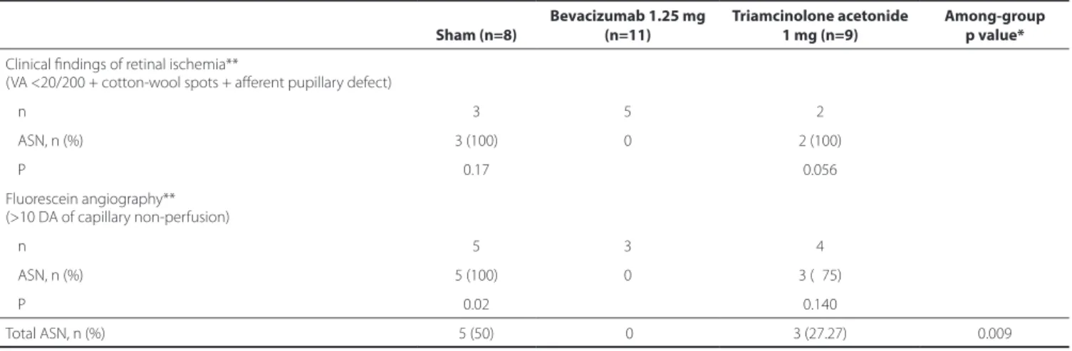
We also analyzed the presence of clinically diagnosed retinal ischemia (BCVA<20/200, APD, and cotton-wool spots). At baseline, 10 eyes presented clinical indings suggestive of retinal ischemia. Of these, ive were randomized to the IVB group, and none developed ASN during the follow-up period. The remaining ive eyes with ische-mia developed ASN, three and two of which were randomized to the sham (p=0.17) and IVTA groups (p=0.056). Two patients randomized to sham treatment did not exhibit baseline retinal ischemia, but ASN developed during the follow-up period.
Twelve eyes had more than 10 disc areas of retinal capillary non-perfusion on the baseline FA images. Of these, eight (66.67%) develo-ped ASN (ive eyes in the sham group [p=0.02] and three eyes in the triamcinolone group [p=0.14]). Four eyes displayed the angiographic criteria of ischemia, but ASN did not develop (three eyes randomized to IVB and one eye randomized to IVTA) (Table 3).
F
UNCTIONALOUTCOMESATMONTH12
At months 6 and 12, the mean logMAR BCVA levels were 0.96 ± 0.67 (p=0.41) and 0.99 ± 0.53 (p=0.44), respectively. The mean change in BCVA during the irst 12 months in the groups is shown in igure 2. BCVA difered signiicantly (p<0.05) among the groups only at month 1. Macular ischemia was observed in 13 eyes, including four (40%), ive (45.45%), and four (28.57%) eyes randomized to sham, IVTA, and IVB treatment (p=0.84), respectively.
A
NATOMICOUTCOMESATMONTH12
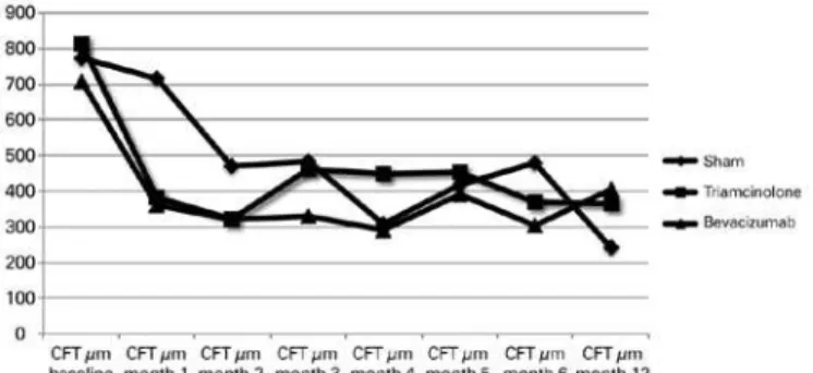
At months 6 and 12, the mean CFTs were 345.4 ± 182.87 (p=0.39) and 346.17 ± 195.92 (p=0.35), respectively. The mean changes in CFT on SD-OCT during the irst 12 months in the groups are shown in igure 3. CFT did not difer signiicantly among the groups during this period.
Safety outcomes at month 12
Seven (20%) eyes developed or exhibited worsening of cataracts (p=0.31). Ten patients required topical antiglaucomatous drops for increased IOP, none of whom had uncontrolled IOP. The diferences among the groups did not reach signiicance (p=0.41). One patient discontinued follow-up 3 months after inferior paresis that required hospitalization. The patient was diagnosed with Miller Fisher syndro-me. Another patient with cardiomyopathy related to Chagas disease required pacemaker implantation during follow-up.
All patients with ANS were referred for scatter PRP. NVG develo-ped in one patient despite laser treatment, and surgery was needed to control IOP.
DISCUSSION
Table 2. Patient demographics and baseline ocular characteristics
Sham (n=10)
Bevacizumab 1.25 mg (n=14)
Triamcinolone acetonide 1 mg (n=11)
Among-group p value*
Age (years)
Mean (SD) 55.6 (14.41) 61.86 (12.62) 60 (12.63) 0.450
Range 31-83 37-89 45-80
Gender, n (%) 1.000
Male 6 (60%) 9 (64.29%) 7 (63.64%)
Female 4 (40%) 5 (35.71%) 4 (36.36%)
Race, **n (%) 0.460
White 6 (60%) 7 (50.00%) 9 (81.82%)
Black 2 (20%) 4 (28.57%) 2 (18.18%)
Asiatic 1 (10%) 0 0
Other 1 (10%) 3 (21.43%) 0
Time of symptoms 0.482
Mean (SD) 31.9 (18.60) 25.42 (22.07) 37.54 (30.11)
Range 4-60 3-60 3-85
BCVA (logMAR) 0.275
Mean (SD) 1.64 (0.44) 1.32 (0.53) 1.40 (0.62)
Range 0.9-2.2 0.3-1.79 0.5-2.2
VA<20/200, n (%) 9 (90%) 11 (78.57%) 7 (63.64%) 0.418
Cotton wool spots, n (%) 6 (60%) 08 (57.14%) 7 (63.64%) 1.000
Aferent pupillary defect, n (%) 6 (60%) 07 (50.00%) 2 (20.00%) 0.029
Lens status, n (%) 1.000
Phakic 10 (100%) 13 (92.86%) 11 (100%)
Pseudophakic 0 01 (07.14%) 0
>10 DA of capillary non-perfusion, n (%) 0.357
Yes 5 (55.56%) 3 (21.43%) 4 (36.36%)
No 3 (33.33%) 4 (28.57%) 4 (36.36%)
Undetermined 1 (11.11%) 7 (50.00%) 3 (27.27%)
CFT (SD-OCT), µm 0.500
Mean (SD) 774.87 (276.85) 706 (261.93) 813 (152.90)
Range 252-1059 262-1146 555-999
*= P-values less than 0.05 were considered statistically signiicant; **= multiracial patients were counted in each race category that they indicated. SD=standard deviation; BCVA=best correct visual acuity; VA=visual acuity; CFT=central foveal thickness; SD-OCT=spectral domain optical coherence tomography.
Table 3. Primary outcome
Sham (n=8)
Bevacizumab 1.25 mg (n=11)
Triamcinolone acetonide 1 mg (n=9)
Among-group p value*
Clinical indings of retinal ischemia**
(VA <20/200 + cotton-wool spots + aferent pupillary defect)
n 3 5 2
ASN, n (%) 3 (100) 0 2 (100)
P 0.17 0.056
Fluorescein angiography** (>10 DA of capillary non-perfusion)
n 5 3 4
ASN, n (%) 5 (100) 0 3 (075)
P 0.02 0.140
Total ASN, n (%) 5 (50) 0 3 (27.27) 0.009
Figure 3. Mean change from the baseline central foveal thickness (CFT) over time to month 12. CFT did not difer signiicantly among the groups over the 12 months.
Figure 2. Mean change from baseline best-corrected visual acuity (BCVA) in the study over time to month 12. Statistically signiicant BCVA logarithm of the minimum angle of resolution (logMAR) (*p<0.05) diference was found at Month enter groups.
related to CRVO are ASN and NVG. The cumulative incidence of NVG is 40% in ischemic CRVO and 10% in nonischemic CRVO(6). Despite the fact that many studies reported the beneits of intravitreal medi-cations for improving BCVA and macular edema, there is little infor-mation about the impact of treatment on the natural history of ASN.
This study was a prospective analysis of diferent treatments for macular edema after CRVO with focus on preventing ASN and NVG.
Diferentiating between ischemic and nonischemic CRVO may be challenging in the early stages. Clinical features such as initial BCVA worse than 20/200, APD, and cotton-wool spots are suggestive of ischemic CRVO and can predict prognosis(3,5,22-25). The presence of extensive nonperfused capillary areas in the FA images is a good indicator of retinal ischemia(5,6), but this can be diicult to assess while setting CRVO with substantial intraretinal hemorrhages. Although electroretinogram (ERG) is a good indicator of retinal ischemia in CRVO(23,26-28) and is predictive of iris neovascularization(29,30), it is ex-pensive and not always available in daily clinical practice. Considering the diiculty of performing ERG, we correlated the clinical data that suggested ischemic CRVO with the development of ASN. Thus, it is possible to assess which patient groups may beneit from each ma-cular edema treatment to prevent this complication.
In this study, ASN developed in eight (22.86%) patients. Ramezani et al. reported an incidence as high as 50% at 6 months after CRVO in a study in which the CRVO subtype was unclassiied(30). In the CVOS, which considered eyes initially categorized as nonperfused or inde-terminate, 35% of eyes developed ASN, compared with 10% of eyes initially categorized as perfused(5).
Five (62.5%) eyes that developed ASN were randomized to sham treatment, and three (37.5%) eyes were randomized to the IVTA group. No eyes in the IVB group developed ASN during the irst 6 months of follow-up (p=0.009).
In this study, ASN developed after an average of 59.75 ± 42.79 (65.6 ± 54.22 days in the sham group; and 50 ± 17.32 days in the triam cinolone group).
Twelve patients had baseline FA images classiied as ischemic. Among these patients, ive, four, and three eyes were randomized to sham, IVTA, and IVB treatment, the last of which was the only group with an ischemic angiographic pattern that did lead to the deve-lopment of ASN.
The CRUISE study reported iris neovascularization in only 12 of 390 eyes and NVG in two eyes. Only three patients treated with rani-bizumab in that study developed iris neovascularization, and none developed NVG; however, the study excluded patients with APD and included patients with BCVAs ranging from 20/40 to 20/320. In this study, BCVA and APD were not the exclusion criteria, and our sample probably included patients with more severe retinal ischemia com-pared to the CRUISE study.
The functional and anatomic outcomes in this study were worse than the SCORE and CRUISE results. BCVA in patients treated with anti-VEGF injections was signiicantly (p<0.05) better than that in the other groups only at month 1. CFT measured on SD-OCT images in these patients was not signiicantly better than that in the other groups. These results are probably related to the small sample size and the inclusion of patients with a worse prognosis compared to patients in other trials.
At the end of the 12-month follow-up period, we found similar ocular adverse events (cataract and ocular hypertension) rates com-pared with previous studies(11). Endophthalmitis did not develop during the study. No SAEs reported were related with the use of the study medications.
Although this was a prospective, randomized, sham-controlled study, our study had limitations, including the small sample size in each group, which might have compromised the statistical power to detect diferences.
CONCLUSION
In conclusion, early treatment with IVB decreases the rates of ASN and NVG after CRVO in eyes with clinical signs suggestive of ischemia. Multicenter studies with larger samples of patients should be performed to conirm these indings.
REFERENCES
1. Cheung N, Klein R, Wang JJ, Cotch MF, Islam AF, Klein BE, et al. Traditional and novel cardiovascular risk factors for retinal vein occlusion: the multiethnic study of athe-rosclerosis. Invest Ophthalmol Vis Sci. 2008;49(10):4297-302.
2. Klein R, Klein BE, Moss SE, Meuer SM. The epidemiology of retinal vein occlusion: the Beaver Dam Eye Study. Trans Am Ophthalmol Soc. 2000;98:133-41; discussion 141-3. 3. Rogers S, McIntosh RL, Cheung N, Lim L, Wang JJ, Mitchell P, Kowalski JW, Mguyen
H, Wong TY; International Eye Disease Consortium. The prevalence of retinal vein occlusion: pooled data from population studies from the United States, Europe, Asia, and Australia. Ophthalmology. 2010;117(2):313-9.
4. Ehlers J, Fekrat S. Retinal vein occlusion: beyond the acute event. Surv Ophthalmol. 2011;56(4):281-99.
5. Central Vein Occlusion Study Group. Natural history and clinical management of central retinal vein occlusion. Arch Ophthalmol. 1997;115(4):486-91. Erratum in: Arch Ophthalmo.l 1997;115(10):1275.
6. Hayreh SS, Rojas P, Podhajsky P, Montague P, Woolson RF. Ocular neovascularization with retinal vascular occlusion-III. Incidence of ocular neovascularization with retinal vein occlusion. Ophthalmology. 1983;90(5):488-506.
7. Central Vein Occlusion Study Group. Evaluation of grid pattern photocoagulation for macular edema in central vein occlusion. The Central Vein Occlusion Study Group M report. Ophthalmology. 1995;102(10):1425-33.
8. Felinski EA, Antonetti DA. Glucocorticoid regulation of endothelial cell tight junc-tion gene expression: novel treatments for diabetic retinopathy. Curr Eye Res. 2005; 30(11):949-57.
factor up-regulation in human central retinal vein occlusion. Ophthalmology. 1998; 105(3):412-6.
11. Ip MS, Scott IU, VanVeldhuisen PC, Oden NL, Blodi BA, Fisher M, Singerman LJ, To-letino M, Chan CK, Gonzalez VH; SCORE Study Research Group. A randomized trial comparing the eicacy and safety of intravitreal triamcinolone with observation to treat vision loss associated with macular edema secondary to central retinal vein occlusion: the Standard Care vs Corticosteroid for Retinal Vein Occlusion (SCORE) study report 5. Arch Ophthalmol. 2009;127(9):1101-4. Erratum in: Arch Ophthalmol. 2009;127(12):1648.
12. Haller JA, Bandello F, Belfort R Jr, Blumenkranz MS, gillies M, Heier J, Loewenstein A, Yoon YH, Jacques ML, Jiao J, Li Y, WHitcup SM; OZURDEX GENEVA Study Group. Randomized, sham-controlled trial of dexamethasone intravitreal implant in patients with macular edema due to retinal vein occlusion. Ophthalmology. 2010;117(6):1134-46. Comment in: Ophthalmology. 2010;117(6):1061-3.
13. Brown DM, Campochiaro PA, Singh RP, Li, Gray, Saroj N, Rundle AC, Rubio RG, Murahashi WY; CRUISE Investigators. Ranibizumab for macular edema following central retinal vein occlusion: six-month primary end point results of a phase III study. Ophthalmology. 2010;117(6):1124-33. Comment in: Ophthalmology. 2010; 117(6):1061-3.
14. Heier JS, Campochiaro PA, Yau L, Li Z, Saroj N, Rubio RG, et al. Ranibizumab for ma-cular edema due to retinal vein occlusions-long-term follow-up in the HORIZON Trial. Ophthalmology. 2012;119(4):802-9.
15. Matsumoto Y, Freund KB, Peiretti E, Cooney MJ, Ferrara DC, Yannuzzi LA. Rebound macular edema following bevacizumab (Avastin) therapy for retinal venous occlusive disease. Retina. 2007;27(4):426-31.
16. Rabena MD, Pieramici DJ, Castellarin AA, Nasir MA, Avery RL. Intravitreal bevacizumab (Avastin) in the treatment of macular edema secondary to branch retinal vein occlu-sion. Retina. 2007;27(4):419-25.
17. Spandau UH, Ihlof AK, Jonas JB. Intravitreal bevacizumab treatment of macular edema due to central retinal vein occlusion. Acta Ophthalmol. 2006;84(4):555-6. 18. Rosenfeld PJ, Fung AE, Puliaito CA. Optical coherence tomography indings after an
intravitreal injection of bevacizumab (Avastin) for macular edema from central retinal vein occlusion. Ophthalmic Surg Lasers Imaging. 2005;36(4):336-9.
19. Ferrara DC, Koizumi H, Spaide RF. Early bevacizumab treatment of central retinal vein occlusion. Am J Ophthalmol. 2007;144(6):864-71.
20. Figueroa MS, Contreras I, Noval S, Arruabarrena C. Results of bevacizumab as the primary treatment for retinal vein occlusion. Br J Ophthalmol. 2010;94(8):1052-6. 21. Rosenfeld PJ, Brown DM, Heier JS, Boyer DS, Kaiser PK, Chung CY, Kim RY; MARINA
Study Group. Ranibizumab for neovascular age-related macular degeneration. N Engl J Med. 2006;355(14):1419-31. Comment in: N Engl J Med. 2006;355(14):1409-12; N Engl J Med. 2007;356(7):748-9; author reply 749-50.
22. Hayreh SS, Klugman MR, Beri M, Kimura AE, Podhajsky P. Diferentiation of ischemic from non-ischemic central retinal vein occlusion during the early acute phase. Graefes Arch Clin Exp Ophthalmol. 1990;228(3):201-17.
23. Central Vein Occlusion Study Group. A randomized clinical trial of early panretinal photocoagulation for ischemic central vein occlusion: the Central Vein Occlusion Study Group N Report. Ophthalmology. 1995;102(10):1434-44.
24. Hayreh SS. Management of central retinal vein occlusion. Ophthalmologica. 2003; 217(3):167-88.
25. Bresnick GH, Wis M. Following up patients with central retinal vein occlusion. Arch Ophthalmol. 1988;106(3):324-6.
26. Hayreh SS, Klugman MR, Podhajsky P, Kolder HE. Electroretinography in central retinal vein occlusion. Correlation of electroretinographic changes with pupillary abnorma-lities. Graefe’s Arch Clin Exp Ophthalmol. 1989;227(6):549-61.
27. Sabates R, Hirose T, McMeel JW. Electroretinography in the prognosis and classiica-tion of central retinal vein occlusion. Arch Ophthalmol. 1983;101(2):232-5. 28. Breton ME, Quinn GE, Keene SS, Dahmen JC, Brucker AJ. Electroretinogram
para-meters at presentation as predictors of rubeosis in central retinal vein occlusion patients. Ophthalmology. 1989;96(9):1343-52. Comment in: Ophthalmology. 1990; 97(2):151.
29. Matsui Y, Katsumi O, McMeel JW, Hirose T. Prognostic value of electroretinogram (ERG) in central retinal vein obstruction (CRVO). Graefe’s Arch Clin Exp Ophthalmol. 1994;232(2):75-81.