2255-4823/$ – see front matter © 2013 Elsevier Editora Ltda. All rights reserved.
ASSOCIAÇÃO MÉDICA BRASILEIRA
REVISTA DA
Volume 58 • Número 6 • Novembro/Dezembro 2012 • ISSN 0104-4230 • ISSN 1806-9282 (On-line)
www.ramb.org.br
ARTIGOS
ARTIGOS ORIGINAIS _____________Qualidade da informação da internet disponível para pacientes em páginas em português ___________________________________________________________645 Acesso a informações de saúde na internet: uma questão de saúde pública? ______650 Maus-tratos contra a criança e o adolescente no Estado de São Paulo, 2009_______659 Obesidade e hipertensão arterial em escolares de Santa Cruz do Sul – RS, Brasil ___666 Bone mineral density in postmenopausal women with and without breast cancer ___673 Prevalence and factors associated with thoracic alterations in infants born prematurely __________________________________________________679 Análise espacial de óbitos por acidentes de trânsito, antes e após a Lei Seca, nas microrregiões do estado de São Paulo ___________________________________685 Sobrevida e complicações em idosos com doenças neurológicas em nutrição enteral ______________________________________________________691 Infliximab reduces cardiac output in rheumatoid arthritis patients without heart failure ______________________________________________________698 Análise dos resultados maternos e fetais dos procedimentos invasivos genéticos fetais: um estudo exploratório em Hospital Universitário _______________703 Frequência dos tipos de cefaleia no centro de atendimento terciário do Hospital das Clínicas da Universidade Federal de Minas Gerais __________________709 ARTIGO DE REVISÃO ______________Influência das variáveis nutricionais e da obesidade sobre a saúde e o metabolismo __714
EDITORIAL
Conclusão: como exibir a cereja do bolo 633
PONTO DE VISTA
Os paradoxos da medicina contemporânea 634
IMAGEM EM MEDICINAObstrução duodenal maligna: tratamento endoscópico paliativo utilizando prótese metálica autoexpansível 636
Gossypiboma 638
DIRETRIZES EM FOCO
Hérnia de disco cervical no adulto: tratamento cirúrgico 639
ACREDITAÇÃOAtualização em perda auditiva: diagnóstico radiológico 644 SEÇÕES ____________________________
•
•
•
30/11/12 11:24
www.ramb.org.br
Revista da
ASSOCIAÇÃO MÉDICA BRASILEIRA
REV ASSOC MED BRAS. 2013;59(4):318-320
Image in Medicine
Proteus syndrome: case report
Síndrome de Proteus: relato de caso
Letícia Silva Sene*, Polyane de Oliveira Sales, Rubens Chojniak
Department of Imaging, Hospital A. C. Camargo, São Paulo, SP, Brazil* Corresponding author.
E-mail: [email protected] (L.S. Sene).
Proteus syndrome is a rare hamartomatous syndrome, a congenital disorder of genetic origin. It was first described in 1979 by Cohen and Hayden1 as a hamartoma syndrome,
and was subsequently named “Proteus syndrome” in 1983 by Wiedeman.2 The syndrome has characteristic malformations
caused by an excess and multifocal growth of tissue derived from all three germ layers, which determine partial gigantism of the limbs, pigmented nevi, subcutaneous tumors, macrocephaly, and visceromegalies, but normal mental development.2-4 Lindhurst et al. confirmed that the syndrome
was genetically inherited in 2011, after performing the genetic sequencing of 29 patients with the syndrome and finding a somatic mutation in the AKT1 oncogene, thus demonstrating the presence of somatic mosaicism for a mutation that is lethal in the non-mosaic state. It was also demonstrated that a dysfunction in the PI3K-AKT pathway is related to the overgrowth.5
This article reports a case of Proteus syndrome in a 24-year-old female patient. This is a unique case in the family, whose onset started at birth with epidermal nevi located in the right upper limb and trunk. During childhood, the patient experienced disproportionate growth of the limbs, which currently show hemihypertrophy of the left leg due to lipomatous overgrowth and of the right arm due to a vascular malformation, shown in Figures 1 and 2, respectively. This asymmetric hemihypertrophy resulted in scoliosis, with consequent upper and lower back pain.
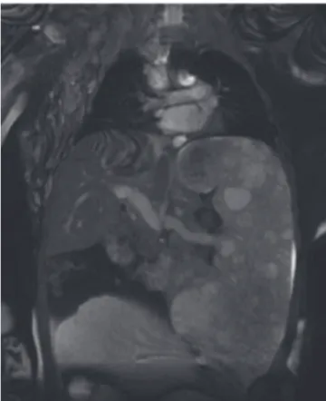
The arm shows a few lymph vesicles and hardened nodules – likely foci of thrombosis in this limb, which are characterized by foci of vascular channels in Figure 2. The patient presents splenomegaly as a visceral abnormality, shown in Figure 3. Neuropsychomotor development is normal. The syndrome diagnosis was established by the abovementioned clinical findings associated with lesion presentation pattern, which are: mosaic distribution, progressive course, and sporadic occurrence. The patient came to the clinic complaining of heaviness in the right arm, and is being followed by a multidisciplinary team that includes genetic services, plastic surgery, orthopedics, vascular surgery, and diagnostic imaging. The proposed surgical treatment consists of partial correction of the deformities, including amputation of the right arm and splenectomy. The patient and family are considering the possibility of having them performed, as they have been informed of the surgical risks of bleeding and vascular complications.
REV ASSOC MED BRAS. 2013;59(4):318-320
319
the following: epidermal nevi; disproportionate overgrowth of limbs, vertebrae, or viscera; macrocephaly or hyperostosis; bilateral ovarian cystadenoma or monomorphic adenoma of the parotid gland; or three of the following adipose tissue dysregulations, vascular malformations, or facial phenotypes.3
The visceral abnormalities are less common than the musculoskeletal; the following have been reported:
Fig. 3 – Magnetic resonance imaging of the upper abdominal region with coronal T2‑weighted sequence, showing splenomegaly with multiple nodular images and hyperintense signal on T2, characteristics of angiomatosis. Fig. 1 − Magnetic resonance imaging of lower limbs with
coronal T1‑weighted sequences showing diffuse left leg enlargement due to extensive proliferation of adipose tissue associated with atrophy of adjacent muscle groups.
Fig. 2 − Magnetic resonance imaging of the right arm with coronal T1‑weighted sequences before and after administration of paramagnetic contrast, showing diffuse limb enlargement due to the presence of multiple images with low signal on T1 and intense enhancement after contrast, characteristics of hemangioma associated with adjacent muscle atrophy.
splenomegaly, macrocephaly, white matter abnormalities, nephromegaly, and cystic and emphysematous lung alterations.4 Most patients have normal psychomotor
development. Life expectancy is 9 months to 29 years, according to the severity of the abnormalities.4 The fourth leading
320
REV ASSOC MED BRAS. 2013;59(4):318-320malformations, surgical convalescence, and (in extreme cases of deformity) by restricted mobility.6 Benign neoplasms
associated with the syndrome include lipomas, ovarian cystadenomas, and monomorphic adenoma of the parotid gland; malignant neoplasms include papillary adenocarcinoma of testicles, mesothelioma of the tunica vaginalis, and peritoneal mesothelioma.3,4 Differential diagnoses include
vascular and pigmented syndromes and lipomatoses, mainly Klippel-Trenaunay syndrome and hemihyperplasia/ lipomatosis syndrome.3,4,6
Treatment is multidisciplinary, including clinical and psychological support. Proteus syndrome results in significant social stigma, due to its rarity and disfiguring features; thus, these patients must undergo psychological counseling.7 Genetic counseling provides information
on the nature, inheritance, and implications of genetic disorders to help individuals and families to make medical and personal decisions. Skeletal overgrowth can result in biomechanical dysfunction and functional limitation; the correction of these conditions involves epiphysiodesis, limb shortening, reduction of asymmetries, stretching, arthrodesis, arterial ligation, and even amputation; however, deformity recurrence is common.8 The most urgent and life-threatening
complications include deep vein thrombosis and pulmonary embolism, which may have a late diagnosis due to their very low incidence in pediatric patients. Patients should be treated with anticoagulants.9
R E F E R E N C E S
1. Cohen MM Jr, Hayden PW. A newly recognized hamartomatous syndrome. Birth Defects Orig Artic Ser. 1979;15(5B):291-6. 2. Wiedemann HR, Burgio GR, Aldenhoff P, Kunze J, Kaufmann HJ,
Schirg E. The Proteus syndrome. Partial gigantism of the hands and/or feet, nevi, hemihypertrophy, subcutaneous tumors, macrocephaly or other skull anomalies and possible accelerated growth and visceral affections. Eur J Pediatr. 1983;140:5-12. 3. Biesecker LG, Happle R, Mulliken JB, Weksberg R, Graham JM Jr,
Viljoen DL, et al. Proteus syndrome: diagnostic criteria, differential diagnosis, and patient evaluation. Am J Med Genet. 1999;84:389-95. 4. Jamis-Dow CA, Turner J, Biesecker LG, Choyke PL. Radiologic
manifestations of Proteus syndrome. RadioGraphics. 2004;24: 1051-68.
5. Lindhurst MJ, Sapp JC, Teer JK. A mosaic activating mutation in AKT1 associated with the Proteus Syndrome. N Engl J Med. 2011;365:611-9.
6. Demir MK. Case 131: Proteus syndrome. Radiology. 2008;246: 974-9.
7. Turner J, Biesecker B, Leib J, Biesecker L, Peters KF. Parenting children with Proteus syndrome: experiences with, and adaptation to, courtesy stigma. Am J Med Genet A. 2007;143A:2089-97 8. Cruz R, Nunes ALS, Fortuna CMM, Pimentel HM, Teixeira E.
Síndrome de Proteus: relato de dois casos e revisão da literatura. Rev Bras Ortop. 1999;34:299-303.