19 artigo 469
CAsE REPORt
1 – Attending Physician in the Shoulder and Elbow Group, Institute of Orthopedics, HC/FMUSP, São Paulo, SP, Brazil.
2 – Collaborating Professor in the School of Medicine, University of São Paulo, and Head of the Shoulder and Elbow Group, Institute of Orthopedics, HC/FMUSP, São Paulo, SP, Brazil.
3 – Trainee in the Shoulder and Elbow Group, Institute of Orthopedics, HC/FMUSP, São Paulo, SP, Brazil. 4 – Pathologist in the Institute of Orthopedics, HC/FMUSP, São Paulo, SP, Brazil.
Work performed in the Medical Investigation Laboratory for the Musculoskeletal System (LIM-41), Department of Orthopedics and Traumatology, FMUSP, São Paulo, SP, Brazil. Correspondence: Flavia de Santis Prada, Rua Adma Jafet 74, cj. 93, Bela Vista, 013010-050 São Paulo, SP, Brazil. E-mail: [email protected]
Work received for publication: December 23, 2010; accepted for publication: July 26, 2011.
LIPOmA ARBORESCENS: RARE CASE OF ROTATOR CuFF
TEAR ASSOCIATED WITH THE PRESENCE OF LIPOmA
ARBORESCENS IN THE SuBACROmIAL-SuBDELTOID
AND gLENOHumERAL BuRSA
Eduardo Benegas1, Arnaldo Amado Ferreiro Neto2, Daniel Sabatini Teodoro3, Marcos Vinícius Muriano da Silva3, Augusto Medaglia de Oliveira3, Renée Zon Filippi4, Flávia de Santis Prada1
ABstRACt
Lipoma arborescens is a rare intra-articular disease that is usually monoarticular and is characterized by extensive proliferation of the synovial villi and hyperplasia of the subsynovial fat. The synovial tissue is progressively replaced by mature fat cells in the synovial membrane. The present study reports a case of a rare condition of lipoma arborescens that was simultaneously intra-articular (glenohumeral joint) and in the subacromial-subdeltoid bursa, in association
INtRODUCtION
Lipoma arborescens is a rare condition in which proliferation of the synovial villi and hyperplasia of the synovial fat are observed. The joint affected presents increased volume, pain, moderate edema and loss of range of motion. This condition usually affects only one joint, and the knee is the joint most frequently affected(1). The imaging examination most
recommended is magnetic resonance imaging, and the treatment may consist of open or arthroscopic synovectomy(1-3). Although hypotheses suggesting
that lipoma arborescens may result from trauma, trauma-related ailments, development, inflammatory causes or neoplastic causes have been put forward, its etiology remains unknown. In cases of this clinical and imaging picture, other synovial conditions need
to be considered as differential diagnoses: synovial chondromatosis, pigmented villonodular synovitis, synovial hemangiomatosis and rheumatic disease.
Increased shoulder volume may occur through trau-ma, an arthritic condition or a tumor. Few cases in which the increased volume was caused by lipoma arborescens have been reported in the literature(4). The present study
reports a rare case of lipoma arborescens of the shoulder that simultaneously affected the subacromial-subdeltoid bursa and glenohumeral joint, and was also associated with injury to the tendon of the supraspinatus muscle.
CAsE REPORt
A 65-year-old male patient sought our services with low to moderate-intensity pain in his right shoul-der that he had had intermittently at night for eight
Rev Bras Ortop. 2012;47(4):517-20 The authors declare that there was no conflict of interest in conducting this work
this article is available online in Portuguese and English at the websites: www.rbo.org.br and www.scielo.br/rbort
with a torn supraspinatus tendon. The clinical, histological and radiographic presentations and treatment are discussed here. The description of this case includes radiographic and magnetic resonance evaluations and pathological examination. Although lipoma arborescens is a rare condition, it should be taken into consideration in cases presenting synovial hyperproliferation and synovial fat replacement.
518
were negative. He did not present any abnormalities on laboratory tests and, in particular, the inflamma-tory markers (VHS and PCR) and rheumatic markers (rheumatoid factor, Waaler-Rose and VDRL) were shown to be normal.
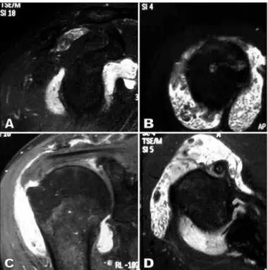
Simple radiography did not show any abnormali-ties, except for increased soft-tissue volume. Magne-tic resonance imaging was then requested, and this showed tearing of the full thickness of the anterior portion of the tendon of the supraspinatus muscle and significant glenohumeral synovitis and synovitis of the subacromial bursa, with signs of fatty metaplasia (Figure 1 A-D).
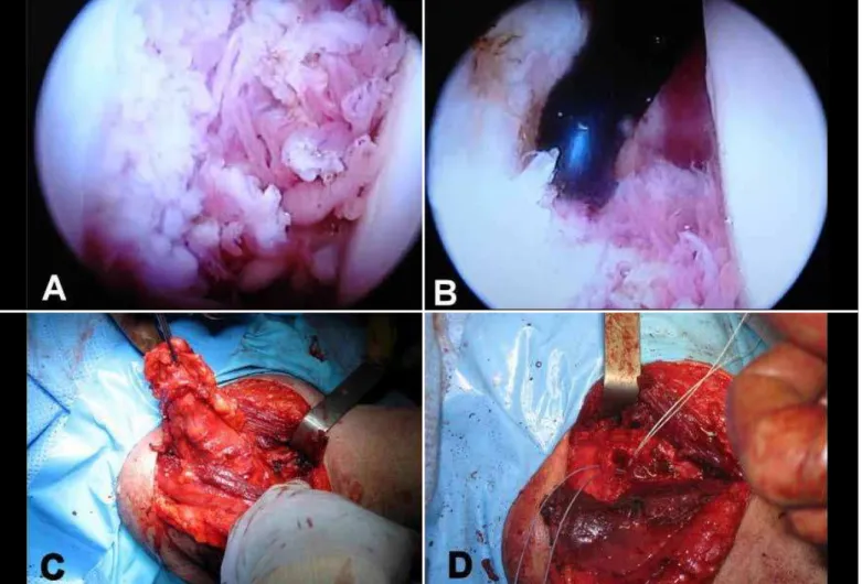
The therapeutic proposal indicated was arthroscopic surgery (Figure 2) for synovectomy of the synovial process/component, followed by an open procedure to excise the lipoma from the bursa and repair the tendon of the supraspinatus muscle (Figure 2). The surgery was performed on October 23, 2008. After the arthroscopy, an expanded deltopectoral access was constructed, with months. The pain worsened in cold environments (i.e.
with air conditioning) and with movement, and was relieved through using non-steroidal anti-inflamma-tory drugs (NSAIDs). The pain had been increasing in frequency and, over the last three months, the patient had noticed an increase in the volume of the right shoulder, which had progressed up to the day of the consultation. He did not have any history of trauma. His personal history included a renal tumor that had been treated and cured four years previously, and also systemic arterial hypertension that was properly under control. Orthopedic physical examination showed bi-lateral eutrophic musculature with increased volume in the right shoulder, without pain on palpation but slight pain on mobilization, and active and passive ranges of motion without limitations (elevation 180º, abduction 180º, lateral rotation 60º and medial rota-tion as far as T7). The patient presented a positive Jobe test for pain and a negative test for strength. The tests for the infraspinatus and subscapularis muscles
Rev Bras Ortop. 2012;47(4):517-20
Figure 1 – (A) Oblique sagittal slice showing juxta-glenoid intra-articular anterior and posterior involvement. (B) Axial slice through the upper portion demonstrating a lesion
519
deinsertion of the anterior portion of the deltoid from the acromion, in order to view the posterior part of the lipoma and to identify and protect the deep branch of the axillary nerve. The rotator cuff was sutured in the usual manner. Intraoperative frozen-section examination confirmed the diagnosis of lipoma arborescens. The result from the final anatomopathological examination was that this was a case of lipoma arborescens, showing foci of cartilaginous metaplasia in association with moderate-grade hyperplastic synovitis with numerous fibrin bodies predominating in the intra-articular component of the lesion. Currently, four months after the operation, the patient presents good shoulder function, without any neurological sequelae.
DIsCUssION
Lipoma arborescens is characterized by diffuse villous proliferation from the synovial membrane and replacement of the subsynovial tissue with mature adipocytes. It can also be named lipomatous villous proliferation from the synovial membrane, which avoids confusion with a neoplastic disease(1).
Its etiology is unknown, but the best-accepted hy-pothesis, based on histological findings of mononu-clear infiltrate over the synovial membrane, is that it is a nonspecific reaction to chronic inflammation. It has also been seen to be associated with trauma,
de-Figure 2 – (A and B) Intra-articular appearance of the lipoma arborescens, seen by
means of arthroscopy. (C) Expanded deltopectoral access and macroscopic appearan-ce of the lipoma at the start of the excision. (D) Macroscopic appearanappearan-ce of the tumor.
Rev Bras Ortop. 2012;47(4):517-20 LIPOMA ARBORESCENS: RARE CASE OF ROTATOR CUFF TEAR ASSOCIATED WITH THE PRESENCE OF LIPOMA ARBORESCENS
IN THE SUBACROMIAL-SUBDELTOID AND GLENOHUMERAL BURSA
generative joint disease, rheumatoid arthritis, psoriatic arthritis and diabetes mellitus(1-3,5).
Lipoma arborescens is rare, with fewer than 100 cases described in the literature, in the English lan-guage. It typically affects patients between their fifth and seventh decades of life, although cases in young adults and during childhood have also been repor-ted(5-7). Men and women seem to be equally affected(2).
The disease generally only affects a single joint, and the knee is the location most commonly affected, particularly the suprapatellar bursa(1). In the literature,
there are reports that the wrist, elbow, shoulder, hip and ankle can also be affected, and that there may be bilateral involvement or multi-joint disease(4,5,8-10).
Patients with lipoma arborescens typically present with painless tumefaction of the joint affected, which begins insidiously and evolves over several months or years. Pain may arise through this evolution, and it is progressive, accompanied by joint effusion, with intermittent exacerbations. Mechanical symptoms may occur, such as limitation of the range of motion, blockage, rebound and crepitation. The hemogram, erythrocyte sedimentation rate and levels of C-reactive protein, uricemia, rheumatoid factor, Waaler-Rose and VDRL are usually normal. The synovial fluid is clear and citrus-yellow, presents normal viscosity and is negative for cells and crystals, and its cultures are negative.
Regarding imaging examinations, simple radio-graphy is non-specific, and may not present any ab-normalities. On the other hand, it may show tume-faction of the soft tissue. It is important to rule out bone tumors.
Ultrasonography shows hyperechogenic villous proliferations suggestive of fat.
Computed tomography shows low-density villono-dular masses, absence of intravenous contrast uptake and, if present, intra-articular effusion. In addition, it enables confirmation of whether the lipoma arborescens is intra-articular or not, the synovial origin and the size and extent of the lesions(2). However, it is an
examina-tion with low specificity.
520
The treatment consists of open or arthroscopic synovectomy. If complete resection of the lesion is achieved, the prognosis is good, with cure achieved in almost all cases. Only one case of recurrence of the lesion after synovectomy has been reported in the literature. Synovectomy using radioactive compounds such as yttrium-90, or with steroids or chemical com-pounds such as osmic acid, has been reported as an alternative to surgical treatment(3,11), but there is no
long-term follow-up that proves its efficacy.
Cases affecting the shoulder have sometimes been described in the literature. Most of the reports are on cases affecting the subdeltoid bursa(4), and some
have been associated with rotator cuff injuries. So far, two cases of lipoma arborescens of the glenohumeral joint have been described in the literature, of which only one was treated surgically. Our case portrayed an occurrence of lipoma arborescens that was simul-taneously bursal and intra-articular, associated with an injury to the tendon of the supraspinatus muscle. Up until now, no other case of lipoma arborescens with this pattern has been reported.
sequences and low signal in fat-suppression and STIR sequences; potential chemical-shift artifact at the fat--fluid interface; and absence of gadolinium uptake by the lesion. In addition, MRI may demonstrate certain associated conditions, such as joint effusion and dege-nerative joint abnormalities, which are common in pa-tients within the age group affected by the disease(1,2).
The findings in the anatomopathological examina-tion consist of hypertrophic synovial villi distended by mature adipose tissue, and in some cases, cartila-ginous and bone metaplasia. There may also be mo-derate infiltration of mononuclear inflammatory cells in the synovial membrane, and also perivascular focal infiltrate(2). An association between lipoma
arbores-cens and chronic joint pathological conditions such as joint effusion, synovial cysts, degenerative joint alterations, bone erosion and synovial chondromatosis has been described, thus corroborating the hypothesis that this entity is a reactive process in the synovium.
Differential diagnoses are made with pigmented villonodular synovitis, synovial osteochondromatosis and synovial hemangioma.
Rev Bras Ortop. 2012;47(4):517-20
REFERENCEs
1. Adelani MA, Wupperman RM, Holt GE. Benign synovial disorders.J Am Acad Orthop Surg. 2008;16(5):268-75.
2. Bernardo A, Bernardes M, Brito I, Vieira A, Ventura F. [Synovial lipoma rbores-cens]. Acta Med Port. 2004;17(4):325-8.
3. Davies AP, Blewitt N. Lipoma arborescens of the knee. Knee. 2005;12(5):394-6.
4. Dawson JS, Dowling F, Preston BJ, Neumann L. Case report: lipoma arbores-cens of the sub-deltoid bursa. Br J Radiol. 1995;68(806):197-9.
5. Bejia I, Younes M, Moussa A, Said M, Touzi M, Bergaoui N. Lipoma arbores-cens affecting multiple joints. Skeletal Radiol. 2005;34(9):536-8.
6. Cil A, Atay OA, Aydingöz U, Tetik O, Gedikoğlu G, Doral MN. Bilateral lipoma arborescens of the knee in a child: a case report. Knee Surg Sports Traumatol Arthrosc. 2005;13(6):463-7.
7. Donnelly LF, Bisset GS 3rd, Passo MH. MRI findings of lipoma arborescens of the knee in a child: case report. Pediatr Radiol. 1994;24(4):258-9.
8. Babar SA, Sandison A, Mitchell AW. Synovial and tenosynovial lipo-ma arborescens of the ankle in an adult: a case report. Skeletal Radiol. 2008;37(1):75-7.
9. Dinauer P, Bojescul JA, Kaplan KJ, Litts C. Bilateral lipoma arborescens of the bicipitoradial bursa. Skeletal Radiol. 2002;31(11):661-5.
10. Doyle AJ, Miller MV, French JG. Lipoma arborescens in the bicipital bursa of the elbow: MRI findings in two cases. Skeletal Radiol. 2002;31(11):656-60.