11 artigo 391
OrigiNAl ArticlE
The authors declare that there was no conflict of interest in conducting this work
1 –Head of the Spinal Pathology Group, Department of Orthopedics, Hospital Santa Marcelina, Eastern Zone, São Paulo, Brazil. 2 – Attending Physician, Spinal Pathology Group, Department of Orthopedics, Hospital Santa Marcelina, Eastern Zone, São Paulo, Brazil. 3 – Head of the Orthopedics and Traumatology Service, Hospital Santa Marcelina, Eastern Zone, São Paulo, Brazil.
Work performed in the Department of Orthopedics and Traumatology, Hospital Santa Marcelina.
Correspondence: Luiz Cláudio Lacerda Rodrigues - Departamento de Ortopedia - Hospital Santa Marcelina - Rua Santa Marcelina, 177 - Itaquera - 08270-070 São Paulo, SP. E-mail: [email protected]
Work received for publication: August 1, 2010; accepted for publication: December 23, 2010.
evaluatiOn Of surgical treatment Of fractures Of
thOracOlumbar spine With third-generatiOn
material fOr internal fixatiOn
Adalberto Bortoletto1, Luiz Cláudio Lacerda Rodrigues2, Marcelo Hide Matsumoto3
AbStrAct
Objective: To evaluate the functional results from patients with surgical fractures in the thoracolumbar spine. Method: A prospective study including 100 patients with spinal fractures in the thoracic and lumbar segments was con-ducted. The lesions were classified in accordance with the AO system, and the patients were treated surgically. The presence of early kyphosis and its evolution after the surgical intervention, and the presence of postoperative pain and its evolution up to the 24th week after the surgery,
were evaluated. We compared our data with the literature. Results: One hundred surgical patients were analyzed, of which 37 were type A, 46 were type B and 17 were Type C. Patients who presented Frankel A kept their clinical
status, but patients with Frankel B or higher evolved with some improvement. The average improvement in pain ba-sed on a visual analog scale was more than four points. All the patients were able to return to their daily routine activities, although we did not take the return to work to be an assessment criterion. Conclusion: Despite controversy regarding the indications for surgery in cases of fractured spine, we believe that the method that we used was satis-factory because of the good results and low complication rate. However, more randomized prospective studies with longer follow-up are needed in order to evaluate this type of fixation.
Keywords - Spinal Fractures/classification; Spinal
Frac-tures/surgery; Spinal Cord Injuries; Treatment Outcome
iNtrOdUctiON
The management for patients with fractures of the thoracolumbar spine is a matter of controversy and has, over recent decades, been the topic of studies and investigations in many specialized centers. This research has produced new concepts and procedures of greater efficiency for early rehabilitation of such patients. Nonetheless, different opinions exist regar-ding the best approach.
The use of third-generation material started with Cotrel and Dubousset(1) in 1985, simultaneously with
dissemination of the concepts of rigid stabilization and three-dimensional correction.
The internal fixator was presented for the first time by Dick in 1987(2). This was based on the Margel
sys-tem and consisted of Shanz pins connected to a bar by a system that allowed movement in the longitudinal direction and also allowed angular movement. Long pins enabled manual reduction, and these were cut after fixation of the system.
residual pain using the VAS classification, in relation
to the presence of kyphosis, the patient’s sex and the
type of trauma.
All the patients were operated by the same team, and the Synthes® third-generation internal fixator system was used for all cases.
rESUltS
From evaluations of neurological condition, none of the cases worsened. All conditions graded as Frankel B, C and D improved. The proportion of cases at L1 level was significantly greater than the cases at other levels (chi-square test with p < 0.001). The incidence at L2 and T12 levels was similar (chi--square test with p = 0.724). There were no signifi-cant differences from T11 with other levels in smaller proportions (p = 0.101).
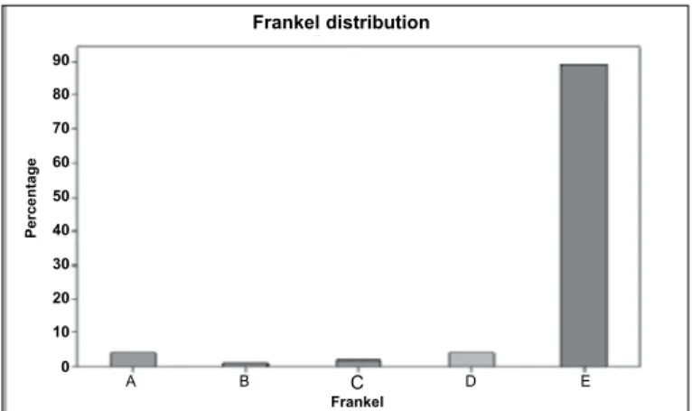
There was no statistical difference between the proportions of type A and type B fractures (chi-square test; p = 0.323), but the percentage of type C fractures was significantly lower (chi-square test; p = 0.001). The proportion of Frankel E was significantly grea-ter (chi-square test; p < 0.001), while there were no significant differences between the others (chi-square test; p = 0.484) (Figure 2).
The mean pain assessment based on VAS was 7 (ranging from 5 to 9) before the operation. One month after the operation, the mean pain level was 6 (ranging from 2 to 7) and, six months after the operation, it was 4 (ranging from 0 to 6). The initial mean kyphosis was 56º (ranging from 29º to 80º). The mean immediate postoperative Cobb was 28º (ranging from 15º to 50º). Six months after the operation, the mean Cobb was 32º (ranging from 20º to 52º).
Distribution according to level
T6 T5 L4 L3 T11 T12 L2 L1
40
30
20
10
0
Pe
rc
e
n
ta
g
e
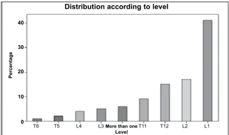
Figure 1 – Distribution of fractures according to level. presence of residual deformity, and the progression
of such deformities was evaluated during the posto-perative follow-up.
MAtEriAlS ANd MEthOdS
This was a prospective study in which all patients with fractures of the thoracolumbar spine who agre-ed to participate were includagre-ed. The evaluation was concluded 24 weeks after assessing the last patient included.
Prospective analysis was conducted on 100 pa-tients who underwent operations between January 2006 and July 2009 at Hospital Santa Marcelina, in the eastern zone of São Paulo, to treat fractures of the thoracolumbar spine. The inclusion criteria were the presence of fractures of the thoracolumbar spine that had occurred less than 10 days earlier, an indica-tion for surgery and signing of an informed consent statement in accordance with the study protocol. All the patients underwent radiographic and computed tomography (CT) examinations. Patients presenting neurological deficits also underwent magnetic reso-nance imaging (MRI). All the patients were followed up for a minimum of 24 weeks.
During the follow-up, the patients underwent ra-diographic examinations in an upright standing po-sition to control for kyphosis. This was assessed by
means of the Cobb method(3) before the operation,
during the immediate postoperative period, one month after the operation and six months after the operation. In measurements for regional evaluations, the para-meters were the end plate of the vertebra above the fractured vertebra and the distal plate of the vertebra below the fractured vertebra.
The classification used was as recommended by AO. It was observed that there were 37 type A fractu-res, 46 type B and 17 type C. The predominant level was L1, in 41 cases, followed by L2 in 17 cases and T12 in 15 cases. The total distribution is represented in Figure 1.
The main cause of the fractures was falls, in 78 cases. Twelve cases resulted from car accidents, nine were due to motorcycle accidents and one case was due to being run over. The Frankel scale was used to assess the neurological condition(4).
The results were assessed based on comparisons of
301
Frankel distribution
C
Frankel
A B D E
90 80 70 60 50 40 30 20 10 0 Pe rc e n ta g e
Figure 2 – Correlation of the patients’ neurological deficits.
There were seven cases of infection, which made it necessary to remove the material in five of these cases. One case of removal of material was in a patient with a Chance fracture who, because of multiple skin abra-sions, was not in a condition to use a vest. We decided to use instrumentation without arthrodesis in this case alone, with removal six months later. The mean num-ber of screws used was eight (ranging from four to 16). In pain data comparisons, we observed that re-peated-measurement ANOVA did not show any sig-nificant differences between the sexes at any time (intergroup comparison; p = 0.461), but there were differences within each sex between times (intragroup comparison; p < 0.001) (Figure 3).
In kyphosis comparisons using repeated-measure-ment ANOVA, there were no differences between the sexes at any time (intergroup comparison; p = 0.958), but there were differences between times (intragroup comparison; p < 0.001). The immediate postoperative Cobb was significantly smaller than the initial Cobb, but then started to increase again after six months (Figure 4).
Figure 3 – Evolution of the painful condition in relation to sex.
Pain boxplot according to sex
Female Male Previous pain 9 8 7 6 5 4 3 2 1 0 Pa in
Female Male Pain after one month
Female Male Pain after six months
Figure 4 – comparison between cobb and sex, and its evolution. Female Male
Initial COBB
Female Male Immediate postoperative
COBB
Female Male COBB after six
months
CObb boxplot according to sex
C O b b 80 70 60 50 40 30 20 10
MATERIAL FOR INTERNAL FIXATION
We found significant differences between the se-xes, i.e. females had significantly more infections than shown by males (chi-square test; p = 0.030; or Fisher exact test; p = 0.043). We found five cases of infection in females.
Comparing ages according to type of fracture, cause of accident and Frankel, we observed the following. There were no significant differences between the mean ages of the three groups according to type of fracture (ANOVA test; p = 0.063). There were no significant difference between the mean ages of the three groups according to the cause of the accident (ANOVA test; p = 0.156). There were no significant differences between the age distributions of the va-rious groups, in comparisons of neurological deficits (Kruskal-Wallis test; p = 0.931). Comparison between age and Cobb angle showed that there was no rela-tionship. There was also no correlation between age
and patients’ pain. There were no significant differen -ces in the proportions of different types of fractures between the different types of causes of accidents (chi-square test; p = 0.987) (Figure 5).
Figure 5 – Comparison between type of fracture and cause of accident.
Distribution of type of fracture according to cause of accident
A B C Car 50 40 30 20 10 0 p e rc e n ta g e w ith in e a c h c a u s e o f a c c id e n t
A B C Motorcycle
There were 89 patients with Frankel E, four with Frankel D, two with Frankel C, one with Frankel B and four with Frankel A. There were significant di-fferences in the proportions of the different types of Frankel between the different types of fractures (chi- square test; p = 0.001). Investigation of where the significant differences were showed the following: Frankel type A was significantly greater in fracture type C (p = 0.017); Frankel type E was significan-tly greater in fracture types A and B than in C (p < 0.001); while the other Frankel types did not present any significant differences (Figure 6).
Based on repeated-measurement ANOVA, diffe-rences between the types of fracture were seen as all times (intergroup comparison; p = 0.001). Type A fractures had significantly lower pain values than shown by type C fractures, at all times (p = 0.001) (Figure 7).
Using the same repeated-measurement tool, diffe-rences between fracture types were seen only during the immediate postoperative period (p = 0.033). Type A fractures only had significantly lower Cobb values than shown by type B fractures during the immediate postoperative period (p = 0.028). Comparing the ti-mes, there were differences between all the times for all the types of fractures (p < 0.001).
There was a significant Spearman correlation for pain at different times, i.e. patients who felt more pain initially continued to feel more pain at other times.
Infection was seen to be present in seven patients (five females and two males). The implants had to be removed in the two male cases and three of the females, and these five patients had to use postope-rative orthoses.
There were no differences between patients with and without infection at any time (intergroup compa-rison; p = 0.121), but we found a difference between the times (intragroup comparison; p < 0.001). The-re weThe-re significant diffeThe-rences between all the times (also p < 0.001) (Figure 8).
Figure 6 – Comparison between cause of accident and evolution of pain.
boxplot of pain according to cause of accident
Pa
in
9
8
7 6
5
4
3 2
1
0
Car Motorcycle Fall Previous pain
Car Motorcycle Fall Pain after one month
Car Motorcycle Fall Pain after six months
Figure 7 – Comparison between the type of fracture and the
evolution of pain.
boxplot of pain according to type of fracture
A B C Previous pain
A B C Pain after one month
A B C Pain after six months
9
8
7
6
5
4
3
2
1
0
Pa
in
Figure 8 – Correlation of Cobb angle with presence or absence of infection at the surgical site.
diScUSSiON
There are few studies evaluating this type of fixation in the literature. This study did not aim to be a guide for treating fractures but, rather, simply presents our results, in which we evaluated the radiological evolution, with periodic measurements of kyphosis and postoperative pain, which were compared
between the sexes, types of fractures and patients’
functional results. No Yes
Initial
No Yes Six months
80
70
60
50
40
30
20
10
C
O
b
b
Cobb boxplot according to infection
303
The first studies using pedicle screws were descri-bed by Fuentes and Defino in 1990(5). However, they
used simple pedicle screws in their 12 patients. They reported that postoperative orthoses did not need to be used for any of their patients. Our range of Cobb angles was 4° during the immediate postoperative pe-riod, for evolution over six months with an internal fi-xator, and remaining one degree lower than the mean shown in the American and European literature(6,7).
Siebenga et al(8) presented some data showing that
instrumentation with pedicle screws preserved the fractured vertebra but was unable to preserve the disc space. They observed that the disc height diminished and that a posterolateral graft aided fusion but did not influence the sequence of events.
Comparing our data with the literature(6,7), there
was no neurological worsening in any case. There were improvements on the Frankel scale among the patients assessed as B, C and D, but we did not ob-serve any improvement in the patients with Frankel A. This shows the efficacy of the instrument and the rigid immobilization that it presents, since stabiliza-tion enables early rehabilitastabiliza-tion and protecstabiliza-tion of the neural tissue, which is no longer in a distressed state (Figures 9A, 9B, 9C and 9D).
Unlike in the data presented by Wood et al(9), who
correlated the presence of kyphosis and the functio-nal results, we did not find any correlation between kyphosis and postoperative pain based on serial VAS assessments, even though the mean loss of kyphosis was 6.7°. Studies showing this distortion were
pu-blished as early as 1987(10-12), but so far no study has
explained this abnormality.
We compared our pain assessments with those of other studies, and noted that reductions in pain have been reported in several studies, with significant de-creases in VAS levels(9,13-16).
Contrary to some studies, our study did not reveal any neurological worsening as reported by others, especially during the immediate postoperative pe-riod(13,17). Our patients who presented Frankel B, C
and D achieved increases of one degree on the Frankel scale.
In the studies by Knop et al(18) and Marti Garin et
al(19), it was reported that the presence of infection
might give rise to the need to remove the synthesis material. In our study, the presence of infection led to removal of the synthesis material in five patients. All of these cases then presented resolution of the secretion. We also had two cases of infection that presented improvements in the infectious process after open cleaning, culturing and correctly applied antibiotic. In one specific case, the material had to be removed because the patient presented a type B21
fracture (Chance), Because of this patient’s poor skin
condition at the time of the trauma, we decided to use instrumentation without arthrodesis in this case alone. This was removed six months later with an excellent clinical and radiological result. All the other patients received a graft from the posterolateral iliac.
Another complication described in the literature is pain at the site of graft harvesting(20,21), with reports
that there is significant incidence of pain at the donor
Figures 9A-d – Case of type C fracture viewed during the immediate postoperative period.
site. We harvested bone grafts from almost all of our patients: they reported the presence of postoperative pain with improvement over the evolution of the ca-ses. The above authors also reported that there was a possibility of infection in the donor region: there was one case that evolved with infection in the iliac crest, for which 42 days of vancomycin was required to resolve the problem.
We did not assess return to work as a characteristic indicating a good result because only 32% of these patients actually returned to work. From the radio-graphic result and the pain assessment, these patients did not present any significant differences from the general mean and were within the maximum and mi-nimum values.
cONclUSiON
It is known that thoracolumbar spinal trauma is related to high-energy traumatic events such as falls
from a height or car accidents. These patients can-not be neglected in the emergency services, since all multiple-trauma patients are potentially spinal trauma patients until such a diagnosis can be ruled out.
For patients presenting fractures of the thoraco-lumbar spine, we suggest that the AO classification should be used for surgical indications, with early surgery for such patients. This diminishes the com-plications relating to hospitalization.
There is some controversy regarding surgical indi-cations, but our favorable results and low complica-tion rate have led us to think better of this procedure and its results. The results with an internal fixator showed significant correction of the kyphosis and a mean loss lower than has been presented in studies using simple pedicle screws. We conclude that inter-nal fixators provide a safe method with good results, although we are aware that further controlled and ran-domized studies are necessary.
rEFErENcES
1. Cotrel Y, Dubousset J. New segmental posterior intrumentation of the spine. Orthop Trans.1985;9:118.
2. Dick W .The “fixateur interne” as a versatile implant for spine surgery. Spine (Phila Pa 1976). 1987;12(9):882-900.
3. Cobb JR. Outline for the study of escoliosis, Am Acad Orthop Surg Instr Cour-seLect 1948;5:261-75.
4. Frankel HL, Hancock DO, Hyslop G, Melzak J, Michaelis LS, Ungar GH, The value of postural reduction in the initial managemnet of closed injuries of the spine with paraplegia and tetraplegia, Part 1. Paraplegia. 1969;7(3):179-92. 5. Fuentes AE, Defino HL. Experiência inicial com o Instrumental de
Cotrel-Du-bousset. Rev Bras Ortop. 1995;30(3):119-24.
6. Stambough JL. Cotrel-Dubosset intrumentation and thoracolumbar spine trau-ma: a review of 55 cases. J Spinal Disord. 1994;7(6):461-9.
7. de Peretti F, Hovorka I, Cambas PM, Nasr JM, Argenson C. Short device fixation and early mobilization for burst fractures of the thoracolumbar junction. Eur Spine J. 1996;5(2):112-20.
8. Siebenga J, Leferink VJ, Segers MJ, Elzinga MJ, Bakker FC, Haarman HJ, et al. Treatment of traumatic thoracolumbar spine fractures: a multicenter prospective randomized study of operative versus nonsurgical treatment. Spine (Phila Pa 1976). 2006;31(25):2881-90.
9. Wood K, Buttermann G, Mehbod A, Garvey T, Jhanjee R, Sechriest V, et al. Operative compared with nonoperative treatment of a thoracolumbar burst fracture without neurological deficit: a prospective, randomized study. J Bone Joint Surg Am. 2003;85:773–81.
10. Resch H, Rabl M, Klampfer H, Ritter E, Povacz P. [Surgical vs. conserva-tive treatment of fractures of the thoracolumbar transition]. Unfallchirurg. 2000;103(4):281–8.
11. Briem D, Linhart W, Lehmann W, Bullinger M, Schoder V, Meenen NM, et al. Investigation of the health-related quality of life after a dorsoventral stabilization of the thoracolumbar junction. Unfallchirurg. 2003;106(8):625–32.
12. Butler JS, Walsh A, O’Byrne J. Functional outcome of burst fractures of the first lumbar vertebra managed surgically and conservatively. Int Orthop. 2005;29(1):51–4.
13. Denis F, Armstrong GW, Searls K, Matta L. Acute thoracolumbar burst fractu-res in the absence of neurologic deficit. A comparison between operative and nonoperative treatment. Clin Orthop Relat Res. 1984;(189):142-9.
14. Domenicucci M, Preite R, Ramieri A, Ciappetta P, Delfini R, Romanini L. Thora-columbar fractures without neurosurgical involvement: surgical or conservative treatment? J Neurosurg Sci. 1996;40(1):1-10.
15. Leferink VJ, Keizer HJ, Oosterhuis JK, van der Sluis CK, ten Duis HJ. Func-tional outcome in patients with thoracolumbar burst fractures treated with dor-sal instrumentation and transpedicular cancellous bone grafting. Eur Spine J. 2003;12(3):261-7.
16. Dai LD. Low lumbar spinal fractures: management options. Injury. 2002;33(7):579-82.
17. Mumford J, Weinstein JN, Spratt KF, Goel VK. Thoracolumbar burst fractures. The clinical efficacy and outcome of nonoperative management. Spine (Phila Pa 1976). 1993;18(8):955-70.
18. Knop C, Blauth M, Bastian L, Lange U, Kesting J, Tscherne H. [Fractures of the thoracolumbar spine. Late results of dorsal instrumentation and its conse-quences]. Unfallchirurg. 1997;100(8):630-9.
19. Marti Garin D, Villanueva Leal C, Bago Granell J. Stabilization of the lower thoracic and lumbar spine with the internal spinal skeletal fixation system and a cross-linkage system. First results of treatment. Acta Orthop Belg. 1992;58(1):36-42.
20. Ebraheim NA, Elgafy H, Xu R. Bone-graft harvesting from iliac and fibular donor sites: techniques and complications. J Am Acad Orthop Surg. 2001;9(3):210-8. 21. Hill NM, Horne JG, Devane PA. Donor site morbidity in the iliac crest bone graft.