Asymptomatic vertebral fractures in patients with low bone
mineral density
CAIO CESAR LEITEDE NEGREIROS1, MARINA GUARESCHI BERIGO2, ROBSON LUIZ DOMINONI3*, DEISI MARIA VARGAS4
1Medical Student. Fundação Universidade Regional de Blumenau (Furb), Blumenau, SC, Brazil 2Medical Student, Furb, Blumenau, SC, Brazil
3Rheumatologist. – Professor at the Department of Medicine. MSc Student, Masters Program in Collective Health, Furb, Blumenau, SC, Brazil 4PhD. – Professor at the Department of Medicine, and Lecturer of the Masters Program in Collective Health, Furb, Blumenau, SC, Brazil
S
UMMARYStudy conducted at Fundação Universidade Regional de Blumenau (Furb), Blumenau, SC, Brazil
Article received: 5/28/2014
Accepted for publication: 9/9/2014
*Correspondence:
Address: Universidade Regional de Blumenau. Campus III Rua São Paulo, 2171, A-302 Itoupava Seca Blumenau – SC CEP 89030-001 [email protected]
http://dx.doi.org/10.1590/1806-9282.62.02.145
Objective: Vertebral fracture assessment (VFA) is a test technique that can be used to detect asymptomatic vertebral fractures (AVF). It uses dual energy X-ray absorp-tiometry (DXA) and can be performed concurrently with bone densitometry. This study aims to assess the prevalence of AVF in patients with low bone mass. Methods: Cross-sectional study including 135 individuals with low bone min-eral density (BMD) with a T-score < -2.0 standard deviation (SD) in a densitom-etry clinic located in the city of Blumenau (state of Santa Catarina). Anthropo-metric, clinical and lifestyle variables were obtained from history-taking and physical examination. Densitometric variables were obtained by bone mineral densitometry and VFA (Explorer, Hollogic®). Vertebral fractures were classified according to the Genant criteria. Student’s t, chi-square and logistic regression were performed for statistical analysis.
Results: AVFs occurred in 24.4% of the subjects. They were older compared to those without AVF (65±9.25 versus 60.1±8.66; p=0.005), and had a history of low-impact fractures (38.24% versus 19.8%; OR 2.5; p=0.03). Half of the patients that reported steroid therapy had AVFs, compared to one fifth of those who did not use steroids (50% versus 21.49%; OR 3.6; p=0.01).
Conclusion: Asymptomatic vertebral fractures were present in approximately one fourth of patients. The risk factors associated were history of low-impact fracture, use of steroids and age > 61 years.
Keywords: Bone density, spinal fractures, osteoporotic fractures, photon ab-sorptiometry, osteoporosis.
INTRODUCTION
Osteoporosis (OP) is a systemic disease characterized by low bone mass and deterioration of bone tissue micro-architecture, with a consequent increase in fragility.1 Di-agnosis is performed based on the assessment of bone mineral density (BMD) or the occurrence of low-impact fractures in the hip or vertebrae in adulthood. Bone den-sitometry (BD) is currently the reference examination for diagnosing decreased BMD, and it is done using dual en-ergy X-ray absorptiometry (DXA).2
OP can be primary or secondary. Primary OP is de-fined as the absence of an identifiable secondary cause. Secondary OP occurs when disease, deficiency or drug is found to be the cause. They include: hypercortisolism (endogenous or exogenous), hyperparathyroidism,
hy-perthyroidism, acromegaly, hematopoietic malignancies, primary biliary cirrhosis, inflammatory bowel disease, celiac disease, gastrectomy, homocystinuria, hemochro-matosis and inflammatory rheumatic diseases. Fractures are the main clinical manifestations, and vertebral, fem-oral and forearm are the most frequent. They may be symptomatic, i.e. painful, or asymptomatic.3,4
Vertebral fractures are estimated to affect 5% of Cau-casian women older than 50 years, and 25% of women older than 80 years.5 However, about 25 to 35% of these fractures are asymptomatic.6
more likely to experience a new fracture, compared to those with normal BMD and no fractures. A patient with multiple vertebral fractures and low BMD has 75 times greater risk of new fractures.5,9,10
The gold standard for diagnosing vertebral fractures is lateral radiograph of the thoracic and lumbar spine but they are usually requested only if there are symptoms such as pain. This type of X-ray demands additional time and resources, as well as greater patient exposure to radiation. Given that about a third of fractures manifests with symptoms, and the request of spine radiographs is not a routine for screening, the diagnosis of vertebral fractures is underestimated.11
Vertebral fracture assessment (VFA) is a technique that can be used to detect asymptomatic vertebral frac-tures. VFA uses DXA, and can be performed with densi-tometry, allowing rapid evaluation of the vertebral bod-ies. It can make the diagnosis of vertebral fracture easier and serves as an assessment of risk of future fractures.6 VFA can be performed without the need of referral to an-other service; it involves less exposure to radiation and produces a digital image, which can be stored, enabling analysis at any time for comparison purposes.9
The diagnosis of vertebral fractures using VFA is per-formed when there is a reduction of more than 20% in anterior, middle, and posterior height. The Genant meth-od (semi-quantitative and visual inspection) is the most used to grade fractures: grade zero or normal, if there is no deformity; grade 1 or mild (20 to 25% reduction in ver-tebral height); grade 2 or moderate (25 to 40% reduction in vertebral height); and grade 3 or severe (reduction great-er than 40% in vgreat-ertebral height). Fractures are still classi-fied as wedge, concave or crush according to the greatest reduction in height in the anterior, middle or posterior vertebral body, respectively.8,12
The aim of this study was to determine the preva-lence of asymptomatic vertebral fractures in patients with reduced bone mass using the VFA, and to analyze associated factors.
METHODS
This is an observational and retrospective study in 135 patients aged 18 years or older who underwent spine and proximal femur bone densitometry (Explorer, Hollogic®) and had low bone mass (T score < or equal to -2,00 SD) in at least one bone densitometry site. The VFA exam was performed concurrently with densitometry. The study was conducted at a clinic in the city of Blumenau (state of Santa Catarina) from September 2011 to March 2012. Quantitative variables were: bone density in the prox-imal femur (femoral neck and total femur) and lumbar
spine (L1 to L4) expressed as T-score; age in years; body weight measured in Kg using a Plenna® digital scale; height measured in meters using Tonelli® precision stadiometer; body mass index (BMI); daily calcium intake measured in mg/day based on a dietary record; age at menopause in years. To calculate the daily intake of calcium, the recom-mendations of Associação Brasileira de Avaliação Óssea e Osteo-metabolismo (Abrasso) were used: 1 glass of milk (240 mL) = 300 mg of calcium; 1 glass of yogurt (240 mL) = 400 mg; 1 slice of cheese (28.35 g) = 200 mg; calcium from other sources = 250 mg. The tests were performed by a bone den-sitometry equipment operator certified by the Abrasso and the reports were issued by a clinical densitometrist certi-fied by both the Abrasso and the International Society for Clinical Densitometry (ISCD).
The qualitative variables were gender, presence of OP, presence of obesity, daily calcium intake, history of low-impact fractures after 40 years of age, current smoker, his-tory of steroid therapy, sedentary lifestyle, presence of menopause and fractures on VFA (in the absence of pain or previous vertebral fracture).
For diagnosis of vertebral fractures, Genant method was used. Genant grade 1, 2 and 3 was considered posi-tive for asymptomatic vertebral fracture, regardless of the type, and Genant grade 0 was considered negative.12
Data were organized in descriptive tables containing measures such as frequency, mean, median, standard de-viation and coefficient of variation. Estimates of average and proportion (prevalence) were taken at intervals with 95% confidence. To compare frequencies within the same distribution, chi-square test of adhesion was used. For the association of factors with the occurrence of fractures, chi-square test of independence and odds ratio (OR) were used. In order to compare data between the two groups whose variables were quantitative, Student’s t test was used for independent samples. For multivariate data anal-ysis, binary logistic regression procedures were used. In all the tests, the authors considered significant a p-value < 0.05. Data analysis was carried out using Microsoft
Ex-cel 2010 and EpiInfo 2012 version 7 as software. The study was approved on 12/14/10 by the Research Ethics Committee at Fundação Universidade Regional de Blumenau, protocol number 191/10.
RESULTS
24.44% (n=33) of patients presented asymptomatic ver-tebral fractures.
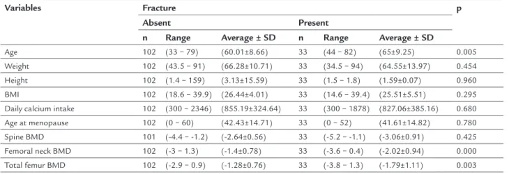
age. In the group with vertebral fractures, age was statis-tically higher (65±9.25 versus 60.01±8.66; p=0.005).
Bone densitometry of the femoral neck and total femur also showed differences. Femoral neck BMD in patients with vertebral fracture was lower compared to the group without fractures (-2.02±0.94 versus -1.4±0.78; p=0.000). Regarding total femur, the group with vertebral fractures also had low-er BMD compared to the group without fractures (-1.79±1.11
versus -1.28±0.76; p=0.003).
Weight, height, BMI, spine BMD, age at menopause and daily calcium intake showed no statistically signifi-cant difference between groups.
Table 2 shows the association between the variables studied and the occurrence of asymptomatic vertebral fracture using bivariate analysis with chi-square test and OR. In the analysis, it is concluded that age over 61 years is associated with increased risk of asymptomatic verte-bral fracture verified by VFA (OR=2.40; 95CI=1.07-5.40; p=0.031). The same is true for steroid therapy (OR=3.65; 95CI=1.18-11.36; p=0.018) and history of low-impact frac-ture (OR=2.50; 95CI=1.07-5.85; p=0.030).
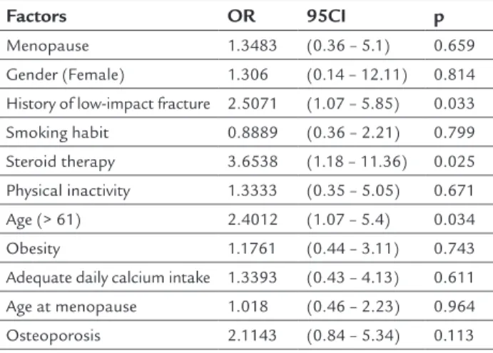
Table 3 shows the multiple logistic regression analy-sis, OR, OR estimate and p-value. Age over 61 years was considered a risk factor for asymptomatic vertebral frac-tures (OR=2.40; 95CI=1.07-5.40; p=0.034), as well as pre-vious steroid therapy (OR=3.65; 95CI=1.18-11.36; p=0.025) and previous low-impact fracture (OR=2.50; 95CI=1.07-5.85; p=0.033).
DISCUSSION
Asymptomatic vertebral fractures were found in 24.4% of participants. These patients were older than those
with-out asymptomatic vertebral fractures (AVF), also present-ing history of low-impact fractures more often. Half of the patients that reported use of steroids had AVFs, com-pared to one fifth of those without steroid therapy (50%
versus 21.49%; OR 3.65; p=0.018).
Greenspan et al. evaluated, using VFA, 482 women with no previous diagnosis of fracture. Vertebral fractures were found in 18.3%.6 These results relate to our study, which found asymptomatic vertebral fractures in approx-imately one quarter of patients.
In our study, the mean age of patients with asymp-tomatic vertebral fracture was higher than in patients without this type of fracture. Steiger et al. studied densi-tometry data of 172 patients aged 50 years or older. They found that 27% had vertebral fractures and that these fractures were more frequent in patients over 65 years old.9 One possible explanation for these results is the cur-rent progressive aging of the population, which increas-es the chance of occurrence of diseasincreas-es related to senincreas-es- senes-cence, such as reduction in BMD.10
Patients who used steroids had a 3.65 times higher risk of vertebral fractures. Of these patients, 50% had as-ymptomatic vertebral fractures. The percentage drops to 21.48% among patients who did not use steroids. It is not yet established whether there is a minimum dose of ste-roids safe for bone tissue. Some authors suggest that a maximum dose of 5 mg per day of prednisone (or equiv-alent) does not have deleterious effects on bone remod-eling. However, in a retrospective study, Van Staa et al. documented increase in the relative risk of vertebral frac-ture with prednisone at doses below 2.5 mg/day, and in-creased relative risk of fracture in the femoral neck at doses
TABLE 1 Comparison of patients with and without asymptomatic vertebral fractures including the variables: age, weight, height, BMI, daily calcium intake, age at menopause, densitometry of the femoral neck and spine, and total femur bone mineral density using Student’s t test.
Variables Fracture p
Absent Present
n Range Average ± SD n Range Average ± SD
Age 102 (33 − 79) (60.01±8.66) 33 (44 − 82) (65±9.25) 0.005
Weight 102 (43.5 − 91) (66.28±10.71) 33 (34.5 − 94) (64.55±13.97) 0.454
Height 102 (1.4 − 159) (3.13±15.59) 33 (1.5 − 1.8) (1.59±0.07) 0.960
BMI 102 (18.6 − 39.9) (26.44±4.01) 33 (14.6 − 39.4) (25.51±5.51) 0.295
Daily calcium intake 102 (300 − 2346) (855.19±324.64) 33 (300 − 1878) (827.06±385.16) 0.680
Age at menopause 102 (0 − 60) (42.43±14.71) 33 (0 − 52) (41.61±14.82) 0.780
Spine BMD 101 (-4.4 − -1.2) (-2.64±0.56) 33 (-5.2 − -1.1) (-3.06±0.91) 0.425
Femoral neck BMD 102 (-3 − 1.3) (-1.4±0.78) 33 (-3.6 − 0.4) (-2.02±0.94) 0.000
Total femur BMD 102 (-2.9 − 0.9) (-1.28±0.76) 33 (-3.8 − 1.3) (-1.79±1.11) 0.003
I − n: Number of patients; SD: standard deviation.
greater than 2.5 mg/day.13-16 In our study, it was not pos-sible to correlate dose of steroids and the presence of frac-tures, as this variable was not quantified.
Femoral neck and total femur BMD were lower in pa-tients with vertebral fractures compared to papa-tients with-out vertebral fractures, while density of the spine was not different. Possible explanations are the fact that age-relat-ed degenerative bone disease at lumbar spine appears ear-lier than proximal femur interfering in BMD quantifica-tion, and that vertebral fractures can falsely increase BMD, since bone mineral content (BMC) would remain the same while occupying a smaller projected area (DMO = BMC/ area). In a study that included 57 menopaused women, 23 had 1 to 3 fractured vertebrae in the lower back. The aver-age increase in BMD per vertebra was 0.07 g/cm2, which led to an increase in T-score from -2.3 to -1.6.17 Thus, the presence of fractures could explain the lack of association between vertebral fracture and bone density in the spine, as the accuracy of the method is impaired in this region.
One of the indications for densitometry is the pres-ence of previous fractures, which increases the risk of new fractures by approximately 25%.10,18 In our study, 25.19% of the participants had a history of low-impact fractures.
TABLE 2 Association between the variables studied and the occurrence of fracture using bivariate analysis with Chi-square test and odds ratio.
Factors Fracture OR 95CI c² p
Present Absent
Menopause No 12 (80%) 3 (20%) 1.3483 (0.36 – 5.1) 0.19480 0.658
Yes 89 (74.79%) 30 (25.21%)
Gender Female 98 (75.38%) 32 (24.62%) 0.7656 (0.08 – 7.1) 0.05553 0.813
Male 4 (80%) 1 (20%)
History of low-impact fracture No 81 (80.2%) 20 (19.8%) 2.5071 (1.07 – 5.85) 4.67979 0.030
Yes 21 (61.76%) 13 (38.24%)
Smoking habit No 75 (75%) 25 (25%) 0.8889 (0.36 – 2.21) 0.06446 0.799
Yes 27 (77.14%) 8 (22.86%)
Steroid therapy No 95 (78.51%) 26 (21.49%) 3.6538 (1.18 – 11.36) 5.52332 0.018
Yes 7 (50%) 7 (50%)
Physical inactivity No 12 (80%) 3 (20%) 1.3333 (0.35 – 5.05) 0.18048 0.670
Yes 90 (75%) 30 (25%)
Age ≤ 61 59 (83.1%) 12 (16.9%) 2.4012 (1.07 – 5.4) 4.61379 0.031
> 61 43 (67.19%) 21 (32.81%)
Obesity No 83 (76.15%) 26 (23.85%) 1.1761 (0.44 – 3.11) 0.10712 0.743
Yes 19 (73.08%) 7 (26.92%)
Adequate daily calcium intake No 90 (76.27%) 28 (23.73%) 1.3393 (0.43 – 4.13) 0.25984 0.610
Yes 12 (70.59%) 5 (29.41%)
Age at menopause > 46 53 (75.71%) 17 (24.29%) 1.0180 (0.46 – 2.23) 0.00198 0.964
≤ 46 49 (75.38%) 16 (24.62%)
Osteoporosis No 37 (84.09%) 7 (15.91%) 2.1143 (0.84 – 5.34) 2.57479 0.108
Yes 65 (71.43%) 26 (28.57%)
I − OR: Odds ratio; CI: confidence interval (estimative for OR with 95% confidence interval) II − c²: Calculated value (Chi-square test for independence)
III − p: Significance value (If p < 0.05, association is significant)
TABLE 3 Factors associated with vertebral fractures (n=135) using bivariate analysis with simple logistic regression.
Factors OR 95CI p
Menopause 1.3483 (0.36 – 5.1) 0.659
Gender (Female) 1.306 (0.14 – 12.11) 0.814
History of low-impact fracture 2.5071 (1.07 – 5.85) 0.033
Smoking habit 0.8889 (0.36 – 2.21) 0.799
Steroid therapy 3.6538 (1.18 – 11.36) 0.025 Physical inactivity 1.3333 (0.35 – 5.05) 0.671
Age (> 61) 2.4012 (1.07 – 5.4) 0.034
Obesity 1.1761 (0.44 – 3.11) 0.743
Adequate daily calcium intake 1.3393 (0.43 – 4.13) 0.611
Age at menopause 1.018 (0.46 – 2.23) 0.964
Osteoporosis 2.1143 (0.84 – 5.34) 0.113
Of these, 38.2% had vertebral fractures. The authors also found that patients with a history of low-impact fractures have 2.5 times more risk of new fractures, compared to those without history of low-impact fractures.
Asymptomatic vertebral fractures are often underdi-agnosed and therefore undertreated.19 The optimization of diagnosis might contribute to reduce the occurrence of new fractures, improving the quality of life of patients and reducing treatment costs.20
Some risk factors for osteoporotic fractures classical-ly described in the literature were not correlated with the presence of fractures in this study.2 For example, in smok-ers the prevalence of vertebral fractures was 22.86%, while in non-smokers it was 25%. Calcium intake and age at menopause were not different between those with and those without vertebral fractures. The reason is that os-teoporosis is a multifactorial disease. Approximately 70% of the factors involved cannot be changed, i.e. are genet-ically defined, while 30% is potentially modifiable and, thus, has to do with environmental issues.21
Mean BMI of patients with vertebral fractures was 25.51 compared to the average of 26.44 in those who did not have fractures, although not statistically significant. Increased mechanical load on the skeleton can cause an adjustment in bone tissue to support greater mechanical strength. In these individuals, there is higher production of estrogen by fat cells, with consequent reduction in bone remodeling. Also, increased insulin resistance and hyperinsulinemia are
observed in this group. Insulin can have a direct effect on bone formation, but it can also induce greater ovarian pro-duction of sex steroids and reduced hepatic propro-duction of the protein that binds to sex hormones, leading to increased blood concentration of estrogens and androgens.22
25% of the sedentary subjects had vertebral fractures, while 20% of the non-sedentary had these fractures. There-fore, there was no association between physical inactivi-ty and vertebral fractures. According to Pinheiro et al., there are no studies to support this variable as a risk fac-tor for vertebral fractures in the Brazilian population.21 The practice of physical activity would impact the increase in BMD, if practiced until reaching a peak in which the individual is subject to gain bone mass. From a certain age, around 30 years, people would only lose bone mass, and physical activity would no longer be relevant.23
CONCLUSION
A quarter of the individuals had asymptomatic vertebral fractures, which were associated with use of steroids, his-tory of low-impact fractures and age. The evaluation of
the spine using VFA concomitant with bone densitome-try would be useful in the early diagnosis of vertebral frac-tures, leading to initiation of treatment, reduced compli-cations and costs, improved prognosis and a better quality of life for patients.
RESUMO
Fraturas vertebrais assintomáticas em indivíduos com baixa massa óssea
Objetivos: vertebral fracture assessment (VFA) é uma técni-ca de exame que pode ser aplitécni-cada na detecção de fratu-ras vertebrais assintomáticas (FVA). Utiliza absorciome-tria de raios-X de dupla energia (DXA) e pode ser realizada concomitantemente ao exame de densitometria óssea. Este estudo visa a avaliar a prevalência de FVA em indivíduos com baixa massa óssea.
Métodos: estudo transversal realizado em 135 indivíduos, com baixa densidade mineral óssea (DMO), com T-score < -2,0 desvio padrão (DP), em uma clínica de densitome-tria de Blumenau (SC). As variáveis antropométricas, clí-nicas e referentes ao estilo de vida foram obtidas por anam-nese e exame clínico; as variáveis densitométricas foram obtidas por DMO e VFA (aparelho modelo Explorer, mar-ca Hollogic®). As fraturas vertebrais foram classificadas de acordo com os critérios de Genant. Os testes estatísticos foram t de student, qui-quadrado e regressão logística. Resultados: FVA ocorreram em 24,4% dos indivíduos. A ida-de ida-desses indivíduos foi superior à dos indivíduos sem FVA (65±9,25 vs. 60,1±8,66; p=0,005), assim como o anteceden-te de fratura por baixo impacto (38,24% vs.19,8%; OR 2,5; p=0,03). A metade dos indivíduos que relataram corticote-rapia possuíam FVA, contrastando com um quinto dos in-divíduos sem corticoterapia (50% vs. 21,49%; OR 3,6; p=0,01). Conclusão: fraturas vertebrais assintomáticas estiveram presentes em aproximadamente um quarto dos pacien-tes. Os fatores de risco associados foram história de fra-tura por baixo impacto, corticoterapia e idade > 61 anos.
Palavras-chave: densidade óssea, fraturas da coluna ver-tebral, fraturas por osteoporose, absorciometria de fóton, osteoporose.
REFERENCES
1. Ahmed SF, Elmantaser M. Secondary osteoporosis. Endocr Dev. 2009; 16:170-90.
3. Pinto Neto AM, Soares A, Urbanetz AA, Souza ACA, Ferrari AEM, Amaral B, et al. Consenso brasileiro de osteoporose 2002. Rev Bras Reumatol. 2002; 42(6):343-54.
4. Cubas ER, Boeving A, Marcatto C, Santos CMC, Borba CZV, Kulak CAM. Principais causas de diminuição da massa óssea em mulheres na pré-menopausa encaminhadas ao ambulatório de doenças ósteo-metabólicas de um Hospital Terciário de Curitiba. Arq Bras Endocrinol Metab. 2006; 50(5):914-9. 5. Lewiecki EM, Laster AJ. Clinical review: clinical applications of vertebral
fracture assessment by dual-energy x-ray absorptiometry. J Clin Endocrinol Metab. 2006; 91(11):4215-22.
6. Greenspan SL, Von Stetten E, Emond SK, Jones L, Parker RA. Instant vertebral assessment: a noninvasive dual x-ray absorptiometry technique to avoid misclassification and clinical mismanagement of osteoporosis. J Clin Densitom. 2001; 4(4):373-80.
7. Chapurlat RD, Duboeuf F, Marion-Audibert HO, Kalpakçioglu B, Mitlak BH, Delmas PD. Effectiveness of instant vertebral assessment to detect prevalent vertebral fracture. Osteoporos Int. 2006; 17(8):1189-95. 8. Ginski TP, Newman ED, Hummel JL, Hummer M. Development and
evaluation of a vertebral fracture assessment program using IVA and its integration with mobile DXA. J Clin Densitom. 2006; 9(1):72-7. 9. Steiger P, Cummings SR, Genant HK, Weiss H. Morphometric X-ray absorptiometry
of the spine: correlation in vivo with morphometric radiography. Study of Osteoporotic Fractures Research Group. Osteoporos Int. 1994; 4(5):238-44. 10. Lipschitz S; National Osteoporosis Foundation of South Africa. Instant
vertebral assessment/lateral vertebral assessment: an integral and essential part of osteoporosis assessment. S Afr Med J. 2004; 94(12):967-8. 11. Damiano J, Kolta S, Porcher R, Tournoux C, Dougados M, Roux C. Diagnosis
of vertebral fractures by vertebral fracture assessment. J Clin Densitom. 2006; 9(1):66-71.
12. Genant HK, Wu CY, Van Kuijk C, Nevitt MC. Vertebral fracture assessment using a semiquantitative technique. J Bone Miner Res. 1993; 8(9):1137-48. 13. Patrício JP, Oliveira P, Faria MT, Pérez MB, Pereira J. Osteoporose induzida
por corticoides. Arq Med. 2006; 20(5-6):173-8.
14. Van Staa TP. The pathogenesis, epidemiology and management of glucocorticoid-induced osteoporosis. Calcif Tissue Int. 2006; 79(3):129-37. 15. Van Staa TP, Leufkens HG, Cooper C. The epidemiology of corticoste-roid-induced osteoporosis: a meta-analysis. Osteoporos Int. 2002; 13(10):777-87.
16. Van Staa TP, Leufkens HG, Abenhaim L, Zhang B, Cooper C. Use of oral costicosteroids and risk of fractures. J Bone Miner Res. 2000; 15(6):993-1000. 17. Ryan PJ, Evans P, Blake GM, Fogeman I. The effect of vertebral collapse on spinal bone mineral density measurements in osteoporosis. Bone Miner. 1992; 18(3):267-72.
18. Brandão CMA, Camargos BM, Zerbini CA, Plapler PG, Mendonça LMC, Albergaria B, et al. Posições oficiais 2008 da Sociedade Brasileira de Densitometria Clínica (SBDens). Arq Bras Endocrinol Metab. 2009; 53(1):107-12.
19. Freedman BA, Potter BK, Nesti LJ, Giuliani JR, Hampton C, Kuklo TR. Osteoporosis and vertebral compression fractures-continued missed opportunities. Spine J. 2008; 8(5):756-62.
20. Cauley JA. Public health impact of osteoporosis. J Gerontol A Biol Sci Med Sci. 2013; 68(10):1243-51.
21. Pinheiro MM, Camargos BM, Borba VZC, Lazaretti-Castro M. FRAX TM: construindo uma ideia para o Brasil. Arq Bras Endocrinol Metab. 2009; 53(6):783-90.
22. Bandeira F. A obesidade realmente fortalece os ossos? Arq Bras Endocrinol Metab. 2007; 51(6):895-97.