04 artigo 666
ORIGINAL ARTICLE
1 – Assistant Professor and Head of the Shoulder and Elbow Group, Department of Orthopedics and Traumatology, School of Medical Sciences, Santa Casa de São Paulo, São Paulo, Brazil.
2 – Resident in the Department of Orthopedics and Traumatology, School of Medical Sciences, Santa Casa de São Paulo, São Paulo, Brazil.
3 – Assistant Professor and Attending Physician in the Shoulder and Elbow Group, Department of Orthopedics and Traumatology, School of Medical Sciences, Santa Casa de São Paulo, São Paulo, Brazil.
4 – Attending Physician in the Shoulder and Elbow Group, Department of Orthopedics and Traumatology, School of Medical Sciences, Santa Casa de São Paulo, São Paulo, Brazil.
5 – Trainee in the Shoulder and Elbow Group, Department of Orthopedics and Traumatology, School of Medical Sciences, Santa Casa de São Paulo, São Paulo, Brazil. 6 – Adjunct Professor, Academic Consultant and Member of the Shoulder and Elbow Group, Department of Orthopedics and Traumatology, School of Medical Sciences,
Santa Casa de São Paulo, São Paulo, Brazil.
Work performed in the Department of Orthopedics and Traumatology, School of Medical Sciences, Santa Casa de São Paulo (DOT-FCMSCSP), “Fernandinho Simonsen” Wing, São Paulo, Brazil. Director: Prof. Dr. Osmar Avanzi.
Correspondence: R. Dr. Cesário Mota Jr. 112, Vila Buarque, 01221-020 São Paulo, SP.E-mail: [email protected]
Work received for publication: February 1, 2011; accepted for publication: March 8, 2012.
EVALUATION OF THE COMPLICATIONS OF SURGICAL TREATMENT
OF FRACTURES OF THE PROXIMAL EXTREMITY OF THE
HUMERUS USING A LOCKING PLATE
Alberto Naoki Miyazaki1, José Renato Depari Estelles2, Marcelo Fregoneze3, Pedro Doneux Santos4, Luciana Andrade da Silva4,
Guilherme do Val Sella4, Fábio Eduardo Ishioka5, João Polydoro Rosa5, Sergio Luiz Checchia6
ABSTRACT
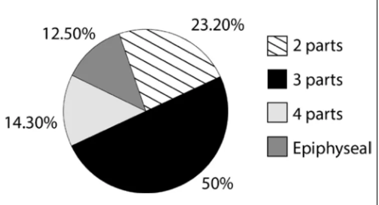
Objective: To evaluate the complications from surgical treatment using a locking plate among patients with fractures of the proximal extremity of the humerus. Methods: Between July 2004 and December 2009, 56 patients with fractures of the proximal extremity of the humerus were treated using the PHILOS® plate. There were 19 male patients and 37 female patients, with a mean age of 62 years (range: 30 to 92 years). All the cases had a mean postoperative follow-up period of 12 months. Thirteen fractures were classified as presenting in two parts, 28 as three, eight as four and seven as epiphyseal fractures. Results: Among the patients operated, 26 were considered to have achieved excellent results, twelve good, ten fair and eight poor, according to the UCLA score. Thirty complications occurred in 20 patients
INTRODUCTION
The frequency of fractures of the proximal extremi-ty of the humerus has been increasing as the population ages. When these fractures occur in elderly patients, they are associated with osteoporosis(1). Thus, choosing
the treatment to use will depend not only on the type of fracture and the surgeon’s experience, but also on the patient’s bone quality, age and degree of activity(2).
Several fixation methods have been described in
(35.7%), among which the most frequent complication was inadequate reduction of the fracture, which occurred in eight cases. Subacromial impact, caused by the plate, occurred in seven cases, while inadequate fixation occurred in six cases. Other complications such as pseudarthrosis, adhesive capsulitis, avascular necrosis, loss of varus reduction and infection were also seen. Conclusion: The functional results from treating fractures of the proximal extremity of the humerus using a locking plate depended on correct anatomical reduction of the fracture and stable fixation of the implant. Complications still occur frequently, particularly because of intraoperative technical difficulty, fracture severity and possible inexperience of the surgeon.
Keywords – Shoulder Fractures; Fracture Fixation, Inter-nal; Bone Plates
The authors declare that there was no conflict of interest in conducting this work
This article is available online in Portuguese and English at the websites: www.rbo.org.br and www.scielo.br/rbort
the literature for treating these fractures: percutaneous fixation with Kirschner wires, angled plates, tension bands, tie bands, intramedullary nails, T-shaped plates and, most recently, “locking plates”(3-8).
The aim in the constant evolution of synthesis materials has been to diminish the incidence of complications(9). Despite the diversity and technical
569
One patient was excluded from the study because this individual was lost from the follow-up. Nineteen patients (34%) were male and 37 (66%) were female.
The inclusion criteria for this study were that the patients should be over 18 years of age, with acute fractures of the proximal extremity of the humerus that required surgical stabilization, and with a mini-mum postoperative follow-up of 12 months.
The following were exclusion criteria: fractures of the proximal extremity of the humerus in patients under the age of 18 years, fractures in bones presenting tumors, exposed fractures, fractures without displacement for which conservative treatment was chosen and fractures treated by means of partial replacement arthroplasty.
The patients’ mean age was 62 years, with a range from 30 to 92. The dominant side was affected in 31 patients (55.3%). The mean number of days between the date of the fracture and the date of the surgery was seven days, with a range from zero to 20 days.
All the fractures of the proximal extremity of the humerus were classified in accordance with Neer(16),
as shown in Figure 1.
The patients were evaluated with regard to age, sex, dominance, length of time between the fracture and the surgery, type of fracture, surgical technique, time taken to achieve consolidation and any presence of complications.
All the patients underwent the operation in the deckchair position under general anesthesia and brachial plexus blockade. The bone fragments were dealt with by means of a deltopectoral route. After reduction of the fragments, the fracture was fixed provisionally using metal wires and/or suturing, the reduction was checked again by means of an image intensifier and the plate was placed in the anterolat-eral position of the proximal region of the humerus.
Figure 1 – Percentage distribution of types of fracture of the proximal ex-tremity of the humerus.
EXTREMITY OF THE HUMERUS USING A LOCKING PLATE
consolidation, avascular necrosis, implant failure, adhesive capsulitis, infection, paralysis of the deltoid muscle, inadequate fixation of the plate, migration of the screws and subacromial impact caused by the implant(4,7,9-14). The development of “locking plates” for
the proximal extremity of the humerus has brought a new perspective for treatment of fractures, especially for fractures in three or four parts, epiphyseal fractures in young patients and fractures in bones that have become fragile, for which there is greater technical difficulty in fixation(13). The theory of the mechanical advantage of
“locking plates” is that sufficient stability is achieved without bone-plate contact, which would be necessary if conventional plates were used(15). This stability is
provided by means of locking screws, thereby leading to better results in bones that are porous(7).
Currently, the Philos® plate (Proximal Humerus In-ternal Locked System) developed by the AO-ASIF group is one of the implants used for treating fractures of the proximal extremity of the humerus. It forms part of the latest generation of locking compression plates(10,14).
Fankhauser et al(4) and Duralde and Leddy(9)
showed their results from using “locking plates” for treating fractures of the proximal extremity of the hu-merus. They found complication rates of 20 to 30%, and these complications included pseudarthrosis, in-adequate reduction, infections, subacromial impact, nerve lesions and implant failure.
Complications from using “locking plates” may occur both in relation to the surgical technique (poor positioning of the plate, inappropriate screw size or quality of the reduction) and in relation to problems with the implant and fracture themselves (poor indi-cation of osteosynthesis, pseudarthrosis and osteone-crosis of the humeral head)(13).
The aim of the present study was to assess the complications from treatment among patients who underwent open reduction and internal fixation of fractures of the proximal extremity of the humerus, using the Philos® “locking plate”.
MATERIALS AND METHODS
A guidewire was passed through the upper region of the plate and through the guide for this purpose, in order to check its height in relation to the greater tubercle. The number of screws placed in the humeral head ranged from four to seven (mean of 5.3). The lengths of the screws were checked using an image intensifier at maximum lateral and medial rotations. Using stitches of non-absorbable thread (number 5), the proximal region of the plate was fixed to the ten-dons of the supraspinatus, infraspinatus and subscapu-laris muscles in order to add stability to the fixation. The size of the Philos® plate used ranged from three to nine holes (mean of 3.2). A bone graft was used when the surgeon judged that this was necessary; this occurred in seven patients (12.5%).
The operated shoulder was immobilized for at least four weeks. The patients underwent passive shoulder exercises between the second and sixth postopera-tive weeks, until there was radiographic evidence of consolidation. After consolidation of the fracture, ex-ercises to gain active shoulder mobility were started.
Postoperative outpatient evaluations were per-formed after two, four, six, 12, 24 and 52 weeks. The patients were evaluated and classified using the Uni-versity of California at Los Angeles (UCLA) score(17)
and radiographically regarding the position of fracture consolidation (anatomical or displaced) and the time taken to achieve consolidation.
The statistical assessment of the present study was done using the chi-square and Fisher exact tests. We used the significance level of 5% (0.05) for applying the statistical tests, and differences were considered to be significant when p < 0.05. This study was approved by the Research Ethics Committee. There were no conflicts of interest in the present study.
RESULTS
Among the 57 patients who were treated surgically using a locking plate, 56 were followed up as outtients after the operation for at least 12 months. One pa-tient did not return for outpapa-tient follow-up. The mean UCLA score was 29.5 points, with a range from 12 to 35 points, and the results can be evaluated in Table 1.
The mean elevation of the operated shoulder was 127°, with a range from 70 to 160°. The mean lateral rotation was 42°, with a range from 10 to 70°. The mean medial rotation was at the L2 level, with a range from T5 to S2.
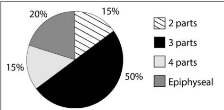
Thirty complications occurred in 20 patients (35.7%) and these are presented in Table 2. Eight pa-tients (40.0%) had more than one complication. The complications taking into account the type of fracture according to the Neer classification(16) can be seen in
Table 3 and Figure 2.
Among the eight cases of inadequate reduction of the fracture, two (25%) were epiphyseal fractu-res, four (50%) were fractures in three parts and two (25%) were fractures in four parts. Among the ina-dequate reductions, six presented varus, one valgus and one anteversion of the humeral head. Inadequate reduction was the most frequent complication, with a statistically significant difference in occurrence be-tween this complication and the others (p < 0.05).
Among the seven cases of complications from the subacromial plate caused by the plate, four occurred in three-part fractures, two in epiphyseal fractures and one in a two-part fracture (p > 0.05).
Out of the six cases of inadequate fixation of the fracture, four had locking screws for the head that were too short (less than one centimeter from the joint surface) and two had screws that were too long. Ina-dequate fixation occurred in four cases of three-part fractures and in two epiphyseal fractures.
Table 1 – Evaluation of the results according to the UCLA score.
Results Number of patients %
Excellent 26 46.4
Good 12 21.4
Fair 10 17.9
Poor 8 14.3
Total 56 100
Fonte: Arquivos médicos da Irmandade Santa Casa de Misericórdia de São Paulo.
Table 2 – Complications relating to fixation of fractures in the proximal third of the humerus using Philos® plates.
Complications Cases %
Inadequate reduction 8 26.7 Impact due to the plate 7 23.3
Inadequate fixation 6 20
Pseudarthrosis 3 10
Adhesive capsulitis 2 6.7
Avascular necrosis 2 6.7
Loss of reduction in varus 1 3.3
Infection 1 3.3
Total 30 100
Source: medical files of Irmandade Santa Casa de Misericórdia de São Paulo.
Table 3 – Complications according to the type of fracture, using the Neer classification.
Type of fracture Number of patients Complications %
2 parts 13 3 23.1
3 parts 28 10 35.7
4 parts 8 3 37.5
Epiphyseal 7 4 57.1
Total 56 20 35.7
571
There were three cases of pseudarthrosis: one pa-tient with an epiphyseal fracture in association with a fracture of the greater tubercle and surgical neck that was poorly reduced, remaining in varus; one pa-tient with a four-part fracture that was inadequately reduced and remained in varus; and one patient with an anatomical reduction and fixation of a three-part fracture but who evolved to pseudarthrosis.
Adhesive capsulitis occurred in two patients, with two and three-part fractures.
There were two cases of necrosis of the humeral head: one in a three-part fracture and the other in a four-part fracture.
One patient with a two-part fracture evolved with loss of the reduction in varus and there was one case of infection in a patient with a three-part fracture.
DISCUSSION
We had 30 complications in 20 patients (35.7%). In the literature, we found complication rates ranging from 3.7 to 33.5%(18,19). In a multicenter prospective
study conducted by Südkamp et al(19), it was observed
that 33.5% of the patients evolved with complications. Among the complications observed by those authors, the following were seen: migration of the screws, su-bacromial impact, pseudarthrosis, loss of reduction, avascular necrosis, neurological lesions, breakage of the implant, inadequate fixation and infection(19). If
we only take into account the patients who presented complications in our study, the mean UCLA score of 29.5 points falls to 22.6. If we only take into ac-count the patients without complications, the mean UCLA score rises to 33.4 points (ranging from 28 to 35 points). Patients with complications evolved with a mean elevation of 118°, while the others presented 132°; mean lateral rotation of 38.5°, versus 43.8°; and
mean medial rotation at L3 level, versus L2.
Inadequate reduction of the fracture was the main complication found in our study, and was the only complication that was statistically significantly diffe-rent among all of them. We had eight cases (14.3%) of poor reduction, which were associated with three-part, four-part and epiphyseal fractures. This rate is in ac-cordance with the literature, which shows rates ranging from 13.7 to 25%(4,12,20). We believe that this high rate
is associated with difficulty in reducing the fracture and keeping it reduced until the plate is positioned and fixed to the bone. Koukakis et al(6) considered that the
greatest challenge in this surgery was to achieve anato-mical reduction of the fracture, especially in three and four-part fractures. Duralde and Leddy(9) affirmed that
in relation to fractures impacted in varus, there is great surgical difficulty in achieving adequate reduction and maintaining this because of the lack of support for the medial region of the metaphysis.
It is known that correct reduction of the greater tuber-cle is also an important parameter for ensuring favorable results(21). Kettler et al(22) found a statistically significant
difference in postoperative evolution between patients with correct reduction of the greater tubercle and those with displacement of the greater tubercle greater than five millimeters. In our study, there was one case (1.7%) in which adequate reduction of the greater tubercle did not occur (Figure 3). Despite this, the patient evolved with consolidation of the fracture, shoulder elevation of 90 degrees, UCLA score of 30 points, absence of pain and satisfaction with the operation.
It is important to emphasize that the locking plate did not contribute towards reduction of the fracture: fixation of the implant should be done with the fractu-red already fractu-reduced, and this is one of the biggest di-fficulties with treatments using this type of implant(9).
Several thin metal wires are placed in order to main-tain the reduction while the plate is being positioned. Since the plate is positioned laterally to the bone, the-re is often gthe-reat difficulty in maintaining this. In our sample, six cases continued to present varus deviation (Figure 3), one case presented inadequate reduction in valgus and one case presented reduction of the hu-meral head with fixation in anteversion.
In the attempt to correct the varus deviation of a frac-ture, overcorrection may occur, thus resulting in reduc-tion in valgus. This may occur due to lack of any anato-mical parameter, because of comminution of the fracture. One patient in our study had a fracture fixed in valgus.
Figure 2 – Complications from surgical treatment in relation to the type of fracture.
Among the cases of inadequate reduction in varus, two cases evolved to pseudarthrosis. We believe that bone failure in the medial portion of the metaphysis may have contributed towards these complications. In these two cases, no grafts were used.
Gardner et al(23) demonstrated recently that
absen-ce of mechanical support in the medial region of the proximal extremity of the humerus, which was caused when there was no cortical contact in the medial re-gion of the metaphysis or in comminutive fractures, contributed towards loss of reduction in varus. Thus, placement of a bone graft in the medial region of the metaphysis is fundamentally important for avoiding loss of reduction(9). Our usual procedure is to place
a bone graft in the cavity between the humeral head and the metaphysis, where the bone failure is, and not necessarily in the medial region of the metaphysis. Moreover, we believe that suturing the tubercles to the plate has an important role in avoiding loosening of the plate. For good fixation, thereby avoiding loss of reduction and pseudarthrosis, another important
factor is the position of the screws in the central, lo-wer and posterior regions of the humeral head, where the trabecular density is greater(24,25). It is important
to emphasize that poor positioning of the humeral head does not allow correct positioning of the screws, given that the screws are placed at a fixed angle.
Pseudarthrosis occurred in 5.3% in our study, while in the literature the rates of pseudarthrosis have ranged from 3 to 5.5%(10,13,21). In the only one of our three
ca-ses of pseudarthrosis in which the cause was not failure of the reduction, the patient was a 47-year-old woman who was a smoker and drinker. It is known that both alcohol consumption and smoking are risk factors for pseudarthrosis(26). A few months after the operation,
thus patient suffered a fracture of the proximal extremi-ty of the humerus on the contralateral side, without dis-placement. She underwent conservative (non-surgical) treatment and also evolved with pseudarthrosis.
The second most frequent complication in our study was subacromial impact caused by the plate, which occurred in seven patients (12.5%). In a review of the literature, we found rates of occurrence of subacromial impact ranging from zero to 10.3%(4,6,12,27). According
to some authors, the plate should be positioned betwe-en five and eight millimeters from the apex of the gre-ater tubercle; if not, a mechanical impact on the acro-mion might occur when the shoulder is raised(9,27,28).
In our study, we observed five cases of impact caused by placement of the plate in positions that were too high, i.e. less than five millimeters from the apex of the greater tubercle, and two cases of impact caused by placement of the plate too laterally (Figure 4). To avoid this complication, we are now placing the guidewire and the plate at positions that are lower than in the recommended technique. In all of our seven cases of impact, there were improvements in the state of pain and shoulder mobility after removal of the implant.
We had two cases in which the screw penetrated the glenohumeral joint. In one of them, a 59-year--old patient with a three-part fracture evolved with osteonecrosis of the humeral head. The rate at which screws are drilled into the joint has ranged from 14 to 16%, and this may have been caused as much by collapse of the subchondral bone as by implant failu-re, screw migration, drilling with the bit into the joint surface or inadequate viewing with the image intensi-fier(19,29,30). Thanasas et al(21) stated that the
commo-nest intraoperative error was to incorrectly choose the
573
size of locking screw, which accounted for between 2 and 17.9% of these complications(14). We had four
cases (7.1%) of short screws and two cases (3.5%) of protruding screws (Figure 3). According to Duralde and Leddy(9), the screws in the humeral head should
be five to ten millimeters from the joint surface. We tended to also tie off the tendons of the rotator cuff muscles at the plate with the aim of augmenting the fracture fixation in order to avoid postoperative loss of the reduction. Loss of the reduction occurred in one case (1.7%) in our study.
Edwards et al(31) demonstrated that in cases of
comminutive fractures of the surgical neck, the bio-mechanical resistance of locking plates (in relation to both their angular and their torsional strength) was
greater than that of locking intramedullary nails. No cases of breakage of the implant occurred in our stu-dy, whereas the rate of occurrence described in other studies was between 0.7 and 3%(4,10,21). This was
pro-bably because in most cases in which the comminu-tion was large, it was decided to place a bone graft together with fixation of the plate. A bone graft was placed in seven patients (12.5%) and, among these, two evolved with complications. The first of these was in a case of a three-part fracture of the greater tubercle and surgical neck, and it evolved with signs of impact caused by the plate, due to high fixation. The second case consisted of a four-part fracture and evolved with avascular necrosis.
Few studies have specifically addressed the
com-Figure 4 – Pre and postoperative radiographic images of the right shoulder of a 71-year-old patient who suffered a two-part fracture of the proximal ex-tremity of the humerus with a metadiaphyseal line. (A) Preoperative corrected frontal view of the fracture. (B) Preoperative scapular lateral view. (C and D) Immediate postoperative views: note the white arrow showing the high position of the plate, very close to the upper margin of the greater tubercle. (E and F) Radiographic views one year after the surgery with the fracture already consolidated. (G and H) Radiographs after removal of the plate, which led to improvement of the painful condition and a gain in mobility.
plications and technical difficulties of surgery for treating fractures of the proximal extremity of the humerus using a “locking plate”. One of the weak points of the present study was that it was retrospec-tive, without a control group.
It is important to emphasize that the functional results from fractures of the proximal extremity of the humerus are less dependent on the choice of implant; rather, they depend on correct anatomical reduction of the fracture and stable fixation of the implant(27). It is
believed that in elderly patients with osteoporosis and in cases of comminutive fractures, “locking plates” ensure greater stability of fixation and fewer risks of loss of reduction.
It has been noted that there is some difficulty in achieving adequate reduction of the fracture in as-sociation with good positioning of the plate on the bone, even taking into account the different fractures
dealt with. Correct reduction and fixation of fractu-res of the proximal extremity of the humerus using this type of synthesis requires technical skills from the surgeon and this, in turn, implies a long learning curve. Südkamp et al(19) concluded that 55% of the
complications encountered were already present at the end of the surgical procedure, and related to in-correct surgical technique. In our study, this occurred in 46.7% of the complications.
CONCLUSION
The Philos® “locking plate” is a treatment option
for fractures of the proximal extremity of the humerus, especially in cases of porous bones and comminutive fractures. Nevertheless, we found a complication rate of around 35.7% with this type of surgery when this osteosynthesis material was used.
REFERENCES
1. Papadopoulos P, Karataglis D, Stavridis SI, Petsatodis G, Christodoulou A. Mid-term results of internal fixation of proximal humeral fractures with the Philos plate. Injury. 2009;(40):1292-6.
2. Iannotti JP, Ramsey ML, Williams GR Jr, Warner JJ. Nonprosthetic manage-ment of proximal humeral fractures. Instr Course Lect. 2004;(53):403-16. 3. Resch H, Hubner C, Schwaiger R. Minimally invasive reduction and
osteosynthe-sis of articular fractures of the humeral head. Injury. 2001;32(Suppl 1):SA25-32. 4. Fankhauser F, Boldin C, Schippinger G, Haunschmid C, Szyszkowitz R. A new
locking plate for unstable fractures of the proximal humerus. Clin Orthop Relat Res. 2005;(430):176-81.
5. Lill H, Hepp P, Korner J, Kassi JP, Verheyden AP, Josten C, Duda GN. Proximal humeral fractures: how stiff should an implant be?: A comparative mechanical study with new implants in human specimens. Arch Orthop Trauma Surg. 2003;(123):74-81. 6. Koukakis A, Apostolou CD, Taneja T, Korres DS, Amini A. Fixation of proximal
humerus fractures using the PHILOS plate: early experience. Clin Orthop Relat Res. 2006;(442):115-20.
7. Brunner F, Sommer C, Bahrs C, Heuwinkel R, Hafner C, Rillmann P, et al. Open reduction and internal fixation of proximal humerus fractures using a proximal humeral locked plate: a prospective multicenter analysis. J Orthop Trauma. 2009;(23):163-72.
8. Checchia SL, Doneux PS, Miyazaki AN, Fregonese M, Silva LA, Lobo AC, et al. Avaliação do tratamento cirúrgico da fratura em duas partes do colo cirúrgico do úmero com placa PFS 80º. Rev Bras Ortop. 2004;39(10):555-67. 9. Duralde XA, Leddy LR. The results of ORIF of displaced unstable proximal hu-meral fractures using a locking plate. J Shoulder Elbow Surg. 2010;19(4):480-8. 10. Björkenheim JM, Pajarinen J, Savolainen V. Internal fixation of proximal humeral fractures with a locking compression plate. A retrospective evaluation of 72 pa-cients followed for a minimum of 1 year. Acta Orthop Scand. 2004;75(6):741-5. 11. Plecko M, Kraus A. Internal fixation of proximal humerus fractures using the
locking proximal humerus plate. Operat Orthop Traumatol. 2005;(17):25-50. 12. Agudelo J, Schürmann M, Stahel P, Helwig P, Morgan SJ, Zechel W, et al.
Analysis of efficacy and failure in proximal humerus fractures treated with locking plates. J Orthop Trauma. 2007;(21):676-81.
13. Clavert P, Adam P, Bevort A, Bonnomet F, Kempf JF. Pitfalls and complica-tions with locking plate for proximal humerus fracture. J Shoulder Elbow Surg. 2010;(19):489-94.
14. Shahid R, Mushtaq A, Northover J, Maqsood M. Outcome of proximal humerus fractures treated by PHILOS plate internal fixation. Experience of a district general hospital. Acta Orthop Belg. 2008;74(5):602-8.
15. Lungershausen W, Bach O, Lorenz CO. [Locking plate osteosynthesis for fractures of the proximal humerus]. Zentralbl Chir. 2003;1289(1):28-33. 16. Neer CS 2nd. Displaced proximal humeral fractures: Part I. Classification and
evaluation. J Bone Joint Surg Am. 1970;52(6):1077-89.
17. Ellman H, Kay SP. Arthroscopic subacromial descompression for chronic
im-pingement. Two five years results. J Bone Joint Surg Br. 1991;73(3):395-8. 18. Fazal MA, Haddad FS. Philos plate fixation for displaced proximal humeral
fractures. J Orthop Surg. 2009;17(1):15-8.
19. Südkamp N, Bayer J, Hepp P, Voigt C, Oestern H, Kääb M, et al. Open reduc-tion and internal fixareduc-tion of proximal humeral fractures with use of the locking proximal humerus plate. Results of a prospective, multicenter, observational study. J Bone Joint Surg Am. 2009;91(6):1320-8.
20. Owsley KC, Gorczyca JT. Displacement/screw cutout after open reduction and locked plate fixation of proximal humeral fractures. J Bone Joint Surg Am. 2008;90(2):233-40.
21. Thanasas C, Kontakis G, Angoules A, Limb D, Giannoudis P. Treatment of proximal humerus fractures with locking plates: a systematic review. J Shoulder Elbow Surg. 2009;(18):837-44.
22. Kettler M, Biberthaler P, Braunstein V, Zeiler C, Kroetz M, Mutschler W. [Treatment of proximal humeral fractures with the PHILOS angular stable plate. Presentation of 225 cases of dislocated fractures]. Unfallchirurg. 2006;109(12):1032-40.
23. Gardner MJ, Weil Y, Barker JU, Kelly BT, Helfet DL, Lorich DG. The importance of medial support in locked plating of proximal humerus fractures. J Orthop Trauma. 2007;21(3):185-91.
24. Cohen M, Amaral MV, Monteiro M, Brandão BL, Motta Filho GR. Osteossíntese das fraturas da extremidade proximal do úmero com sistema de placa de ângulo fixo com parafusos bloqueados: técnica e resultados. Rev Bras Ortop. 2009;44(2):106-11. 25. Tingart MJ, Lehtinen J, Zurakowski D, Warner JJP, Apreleva M. Proximal humeral
fractures: regional differences in bone mineral density of the humeral head affect the fixation strength of cancellous screws. J Shoulder Elbow Surg. 2006;15(5):620-4. 26. Rose PS, Adams CR, Torchia ME, Jacofsky DJ, Haidukewych GG, Steinmann
SP. Locking plate fixation for proximal humeral fractures: initial results with a new implant. J Shoulder Elbow Surg. 2007;16(2):202-7.
27. Handschin AE, Cardell M, Contaldo C, Trentz O, Wanner GA. Functional results of angular-stable plate fixation in displaced proximal humeral fractures. Injury. 2008;39(3):306-13.
28. Monteiro GC, Ejnisman B, Andreoli CV, Pochini AC, Olympio E. Resultados do tratamento das fraturas do terço proximal do úmero com placas de bloqueio. Acta Ortop Bras. 2011;19(2):69-3.
29. Egol KA, Ong CC, Walsh M, Jazrawi LM, Tejwani NC, Zuckerman JD. Early complications in proximal humerus fractures (OTA Types 11) treated with locked plates. J Orthop Trauma. 2008;22(3):159-64.
30. Charalambous CP, Siddique I, Valluripalli K, Kovacevic M, Panose P, Srinivasan M, et al. Proximal humeral internal locking system (PHILOS) for the treatment of proximal humeral fractures. Arch Orthop Trauma Surg. 2007;127(3):205-10. 31. Edwards SL, Wilson NA, Zhang L, Flores S, Merk BR. Two-part surgical neck