Changes in serum cardiac myosin light chain 1 levels in children
with fulminant myocarditis during continuous blood purification
CHUQIAO SHENG1, ZHEN ZHANG1, YONG JIA2, YUMEI LI1*
1Pediatric Intensive Care Unit (PICU), The First Hospital of Jilin University, Changchun, Jilin, China 2College of Public, Hygiene of Dalian Medical University, Dalian, Liaoning, China
S
UMMARYStudy conducted at The First Hospital of Jilin University, Changchun, Jilin, China
Article received: 2/21/2017
Accepted for publication: 3/12/2017
*Correspondence:
Pediatric Intensive Care Unit The First Hospital of Jilin University Changchun, Jilin – China Postal code: 130021 [email protected]
http://dx.doi.org/10.1590/1806-9282.63.10.904
Objective: To investigate the changes in serum cardiac myosin light chain 1 (CMLC-1) levels in children with fulminant myocarditis (FM) during continuous blood purification (CBP), as well as to analyze its correlation with other laboratory indexes.
Method: Twenty-four (24) children with FM who underwent CBP were enrolled. Before and during treatment (48 and 72 hours after treatment, or death), the optical density value of serum CMLC-1 was measured using enzyme-linked immunosorbent assay, and then the serum CMLC-1 concentration was calculated. The correlations between CMLC-1 OD value change and laboratory indexes including creatine kinase-MB (CK-MB), troponin, myohemoglobin and N-terminal pro-brain natriuretic peptide (NT-proBNP) were analyzed.
Results: The serum CMLC-1 concentration significantly increased in the children with FM and decreased obviously during CBP therapy. In the same period, the change of CMLC-1 concentration were positively correlated with creatine kinase-MB (r=0.528), troponin (r=0.726), myohemoglobin (r=0.702), and NT-proBNP levels (r=0.589).
Conclusion: The serum CMLC-1 concentration increases significantly in children with FM, but CBP therapy can effectively control this increase.
Keywords: myocarditis, child, myosins/immunology/metabolism, hemofiltration.
I
NTRODUCTIONCurrently, fulminant myocarditis (FM) is a critical disease that causes death in children and lacks sufficient specific-ity for early diagnosis.1-3 The pathogenesis of FM is
gener-ally believed4 to involve infiltration of a large number of
inflammatory cells and destruction of myocardial cells in the early stages. Then, allergic reaction or autoimmune involvement can lead to further damage to cardiac muscle cells, resulting in localized or diffuse myocarditis.5-7 The
pathogenesis of anti-cardiac antibodies in post-viral auto-immune cases may start with direct viral-induced myocyte damage, with associated release of intracellular proteins.
Autoantibodies potentially pathogenic to various cel-lular components are found in a high percentage of patients with myocarditis. Autoantigens include alpha and beta cardiac myosins. The damage caused by the release of car-diac myosin was included in the course of autoimmune injury. It has been successfully used in the domestic and
foreign application of cardiac myosin for creating a model of autoimmune myocarditis on which to apply treatment interventions.8,9 This shows that cardiac myosin plays an
important role in the damage process. The composition of cardiac myosin includes two heavy chains and two light chains. One essential isoform of myosin light chain is called cardiac myosin light chain (CMLC-1). In recent years, sustained release of CMLC-1 can be detected in patients with myocardial injury. This has aroused our interest; thus, in this study, we focused on CMLC-1.
the serum CMLC-1 level in children with FM before and after CBP treatment. In addition, we investigated the therapeutic effect of CBP on CMLC-1 and found its im-portance in the diagnosis and treatment of FM in children.
M
ETHODClinical data
The children (n=24) were recruited from a study con-ducted by the Department of Pediatric Intensive Care Unit, The First Hospital of Jilin University, during the time period of June 2012 to December 2014. The diag-nostic criteria used were based on details in previous literature.1 Children with congenital cardiovascular
mal-formations were excluded.
Our study was conducted in accordance with the declaration of Helsinki, after approval from the Ethics Committee of Jilin University. Written informed consent was obtained from all participants’ guardians.
Material preparation and experiments
All of the children were treated with CBP.10 We select
CVVHDF/CVVH as the treatment mode since it is the standard mode of CBP for children.11 Fresenius
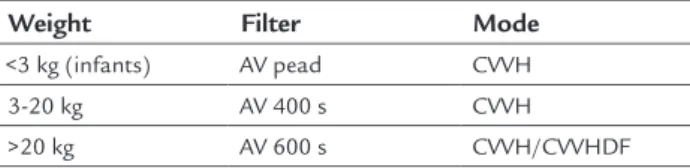
polysul-fone membrane transfusion filters (Fresenius Medical Care AG, Frankfurt, Germany) were used in the continu-ous vencontinu-ous hemodiafiltration/continucontinu-ous veno-venous hemofiltration (CVVHDF/CVVH) mode. Then the filters and mode were selected according to the pa-tient’s weight, as shown in Table 1. Blood samples (3 mL) were collected in coagulation sterile tubes at the initial stage of hospitalization and during CBP treatment (48 and 72 h after treatment, or death).
TABLE 1 Filter and mode selection for different weight ranges.
Weight Filter Mode
<3 kg (infants) AV pead CVVH
3-20 kg AV 400 s CVVH >20 kg AV 600 s CVVH/CVVHDF
CVVH: continuous veno-venous hemodiailtration; CVVH/CVVHDF: continuous veno-venous hemodiailtration/continuous veno-venous hemoiltration.
The collected samples were coagulated at room tempera-ture for 10-20 min and then ultracentrifuged (American Backman L8-80M) for 10 minutes at the speed of 3,000 rpm. The supernatant was labeled after centrifugation, and the samples were maintained at a temperature of -70°C. When all specimens were collected completely, the
samples were thawed at 37°C. The optical density (OD)
value of serum CMLC-1 was measured using
enzyme-linked immunosorbent assay (ELISA; Human CCMLC-1 ELISA Kit, Shanghai Wan Jiang Bio Technology Co., Ltd.), and then the serum CMLC-1 concentration was calculated.
The correlations between CMLC-1 OD value change and laboratory indexes including creatine kinase-MB (CK-MB), troponin, myohemoglobin, and N-terminal pro-brain natriuretic peptide (NT-proBNP) were analyzed.
Result determination
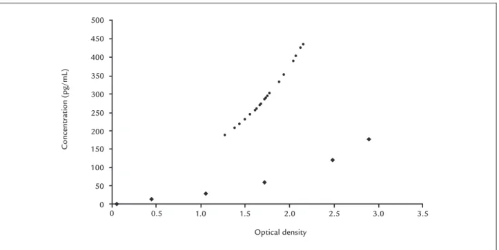
We placed the OD value on the horizontal axis and stan-dard density on the vertical axis, a stanstan-dard curve was drawn (y = 9.8584x3 - 21.182x2 + 41.098x, t = 0.99931) on
a graph paper (Figure 1). The OD values were calculated by using the aforementioned formula, where a higher value indicates positivity.
Statistical analysis
All data were compared using SPSS version 20.0 software (SPSS Inc., Chicago, IL, USA). All normal distribution data were expressed as mean±SD. A paired t-test was used to compare data. Correlation analysis was performed by using the Pearson (normality data) or Spearman correla-tion coefficient (non-normality data). The significance level was set at < 0.05 for all the tests.
R
ESULTSTwenty-four (24) children with FM were enrolled in the study. Length of stay following initiation of treatment ranged from 13 hours to 5 days. Twenty (20) children with FM survived (improvements were seen during treat-ment in heart rate, respiration, CVP) and four patients died, yielding a mortality rate of 16.67%. Because of the small number of cases, death had no statistical significant. Our purpose is to explore the value of serum CMLC-1 level during CBP therapy.
Change in serum CMLC-1 level during the CBP therapy During early hospitalization, serum CMLC-1 OD was measured in all children with FM, and then the substitu-tion curve formula was derived. The CMLC-1 OD were all above the standard curve. It means that the serum CMLC-1 concentration significantly increased in these children. In the early stage of admission, the CMLC-1 OD value was comparable between the children who died and those who survived, but because of the limited number of cases, the difference was not statistically significant.
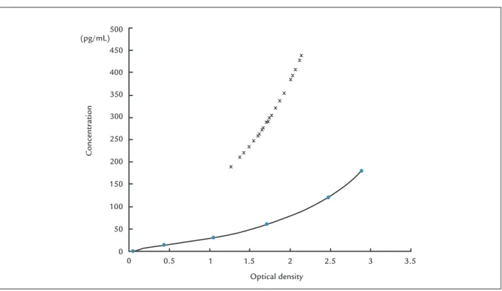
especially at 48 hours after treatment (t=19.58, p<0.01; Table 2). This can be observed in Figure 2.
TABLE 2 Change in CMLC-1 OD values in surviving patients after treatment for up to 48 h.
Time CMLC-1 OD value
Before 1.717±0.25
48 h later 1.309±0.27
t 19.58
p <0.01
In terms of death, because of the small number of deaths, the difference in the time of death was relatively larger (8-192 hours), so the CMLC-1 OD values of the patients who died were not statistically significant.
Concentration changes within the same period of correlation with laboratory results
In the same period, the change of CMLC-1 concentration were positively correlated with creatine kinase-MB (r=0.528), troponin (r=0.726), myohemoglobin (r=0.702), and NT-proBNP levels (r=0.589; Figure 3).
D
ISCUSSIONSystole-diastole is a complex physiological process that is affected by many factors. With the advances in mo-lecular biology, people have a deeper understanding of
this process at a molecular level and increased awareness of contractile and regulatory proteins. In the present study, we found that myosin is a type of contractile protein. Cardiac myosin is located within the sarcomeres, and it is the main unit of the myofibrillar thick wire, along with ATP enzyme activity. It plays an important role in the regulation of myocardial contractility.12,13 Its composition
includes two heavy chains (molecular weight, about 200 kDa) and two light chains (molecular weight, 16-27 kDa).14
The light chains are divided into the basic essential myo-sin light chain and the phosphorylation regulation light chain. The ventricular muscle consists of an essential myosin light chain isoform called cardiac myosin light chain (CMLC-1).
Normal human serum is free of cardiac myosin.15 In
patients with myocardial edema caused by various factors such as necrosis, apoptosis and cardiac myosin in the protease decomposition, high levels of CMLC-1 are
re-leased into the blood through damaged membranes.16 As
a cardiac autoantigen, CMLA-1 antigen presents cells to
generate anti-cardiac myosin antibody (AMA),17 leading
to further immune damage.
Myocardial damage is the result of an autoimmune reaction.18 Cardiac myosin release has now been considered
as a cause of secondary autoimmune injury.8 Neu et al.19
found that cardiac myosin-immunized mice had greater genetic predisposition for autoimmune myocarditis and that cardiac myosin can induce autoimmune myocarditis. FIGURE 1 CMLC-1 optical density value at early admission.
Concentr
ation (pg/mL)
Optical density
0 0.5 1.0 1.5 2.0 2.5 3.0 3.5
500
450
400
350
300
250
200
150
100
50
FIGURE 2 Line diagram of CMLC-1 optical density value in survival.
Concentr
ation
(pg/mL)
Optical density
0 0.5 1 1.5 2 2.5 3 3.5
500
450
400
350
300
250
200
150
100
50
0
Person/case before
after 48h
after 72h
1 2 3 4 5 6 7 8 9 10 11 12 13 14 15 16 17 18 19 20
Optical density
2.5
2
1.5
1
0.5
0
Considered in the absence of virus and other pathogens of infection injury cases, a simple cardiac myosin can lead to the occurrence of myocarditis. The degree and extent of the myocardial damage were positively correlated. There-fore, we believe the concentrations reflect the degree of myocardial injury and thus can be used as a method to measure the area of myocardial injury.
CMLC-1 belongs to the category of middle-sized mo-lecular substances, which can be effectively removed through hemofiltration during CBP treatment. The CMLC-1 anal-ysis in this study revealed that during CBP therapy, serum CMLC-1 level decreased significantly, particularly 48 hours after treatment (p<0.01). CBP treatment can remove inflam-matory mediators, reduce the continuing destruction of myocardial cells, and thereby reduce CMLC-1 release. Re-ducing antigen presentation and autoimmune damage can effectively remove CMLC-1 from the blood.
In the past, we applied cardiac biomarker elevations (e.g., cardiac troponin and B-type natriuretic peptide hor-mone [BNP] levels) to reflect myocardial injury and found a prognostic significance.20-22 However, these tests cannot
be used to establish a diagnosis of myocarditis, as they lack sufficient sensitivity and specificity. Thus, in this study, we focused on the significance of serum CMLC-1 levels in the treatment of children with FM. By monitoring changes in serum CMLC-1, the degree of recovery of myocardial in-jury and the treatment effects were evaluated. During the early stage of admission, CMLC-1 levels were significantly increased. Moreover, the rate of disease progression, sever-ity of clinical manifestations and CMLC-1 levels positively correlated. The more serious the cardiac injury, the worse the cardiac function, whereas the higher the serum CMLC-1 level in the initial stage, the higher the risk of death.
After CBP treatment, the children showed signs of improvement and their serum CMLC-1 levels decreased gradually and showed positive correlation with labora-tory test results. Thus, we considered that serum CMLC-1 may be used as a biological marker to detect the occurrence of diseases such as myocardial infarction or myocarditis, which may have important applications.
However, with the few cases of children with FM in this study and the large difference in the time course of the disease before admission, the correlation between the change in CMLC-1 concentration and deaths was not statistically significant. Further study on the course of treatment is needed.
C
ONCLUSIONIn summary, our study results indicate that the immune system has a great influence on the development of
myo-cardial injury. CMLC-1 levels were significantly increased in the early stage of myocardial injury. CBP treatment effectively decreased these levels and reduced the extent of sustained myocardial damage.
A
CKNOWLEDGMENTSMicroplate reader was performed by Kingmed, provider of experimental technical support.
C
ONFLICT OF INTERESTThe authors declare no conflict of interest.
R
EFERENCES1. Freedman SB, Haladyn JK, Floh A, Kirsh JA, Taylor G, Thull-Freedman J. Pediatric myocarditis: emergency department clinical findings and diagnostic evaluation. Pediatrics. 2007; 120(6):1278-85.
2. Sankar J, Khalil S, Jeeva Sankar M, Kumar D, Dubey N. Short-term outcomes of acute fulminant myocarditis in children. Pediatr Cardiol. 2011; 32(7):885-90.
3. Ghelani SJ, Spaeder MC, Pastor W, Spurney CF, Klugman D. Demographics, trends, and outcomes in pediatric acute myocarditis in the United States, 2006 to 2011. Circ Cardiovasc Qual Outcomes. 2012; 5(5):622-7. 4. Ginsberg F, Parrillo JE. Fulminant myocarditis. Crit Care Clin. 2013;
29(3):465-83.
5. Bratincsák A, El-Said HG, Bradley JS, Shayan K, Grossfeld PD, Cannavino CR. Fulminant myocarditis associated with pandemic H1N1 influenza A virus in children. J Am Col Cardiol. 2010; 55(9):928-9.
6. Lee EY, Lee HL, Kim HT, Lee HD, Park JA. Clinical features and short-term outcomes of pediatric acute fulminant myocarditis in a single center. Korean J Pediatr. 2014; 57(11):489-95.
7. Cooper LT Jr, Hare JM, Tazelaar HD, Edwards WD, Starling RC, Deng MC, et al.; Giant Cell Myocarditis Treatment Trial Investigators. Usefulness of immunosuppression for giant cell myocarditis. Am J Cardiol. 2008; 102(11):1535-9.
8. Wang ZH, Liao YH, Dong JH, Li SL, Wang JP. Experimental study of autoimmune diseases, cardiac myosin-induced. Chinese J Cardiol. 2003; 31:937-40.
9. Cai G, Zhang J, Liu L, Shen Q. Successful treatment of experimental autoimmune myocarditis by adenovirus-mediated gene transfer of antisense CIITA. J Mol Cell Cardiol. 2005; 38(4):593-605.
10. Sheng CQ, Dai XL, Jia Y, Li YM. Clinical analysis of continuous blood purification for the treatment of children with fulminant myocarditis. Int J Clin Exp Med. 2016; 9(6):11788-95.
11. Honoré PM, Jacobs R, Boer W, Joannes-Boyau O, De Regt J, De Waele E, et al. New insights regarding rationale, therapeutic target and dose of hemofiltration and hybrid therapies in septic acute kidney injury. Blood Purif. 2012; 33(1-3):44-51.
12. Tong CW, Nair NA, Doersch KM, Liu Y, Rosas PC. Cardiac myosin-binding protein-C is a critical mediator of diastolic function. Pflugers Arch. 2014; 466(3):451-7.
13. Chang AN, Battiprolu PK, Cowley PM, Chen G, Gerard RD, Pinto JR, et al. Constitutive phosphorylation of cardiac myosin regulatory light chain in vivo. J Biol Chem. 2015; 290(17):10703-16.
14. Gazith J, Himmelfarb S, Harrington WF. Studies on the subunit structure of myosin. J Biol Chem. 1970; 245(1):15-22.
15. Caforio AL, Tona F, Bottaro S, Vinci A, Dequal G, Daliento L, et al. Clinical implications of anti-heart autoantibodies in myocarditis and dilated cardiomyopathy. Autoimmunity. 2008; 41(1):35-45.
16. Samarel AM, Ferguson AG, Vander Heide RS, Davison R, Ganote CE. Release of unassembled rat cardiac myosin light chain 1 following the calcium paradox. Circ Res. 1986; 58(1):166-71.
18. Knowlton KU, Lim BK. Viral myocarditis: is infection of the heart required? J Am Coll Cardiol. 2009; 53(14):1227-8.
19. Neu N, Craig SW, Rose NR, Alvarez F, Beisel KW. Coxsackievirus induced myocarditis in mice: cardiac myosin autoantibodies do not cross-react with the virus. Clin Exp Immunol. 1987; 69(3):566-74.
20. Michtalik HJ, Yeh HC, Campbell CY, Haq N, Park H, Clarke W, et al. Acute changes in N-terminal pro-B-type natriuretic peptide during hospitalization
and risk of readmission and mortality in patients with heart failure. Am J Cardiol. 2011; 107(8):1191-5.
21. Canter CE, Simpson KE. Diagnosis and treatment of myocarditis in children in the current era. Circulation. 2014; 129(1):115-28.