Knockout of p16
INK4apromotes aggregative growth of dermal
papilla cells
YI CHENG1, YANG GAO2, LU ZHAO1*, SHUNQIANG GAO1, GUOQIANG ZHANG1, YAN ZHANG1
1Department of Dermatology, the Fourth Hospital of Hebei Medical University, Shi Jiazhuang, Hebei Province, China 2Department of Interventional Radiology, Hebei Children’s Hospital, Shi Jiazhuang, Hebei Province, China
S
UMMARYStudy conducted at Fourth Hospital of Hebei Medical University and Hebei Children’s Hospital, Shi Jiazhuang, Hebei Province, China
Article received: 3/10/2017 Accepted for publication: 3/11/2017
*Correspondence: Department of Dermatology Fourth Hospital of Hebei
Medical University Address: No.12 Jiankang Road Shi Jiazhuang, Hebei Province – China Postal code: 050011 [email protected]
http://dx.doi.org/10.1590/1806-9282.63.10.883
Objective: Dermal papilla cells (DPCs) are located in the hair follicles and play an important role in hair growth. These cells have the ability to induce hair follicle formation when they display aggregative behavior. DPCs derived from the androgenetic alopecia (AGA) area undergo premature senescence in vitro, associated with p16INK4a expression. The aim of the current study was to investigate the expression of p16INK4a in aggregative and non-aggregative DPCs and the effect of p16INK4a down-regulation in these cells by adenovirus-mediated RNA interference (RNAi).
Method: DPCs were isolated and cultured from healthy human scalp. p16INK4a gene and protein were detected in aggregative and non-aggregative cells. Expression of p16INK4a in DPCs was silenced by infection with rAd5-CDKN1A-1p2shRNA. Cell fate was monitored after infection. The growth of cells was measured by MTT assay. Cell cycle was evaluated by low cytometry (FCM).
Results: DPCs were isolated by digestion and showed aggregative behavior for six passages. The expression of p16INK4a showed a clear upward trend in non-aggregative cells when compared with aggregative group. p16INK4a expression was silenced by rAd5-CDKN1A-1p2shRNA (p<0.05). The p16INK4a-silenced cells grew more rapidly and exhibited a trend towards aggregative growth. There was an increase in the proportion of cells in G1 phase, while those in S phase were reduced after p16INK4a gene silencing (p<0.05).
Conclusion: Our results suggest that p16INK4a plays an important role in the premature senescence and aggregative behavior of DPCs. These observations can lead to novel therapeutic strategies for treatment of AGA.
Keywords: hair follicle, transfection, hair/growth and development.
I
NTRODUCTIONDermal papilla cells (DPCs) are the main mesenchymal cells located at the bottom of the hair follicle (HF) and compose the dermal papilla. The biological characteristic of these cells is the ability to induce HF formation, both in vivo as well as in vitro. DPCs are a cluster of specialized ibroblasts and occur at the central link in the morphol-ogy of the HF and its cyclic growth regulation.1,2 During the initial stage of HF morphogenesis in the course of embryonic development, the ectodermal epithelial cells proliferate and differentiate downward continuously after being stimulated by dermis signaling molecules to form the hair peg. The hair peg provides feedback to the dermis and induces the formation of dermal concentrate
of aggregative growth gradually disappears in vitro, even-tually ceasing entirely. It was reported that when DPCs of low passage were inoculated into a small incision on the mouse auricle, with high-passage cells and ibroblasts as control, clusters of hair ibers grew on the incision containing low passage DPCs. These hair ibers were thicker and longer than naturally occurring hair on the ear, and were similar to the tentacles from where the DPCs were taken. On the other hand, high-passage DPCs and ibroblasts did not show this phenomenon.6 These obser-vations conirm that low passages of cultured DPCs with aggregative growth not only have the ability to induce complete regeneration of hair follicles, but also carry the information needed to determine the nature of the hair, as corroborated by subsequent research.7,8 It should be emphasized that the ability of DPCs to induce HF forma-tion is dependent on their aggregative growth. But the mechanism by which the aggregative behavior disappears is not yet clear.
p16INK4a is a cyclin-dependent kinase (CDK) inhibitor that slows down cell cycle by inhibiting transition from G1 to S phase. Normally, CDK4/6 binds cyclin D to form an active protein complex that phosphorylates retinoblas-toma protein (pRB). Once phosphorylated, pRB disassoci-ates from the transcription factor E2F1, thus liberating E2F1 from its cytoplasm bound state, thereby allowing it to enter the nucleus. In the nucleus, E2F1 promotes tran-scription of target genes that are essential for transition from G1 to S phase.9,10 Tissue ageing causes p16INK4a con-centration to increase dramatically.11 p16INK4a has also been used as a target to delay certain changes related to ageing in mice.12 Recent reports have demonstrated that DPCs taken from male androgenetic alopecia (AGA) patients undergo premature senescence in vitro associated with the expression of p16INK4a. We hypothesized that non-aggrega-tive growth was a feature of ageing and that aggreganon-aggrega-tive growth characteristics were correlated with p16INK4a. We studied p16INK4a expression in DPCs and inhibited p16INK4a expression in DPCs by adenovirus-mediated RNA interfer-ence to explore the possible mechanisms of cultured human DPCs losing aggregative growth characteristics.
M
ETHODEthics statements
The Fourth Hospital of Hebei Medical University insti-tutional review board approved all described studies. The study was conducted according to the Declaration of Helsinki Principles. Informed written consent was ob-tained from all patients.
Isolation and culture of dermal papilla cells
Specimens were taken from the occipital scalp of six male individuals undergoing surgical excision of benign cutane-ous tumors. The patients were not using any hair loss medications when the samples were collected. Dermal papillae were isolated from human scalp hair follicles by digestion with collagenase D and dispase (Sigma, USA) as described earlier.13 The cells isolated were then cultivated in Dulbecco’s modiied Eagle’s medium (DMEM) contain-ing 15% fetal bovine serum (FBS), 100 IU/mL penicillin, 100 µg/mL streptomycin and 0.4 mM L-glutamine (Sigma, USA) in a 95% humidiied atmosphere with 5% CO2 at 37°C.
RT-PCR
p16INK4a expression at the transcriptional level from dif-ferent generations of DPCs was tested by RT-PCR. The ddH2O group was regarded as a negative control. Trizol reagent (Suo Bao Lai Biotechnology Co. Ltd, Shanghai) was used to extract total RNA and reverse transcription of the total RNA was carried out using reverse transcrip-tase and oligo(dT) primers (Tian En Ze technical Co. Ltd, Beijing) according to the manufacturer’s instructions. Glyceraldehyde-3-phosphate dehydrogenase (GAPDH) was used as internal reference. The primer sequences of RT-PCR were as follows:
• p16INK4a forward primer: 5’-CATCCCCGATTGAAA-GAACC-3’;
• reverse primer: 5’- AATGGACATTTACGGTAGTGGG-3’;
• GAPDH forward primer: 5’-TGAACGGGAAGCT-CACTGG-3’;
• reverse primer: 5’-GCTTCACCACCTTCTTGATGTC-3’.
PCR was carried out for 40 cycles (95°C for 10 s, 58°C for 20 s and 72°C for 20 s) according to instructions sup-plied with the Hot Start Fluorescent PCR Core Reagent Kits (Bio Basic) in a real-time luorescent quantitative PCR cycler (Edinburgh biological technology development Co. Ltd, Shanghai).
Immunohistochemistry
with primary antibody diluted in PBS at 4°C. After incu-bation with primary antibodies, cells were washed thrice with PBS. Secondary and tertiary antibodies (Ding Guo Biotechnology Co. Ltd, Beijing) were used as per the manufacturer’s instructions. Immunolocalization was visualized with 3,3’-diaminobenzidine tetrahydrochloride solution (Ding Guo Biotechnology Co. Ltd, Beijing). Cells were counterstained with hematoxylin and mounted with Permount TM Mounting Medium.
Flow cytometry
Flow cytometric analysis was done as described earlier.14 Conluent DPCs were washed with ice cold PBS and then ixed with 0.5% paraformaldehyde for about 5-10 minutes. Cells were then washed with PBS and blocked with 0.1% BSA. Finally, anti-p16INK4a antibodies were added and incubated overnight at 4°C. Cells were washed twice with PBS, trypsinized and resuspended in 250 μL of luores-cence labeled secondary antibody for analysis.
Transfection
p16INK4a gene sequence was obtained from Genbank (NP-00068.1). RNAi adenovirus targeting p16INK4a (rAd5-CD-KN1A-1p2shRNA) and the negative control adenovirus vector (rAd5-HKshRNA-EGFP) were obtained from Wu-han Ximar Biological Co., Ltd. Cells from the 8th genera-tion were seeded in 25 cm2 cell culture lasks. Cells were divided into three groups, non-transfected cells as blank control group (CON), cells transfected by RNAi targeting p16INK4a via rAd5-CDKN1A-1p2shRNA as gene knockout group (shp16), and cells transfected by rAd5-HKshRNA-EGFP as the negative control group (NC). Diluted adeno-virus was added into culture medium according to MOI=100 (concentration of rAd5-CDKN1A-1p2shRNA was 2.4×10⁸/mL, concentration of rAd5-HKshRNA--EGFP was 1.2×10⁹/mL). Cells were incubated for 4 hours
in 37o, 5% CO
2 incubator after transfection, then the medium was replaced by MSCM medium. The cells were inally collected after 48 hours for RNA extraction and detection of protein in order to verify the effects of RNAi.
MTT assay
DPCs of passage 8 were plated in a 96-well plate (1×10⁴ per well), and divided into three groups: shp16, NC and CON.Diluted adenovirus was added into culture medium according to MOI=100 (concentration of rAd5-CDKN1A-1p2shRNA was 2.4×10⁸/mL, concentration of rAd5-HK-shRNA-EGFP was 1.2×10⁹/mL). The serum-free medium was replaced by MSCM culture medium after 4 hours. MTT dye dissolved in 0.15 mL of DMSO was added to
different groups of wells (5 mg/mL) after 24, 48, 72, 96 and 120 hours. Data were analyzed and growth curves of different groups were plotted.
Immunoluorescence
3×10⁵ cells were inoculated in each well of a six-chamber slide and cultured for 24 hours. The samples were prepared as follows: cells were treated with 4% paraformaldehyde for 20 minutes, followed by 0.1% Triton X-100 for 10 min-utes at room temperature, then rinsed with PBS twice. The washed cells were treated with 0.2% BSA for 30 minutes at room temperature, followed by treatment with 1,000x diluted p16INK4a antibody. FITC-labeled secondary antibody was then added and incubated at room temperature for 1 hour. The overnight-treated sample was then treated with 5 μg/mL FITC-phalloidin conjugate for 30 minutes at room temperature, washed twice with PBS and stained for 5 minutes with 250x diluted DAPI (Sigma). The samples were protected from light during the procedure.
Statistical analysis
All values are presented as means ± standard deviations of replicate samples. Experiments were repeated a mini-mum of three times. Differences were assessed using un-paired two-tailed Student’s t test and analysis of variance. In all statistical comparisons, p<0.05 was deined as sig-niicant. SPSS statistics software (Version 15.0) was used for all calculations.
R
ESULTSWe observed that different generations of DPCs con-sistently expressed p16INK4a mRNA, although the expres-sion was very weak. Compared with the passage 5 group, expression of p16INK4a in the passage 6 group was slightly increased. Remarkably, the expression in the passage 8 group was signiicantly elevated when compared with the passage 6 (p<0.01) and passage 5 (p<0.01) groups (Figure 1C). As expected, p16INK4a gene was not detected in the
ddH2O group.
Since the results of Western blot analysis of p16INK4a protein were negative, we investigated expression of p16INK4a protein in different generations by FCM and immunocytochemistry. Weak expression of p16INK4a pro-tein was detected in passage 4 and passage 8 groups by immunocytochemistry. On the other hand, p16INK4a was
undetectable in the negative control group. We observed a higher expression of p16INK4a protein in the passage 8 group when compared to the passage 4 group. Addition-ally, expression of p16INK4a protein was also detected by FCM. The FI value of p16INK4a protein was 1.76±0.05 and 2.03±0.06 in passage 4 and passage 8 respectively, which was not signiicantly different (p>0.05).
It was shown by real-time PCR that 72 hours after transfection with rAd5-CDKN1A-1p2shRNA, rAd5-HK-shRNA-EGFP and non-transfected group (shp16, NC and CON group) as described in materials and methods sec-tion, the level of p16INK4a mRNA in cells of the shp16 group was signiicantly lower than the two control groups (p<0.01). The relative expression of p16INK4a was 1.00 in the CON group, 0.66 in the NC group and 0.04 in the
FIGURE 1 A. Dermal papillae were isolated from hair scalp. B. Dermal papilla cells spread out from dermal papillae like a sunlower after adhesion. C. Expression of p16 mRNA in DPCs. The mRNA in the passage 6 group was slightly increased compared with the passage 5 group, and the mRNA in the passage 8 group was signiicantly elevated compared with passage 6 and passage 5 groups (*p<0.01).
Mean RQ
Passage 5 12
10
8
6
4
2
0
Passage 6 Passage 8
A B
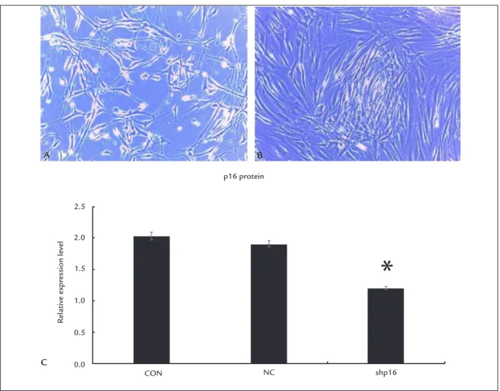
shp16 group. After p16 gene knockout, the DPCs mor-phology tended to fusiform growth, showing a tendency of aggregative growth (Figure 2A and B).
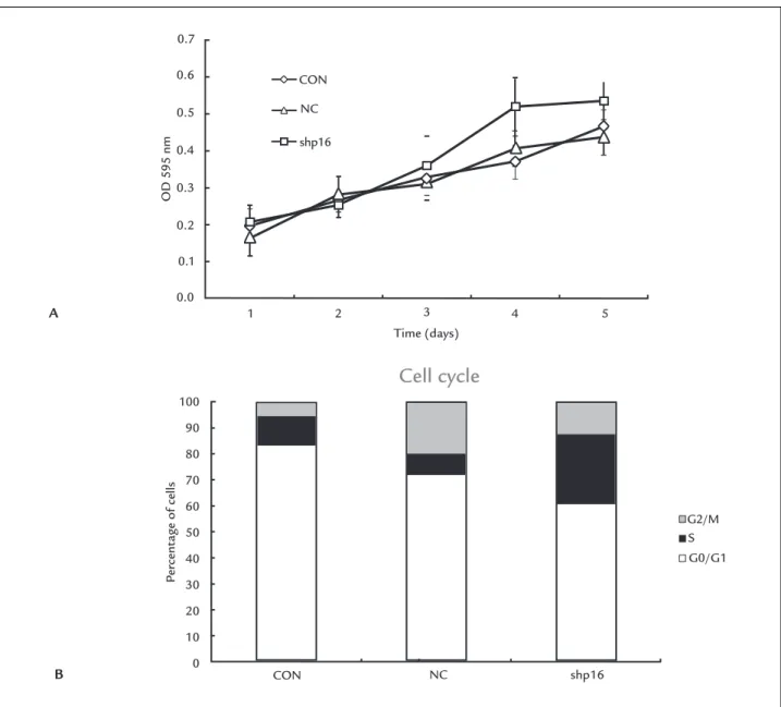
The expression of p16INK4a protein in cells was de-tected by FCM after transfection. The value of FI in each group is shown in Figure 2C. Expression of p16INK4a pro-tein in the shp16 group was signiicantly less than the NC (p<0.05) and CON (p<0.05) groups in the 8th genera-tion. There was no difference in the NC group when com-pared with the CON group in the 8th generation (p>0.05). The OD 595 nm value of cells from different groups from 1st to 5th day in 96 well plates are shown in Table 1 and growth curves were plotted from these values (Figure 3A). It was evident from these curves that cells grew more rapidly after silencing of the p16INK4a gene, but this effect was more obvious after day 3.
TABLE 1 DPC OD 595 nm value in different groups at different times (±S).
NC group
CON group
shp16 group
24h 0.196±0.030 0.164±0.039 0.206±0.047 48h 0.266±0.048 0.283±0.063 0.253±0.033 72h 0.325±0.027 0.314±0.064 0.359±0.081
96h 0.369±0.045 0.406±0.075 0.519±0.079
120h 0.465±0.057 0.437±0.070 0.536±0.052
Changes of the cell cycle in different groups were detect-ed by FCM (Figure 3B). The results revealdetect-ed a statistical-ly signiicant increase in cells in G1 phase, with a con-comitant reduction in cells in S phase after gene silencing.
FIGURE 2 A. DPCs of 8 generation without transfection. B. DPCs of 8 generation transfected by adenovirus vector. After p16 gene knockout, the DPCs morphology tended to fusiform growth, promoting a tendency of aggregative growth. C. p16INK4a protein expression detected by FCM in NC, CON
and shp16 groups. p16INK4a protein expression in shp16 group was signiicantly less than that in the NC and CON groups in the 8th generation (*p<0.05).
p16 protein
CON NC shp16
Relative e
xpr
ession level
2.5
2.0
1.5
1.0
0.5
0.0
A B
CON
1 2 3 4 5
Time (days)
Cell cycle
CON
NC
A
B
NC
shp16
G2/M
G0/G1
P
ercent
age of cells
OD 595 nm
S
shp16 100
90
80
70
60
50
40
30
20
10
0 0.7
0.6
0.5
0.4
0.3
0.2
0.1
0.0
FIGURE 3 A. The growth curves of different groups after transfection. DPCs grew more rapidly after silencing of the p16INK4a gene, and this
effect was more obvious after day 3. B. Cell cycle in NC, CON and shp16 groups detected by FCM is shown. Cells in G1 increased, while those in S phase reduced after gene silencing, the difference was statistically signiicant (p<0.05).
D
ISCUSSIONIn this study, we investigated whether p16INK4a has an effect in premature DPC senescence and aggregative behavior. We found that, compared with the aggregative group, the
expression of p16INK4a in the non-aggregative group in-creased as DPCs showed a gradual aging trend. DPCs grew more rapidly and exhibited a trend of aggregative growth when p16INK4a expression was silenced by rAd5-CDKN1A-1p2shRNA. Upon p16INK4a silencing, there was an increase in cells in G1 phase, with a simultaneous re-duction in cells in S phase. Our results show that p16INK4a
plays an important role in premature senescence and ag-gregative behavior of DPCs.
Previous studies showed that low-passage DPCs could sustain epidermal cell proliferation15 and hair growth-promoting capabilities,16,17 but high-passage DPCs could not. These evidences support the notion that non-aggre-gative DPCs may have a functional defect. The phenom-enon of gradual disappearance of the physiological func-tions of DPCs when aggregative growth characteristics disappear may be associated with changes in cytokines and cell senescence. A published work suggests that ex-pression of a series of cytokines including FGF7, IGF-1, SCF, VEGF etc. changed after different passages of DPCs.14,15 Additionally, research by Bahta et al.18 demon-strated that senescence of balding DPC is associated with increased expression of p16INK4a and pRb but not p53, suggesting that in vitro senescence of balding DPCs is stimulated by environmental stress and not due to repli-cative senescence. A similar observation has been docu-mented19 that oxidative stress as a result of passage-induce telomere shortening, and not replicative senescence, was responsible for the commonly observed senescence of dermal ibroblasts.20
Work by Yang et al.21 showed an increase in expression of p16INK4a in balding DPCs from AGA patients with pre-mature senescence, suggesting that androgen/androgen receptor signaling promotes senescence via the p16INK4a pathway in DPCs. We also found weak but gradually in-creasing expression of p16INK4a in 5, 6, 8 generation DPCs. p16INK4a protein could not be detected by Western blot, possibly owing to its weak expression. Our results suggest that p16INK4a may play an important role during senescence of DPCs and also in the transition from aggregative to non-aggregative growth of DPCs. In order to further clar-ify the biological characteristics of p16INK4a in DPCs, we constructed a shRNA adenovirus vector targeting p16INK4a and stably transfected DPCs. We found that p16INK4a pro-tein expression decreased signiicantly after silencing of the p16INK4a gene, along with an increase in cells in G1 phase and an accompanying reduction in cells in S phase, consistent with mechanism of p16INK4a in the cell cycle. There was a tendency for DPCs to show aggregative growth
after transfection, suggesting that non-aggregative growth is a form of cellular senescence, which can be inluenced by p16INK4a to a certain extent. The expression of
b-galactosidase, pRB and p53 remain to be detected in different passages and evaluated for their inluence on DPCs in the future work.
In conclusion, we present a potential link between aggregative growth of DPCs and p16INK4a, and to a certain degree reveal the mechanism of aggregative growth of DPCs. We also emphasize the beneicial use of knockdown
of the p16INK4a gene, which potentially contributes to the maintenance of aggregative growth and allows more pas-sages of DPCs. Furthermore, this knockdown may provide additional therapies for the treatment of AGA.
A
CKNOWLEDGMENTSThis work was supported by the Chinese Medical Asso-ciation – L’OREAL China hair grant.
R
EFERENCES1. Botchkarev VA, Kishimoto J. Molecular control of epithelial-mesenchymal interactions during hair follicle cycling. J Investig Dermatol Symp Proc. 2003; 8(1):46-55.
2. Roh C, Tao Q, Lyle S. Dermal papilla-induced hair differentiation of adult epithelial stem cells from human skin. Physiol Genomics. 2004; 19(2):207-17.
3. Matsuzaki T, Yoshizato K. Role of hair papilla cells on induction and regen-eration processes of hair follicles. Wound Repair Regen. 1998; 6(6):524-30. 4. Millar SE. Molecular mechanisms regulating hair follicle development. J
Invest Dermatol. 2002; 118(2):216-25.
5. Stenn KS, Cotsarelis G. Bioengineering the hair follicle: fringe beneits of stem cell technology. Curr Opin Biotechnol. 2005; 16(5):493-7.
6. Jahoda CA, Reynolds AJ, Oliver RF. Induction of hair growth in ear wounds by cultured dermal papilla cells. J Invest Dermatol. 1993; 101(4):584-90. 7. Ehama R, Ishimatsu-Tsuji Y, Iriyama S, Ideta R, Soma T, Yano K, et al. Hair
follicle regeneration using grafted rodent and human cells. J Invest Derma-tol. 2007; 127(9): 2106-15.
8. Ito Y, Hamazaki TS, Ohnuma K, Tamaki K, Asashima M, Okochi H. Isolation of murine hair-inducing cells using the cell surface marker prominin-1/ CD133. J Invest Dermatol. 2007; 127(5):1052-60.
9. Rayess H, Wang MB, Srivatsan ES. Cellular senescence and tumor suppressor gene p16. Int J Cancer. 2012; 130(8):1715-25.
10. Hara E, Smith R, Parry D, Tahara H, Stone S, Peters G. Regulation of p16CDKN2 expression and its implications for cell immortalization and senescence. Mol Cell Biol. 1996; 16(3):859-67.
11. Liu Y, Sanoff HK, Cho H, Burd CE, Torrice C, Ibrahim JG, et al. Expression of p16(INK4a) in peripheral blood T-cells is a biomarker of human aging. Aging Cell. 2009; 8(4):439-48.
12. Baker DJ, Wijshake T, Tchkonia T, LeBrasseur NK, Childs BG, van de Sluis B, et al. Clearance of p16Ink4a-positive senescent cells delays ageing-associated disorders. Nature. 2011; 479(7372):232-6.
13. Wu JJ, Liu RQ, Lu YG, Zhu TY, Cheng B, Men X. Enzyme digestion to isolate and culture human scalp dermal papilla cells: a more eficient method. Arch Dermatol Res. 2005; 297(2):60-7.
14. Giri H, Chandel S, Dwarakanath LS, Sreekumar S, Dixit M. Increased endothelial inlammation, sTie-2 and arginase activity in umbilical cords obtained from gestational diabetic mothers. PloS One. 2013; 8(12):e84546. 15. Reynolds AJ, Oliver RF, Jahoda CA. Dermal cell populations show variable competence in epidermal cell support: stimulatory effects of hair papilla cells. J Cell Sci. 1991; 98(Pt 1):75-83.
16. Jahoda CA, Horne KA, Oliver RF. Induction of hair growth by implantation of cultured dermal papilla cells. Nature. 1984; 311(5986):560-2. 17. Hong JB, Chiu HC, Chan JY, Chen RJ, Lin SJ. A woman with iatrogenic
an-drogenetic alopecia responding to finasteride. Br J Dermatol. 2007; 156(4):754-5.
18. Bahta AW, Farjo N, Farjo B, Philpott MP. Premature senescence of balding dermal papilla cells in vitro is associated with p16(INK4a) expression. J Invest Dermatol. 2008; 128(5):1088-94.
19. Upton JH, Hannen RF, Bahta AW, Farjo N, Farjo B, Philpott MP. Oxidative stress-associated senescence in dermal papilla cells of men with androgenetic alopecia. J Invest Dermatol. 2015; 135(5):1244-52.
20. 20. Itahana K, Dimri G, Campisi J. Regulation of cellular senescence by p53. Eur J Biochem. 2001; 268(10):2784-91.